Theulnaorulnar bone(pl.:ulnaeorulnas)[3]is along bonein theforearmstretching from theelbowto thewrist.It is on the same side of the forearm as the little finger, running parallel to theradius,the forearm's other long bone. Longer and thinner than the radius, the ulna is considered to be the smaller long bone of the lower arm. The corresponding bone in thelower legis thefibula.
| Ulna | |
|---|---|
 An example of a human ulna, shown in red (instandard anatomical position). | |
| Details | |
| Pronunciation | /ˈʌlnə/[1][2] |
| Identifiers | |
| Latin | ulna |
| MeSH | D014457 |
| TA98 | A02.4.06.001 |
| TA2 | 1230 |
| FMA | 23466 |
| Anatomical terms of bone | |
Structure
editThe ulna is a long bone found in the forearm that stretches from the elbow to the wrist, and when instandard anatomical position,is found on themedialside of the forearm. It is broader close to the elbow, and narrows as it approaches the wrist.
Close to the elbow, the ulna has a bonyprocess,theolecranon process,a hook-like structure that fits into theolecranon fossaof thehumerus.This preventshyperextensionand forms ahinge jointwith thetrochlea of the humerus.There is also aradial notchfor thehead of the radius,and theulnar tuberosityto which muscles attach.
Close to the wrist, the ulna has astyloid process.
Near the elbow
edit

Near the elbow, the ulna has two curved processes, theolecranonand thecoronoid process;and two concave, articular cavities, thesemilunarand radial notches.[4]
Theolecranonis a large, thick, curved eminence, situated at the upper and back part of the ulna. It is bent forward at the summit so as to present a prominent lip which is received into theolecranon fossa of the humerusin extension of the forearm. Its base is contracted where it joins the body and the narrowest part of the upper end of the ulna. Its posterior surface, directed backward, is triangular, smooth, subcutaneous, and covered by a bursa. Its superior surface is of quadrilateral form, marked behind by a rough impression for the insertion of thetriceps brachii;and in front, near the margin, by a slight transverse groove for the attachment of part of the posterior ligament of the elbow joint. Its anterior surface is smooth, concave, and forms the upper part of the semilunar notch. Its borders present continuations of the groove on the margin of the superior surface; they serve for the attachment of ligaments: the back part of the ulnar collateral ligament medially, and the posterior ligament laterally. From the medial border a part of theflexor carpi ulnarisarises; while to the lateral border theanconeusis attached.
Thecoronoid processis a triangular eminence projecting forward from the upper and front part of the ulna. Its base is continuous with the body of the bone, and of considerable strength. Its apex is pointed, slightly curved upward, and in flexion of the forearm is received into the coronoid fossa of the humerus. Its upper surface is smooth, concave, and forms the lower part of the semilunar notch. Its antero-inferior surface is concave, and marked by a rough impression for the insertion of thebrachialis.At the junction of this surface with the front of the body is a rough eminence, the tuberosity of the ulna, which gives insertion to a part of the brachialis; to the lateral border of this tuberosity the oblique cord is attached. Its lateral surface presents a narrow, oblong, articular depression, the radial notch. Its medial surface, by its prominent, free margin, serves for the attachment of part of theulnar collateral ligament.At the front part of this surface is a small rounded eminence for the origin of one head of theflexor digitorum superficialis;behind the eminence is a depression for part of the origin of theflexor digitorum profundus;descending from the eminence is a ridge which gives origin to one head of thepronator teres.Frequently, theflexor pollicis longusarises from the lower part of thecoronoid processby a rounded bundle of muscular fibers.
Thesemilunar notchis a large depression, formed by the olecranon and the coronoid process, and serving as articulation with the trochlea of the humerus. About the middle of either side of this notch is an indentation, which contracts it somewhat, and indicates the junction of the olecranon and the coronoid process. The notch is concave from above downward, and divided into a medial and a lateral portion by a smooth ridge running from the summit of the olecranon to the tip of the coronoid process. The medial portion is the larger, and is slightly concave transversely; the lateral is convex above, slightly concave below.
The radial notch is a narrow, oblong, articular depression on the lateral side of the coronoid process; it receives the circumferential articular surface of the head of theradius.It is concave from before backward, and its prominent extremities serve for the attachment of the annular ligament.
Body
editThebody of the ulnaat its upper part is prismatic in form, and curved so as to be convex behind and lateralward; its central part is straight; its lower part is rounded, smooth, and bent a little lateralward. It tapers gradually from above downward, and has three borders and three surfaces.
- Borders
- The volar border (margo volaris; anterior border) begins above at the prominent medial angle of thecoronoid process,and ends below in front of thestyloid process.Its upper part, well-defined, and its middle portion, smooth and rounded, give origin to theflexor digitorum profundus;its lower fourth serves for the origin of thepronator quadratus.This border separates the volar from the medial surface.
- The dorsal border (margo dorsalis; posterior border) begins above at the apex of the triangular subcutaneous surface at the back part of theolecranon,and ends below at the back of thestyloid process;it is well-marked in the upper three-fourths, and gives attachment to anaponeurosiswhich affords a common origin to theflexor carpi ulnaris,theextensor carpi ulnaris,and theflexor digitorum profundus;its lower fourth is smooth and rounded. This border separates the medial from the dorsal surface.
- The interosseous crest (crista interossea; external or interosseous border) begins above by the union of two lines, which converge from the extremities of the radial notch and enclose between them a triangular space for the origin of part of thesupinator;it ends below at the head of the ulna. Its upper part is sharp, its lower fourth smooth and rounded. This crest gives attachment to theinterosseous membrane,and separates the volar from the dorsal surface.
- Surfaces
- The volar surface (facies volaris; anterior surface), much broader above than below, is concave in its upper three-fourths, and gives origin to theflexor digitorum profundus;its lower fourth, also concave, is covered by thepronator quadratus.The lower fourth is separated from the remaining portion by a ridge, directed obliquely downward and medialward, which marks the extent of origin of the pronator quadratus. At the junction of the upper with the middle third of the bone is thenutrient canal,directed obliquely upward.
- The dorsal surface (facies dorsalis; posterior surface) directed backward and lateralward, is broad and concave above; convex and somewhat narrower in the middle; narrow, smooth, and rounded below. On its upper part is an oblique ridge, which runs from the dorsal end of the radial notch, downward to the dorsal border; the triangular surface above this ridge receives the insertion of theanconeus,while the upper part of the ridge affords attachment to the supinator. Below this the surface is subdivided by a longitudinal ridge, sometimes called the perpendicular line, into two parts: the medial part is smooth, and covered by theextensor carpi ulnaris;the lateral portion, wider and rougher, gives origin from above downward to the supinator, theabductor pollicis longus,theextensor pollicis longus,and theextensor indicis proprius.
- The medial surface (facies medialis; internal surface) is broad and concave above, narrow and convex below. Its upper three-fourths give origin to theflexor digitorum profundus;its lower fourth is subcutaneous.
Near the wrist
edit
Near the wrist, the ulnar, with two eminences; the lateral and larger is a rounded, articular eminence, termed the head of the ulna; the medial, narrower and more projecting, is a non-articular eminence, theulnar styloid process.
- Theheadof the ulna presents an articular surface, part of which, of an oval or semilunar form, is directed downward, and articulates with the upper surface of thetriangular articular diskwhich separates it from the wrist-joint; the remaining portion, directed lateralward, is narrow, convex, and received into theulnar notch of the radius.
- The styloid process projects from the medial and back part of the bone; it descends a little lower than the head, and its rounded end affords attachment to the ulnar collateral ligament of the wrist-joint.
The head is separated from the styloid process by a depression for the attachment of the apex of the triangular articular disk, and behind, by a shallow groove for the tendon of theextensor carpi ulnaris.
-
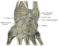 Vertical section through the articulations at the wrist, showing the synovial cavities.
 -
 Bones of left forearm. Anterior aspect.
 -
 Bones of left forearm. Posterior aspect.
 -
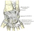 Ligaments of wrist. Anterior view
 -
 Ligaments of wrist. Posterior view.


Microanatomy
editThe ulna is along bone.The long, narrowmedullary cavityof the ulna is enclosed in a strong wall ofcortical tissuewhich is thickest along theinterosseous borderand dorsal surface. At the extremities the compact layer thins. The compact layer is continued onto the back of the olecranon as a plate of close spongy bone with lamellae parallel. From the inner surface of this plate and the compact layer below it trabeculae arch forward toward the olecranon and coronoid and cross other trabeculae, passing backward over the medullary cavity from the upper part of the shaft below the coronoid. Below the coronoid process there is a small area of compact bone from which trabeculae curve upward to end obliquely to the surface of the semilunar notch which is coated with a thin layer of compact bone. The trabeculae at the lower end have a more longitudinal direction.[5]
Development
edit
The ulna isossifiedfrom three centers: one each for the body, the wrist end, and the elbow end, near the top of theolecranon. Ossification begins near the middle of the body of the ulna, about the eighth week of fetal life, and soon extends through the greater part of the bone.
At birth, the ends are cartilaginous. About the fourth year or so, a center appears in the middle of the head, and soon extends into theulnar styloid process.About the tenth year, a center appears in the olecranon near its extremity, the chief part of this process being formed by an upward extension of the body. The upperepiphysisjoins the body about the sixteenth, the lower about the twentieth year.
Function
editJoints
editThe ulna forms part of thewristjoint andelbowjoints. Specifically, the ulna joins (articulates) with:
- trochlea of thehumerus,at the right sideelbowas a hinge joint with semilunartrochlear notchof the ulna.
- theradius,near the elbow as apivot joint,this allows the radius to cross over the ulna inpronation.
- thedistal radius,where it fits into the ulnar notch.
- the radius along its length via theinterosseous membranethat forms asyndesmosisjoint
Muscle attachments
edit

| Muscle | Direction | Attachment |
| Triceps brachii muscle | Insertion | posterior part of superior surface ofOlecranon process(via common tendon) |
| Anconeus muscle | Insertion | olecranon process (lateral aspect) |
| Brachialis muscle | Insertion | anterior surface of thecoronoid process of the ulna |
| Pronator teres muscle | Origin | medial surface on middle portion of coronoid process (also shares origin withmedial epicondyle of the humerus) |
| Flexor carpi ulnaris muscle | Origin | olecranon process and posterior surface of ulna (also shares origin with medial epicondyle of the humerus) |
| Flexor digitorum superficialis muscle | Origin | coronoid process (also shares origin with medial epicondyle of the humerus and shaft of theradius) |
| Flexor digitorum profundus muscle | Origin | anteromedial surface of ulna (also shares origin with theinterosseous membrane) |
| Pronator quadratus muscle | Origin | distalportion ofanteriorulnar shaft |
| Extensor carpi ulnaris muscle | Origin | posteriorborder of ulna (also shares origin withlateral epicondyle of the humerus) |
| Supinator muscle | Origin | proximalulna (also shares origin with lateral epicondyle of the humerus) |
| Abductor pollicis longus muscle | Origin | posterior surface of ulna (also shares origin with the posterior surface of theradius bone) |
| Extensor pollicis longus muscle | Origin | dorsalshaft of ulna(also shares origin with the dorsal shaft of the radius and the interosseous membrane) |
| Extensor indicis muscle | Origin | posterior surface of distal ulna (also shares origin with the interosseous membrane) |
Clinical significance
editFractures
editSpecific types ofulna fractureinclude:
- Monteggia fracture- a fracture of the proximal third of the ulna with the dislocation of thehead of the radius
- Hume fracture- a fracture of theolecranonwith an associatedanteriordislocationof theradial head
Conservative managementis possible for ulnar fractures when they are located in the distal two-thirds, only involve the shaft, with no shortening, less than 10° angulation and less than 50% displacement.[6]In such cases, acastshould be applied that goes above the elbow.[6]
Other animals
edit
In four-legged animals, the radius is the main load-bearing bone of the lower forelimb, and the ulna is important primarily for muscular attachment. In many mammals, the ulna is partially or wholly fused with the radius, and may therefore not exist as a separate bone. However, even in extreme cases of fusion, such as inhorses,the olecranon process is still present, albeit as a projection from the upper radius.[7]
Inbirdsand otherdinosaurs,the ulna forms a surface of attachment for thesecondary feathers.These often leave osteological evidence in the form of quill knobs, allowing for identification of feathers in fossils that otherwise lack integumentary information.[8]
Gallery
edit-
 Position of ulna (red). Animation
 -
 3D image
 -
 Bones of the right arm, showing the ulna, radius, wrist and humerus
 -
 Cross-section through the middle of the forearm, showing the two bones and the muscles, nerves and blood vessels surrounding them.
 -
Ulna anatomy


See also
editReferences
edit![]() This article incorporates text in thepublic domainfrompage 214of the 20th edition ofGray's Anatomy(1918)
This article incorporates text in thepublic domainfrompage 214of the 20th edition ofGray's Anatomy(1918)
- ^OED2nd edition, 1989.
- ^Entry "ulna"inMerriam-Webster Online Dictionary.
- ^"ULNA | Meaning & Definition for UK English".Lexico.com. Archived fromthe originalon January 20, 2021.Retrieved2022-08-24.
- ^"Radius and ulna".Kenhub.Retrieved2024-08-21.
- ^"Ulna".InnerBody.
- ^abSebastian Dawson-Bowling; Pramod Achan; Timothy Briggs; Manoj Ramachandran (2014).Orthopaedic Trauma: The Stanmore and Royal London Guide.CRC Press.ISBN9781444148831.Page 158
- ^Romer, Alfred Sherwood; Parsons, Thomas S. (1977).The Vertebrate Body.Philadelphia, PA: Holt-Saunders International. p. 200.ISBN0-03-910284-X.
- ^Turner, Alan H.; Makovicky, Peter J.; Norell, Mark A. (2007-09-21)."Feather Quill Knobs in the Dinosaur Velociraptor"(PDF).Science.317(5845): 1721.Bibcode:2007Sci...317.1721T.doi:10.1126/science.1145076.ISSN0036-8075.PMID17885130.S2CID11610649.