Amyloidosisis a group of diseases in which abnormalproteins,known asamyloid fibrils,build up in tissue.[4]There are several non-specific and vague signs and symptoms associated with amyloidosis.[5]These include fatigue,peripheral edema,weight loss,shortness of breath,palpitations,andfeeling faint with standing.[5]In AL amyloidosis, specific indicators can include enlargement of the tongue and periorbitalpurpura.[5]In wild-type ATTR amyloidosis, non-cardiac symptoms include: bilateralcarpal tunnel syndrome,lumbarspinal stenosis,biceps tendon rupture,small fiber neuropathy,andautonomic dysfunction.[5]
| Amyloidosis | |
|---|---|
 | |
| Amyloidosis symptoms are often vague and require different physician specialists for diagnosis. Telltale symptoms may include an enlarged tongue (macroglossia) or bruising around the eyes (purpura)[1] | |
| Specialty | Internal medicine |
| Symptoms | Feeling tired, weight loss, swelling of the legs, shortness of breath, bleeding, feeling light headed with standing[2] |
| Usual onset | 55–65 years old[2] |
| Causes | Genetic or acquired[3] |
| Diagnostic method | Tissue biopsy[2] |
| Treatment | Supportive care,directed at the underlying cause,dialysis,organ transplantation[3] |
| Prognosis | Improved with treatment[3] |
| Frequency | 3–13 per million per year (AL amyloidosis)[2] |
| Deaths | 1 per 1,000 people (developed world)[3] |
There are about 36 different types of amyloidosis, each due to a specificprotein misfolding.[6]Within these 36 proteins, 19 are grouped intolocalized forms,14 are grouped assystemic forms,and three proteins can identify as either.[6]These proteins can become irregular due to genetic effects, as well as through acquiredenvironmental factors.[6]The four most common types of systemic amyloidosis arelight chain (AL),inflammation (AA),dialysis-related(Aβ2M), and hereditary and old age (ATTRandwild-type transthyretin amyloid[7]).[2]
Diagnosis may be suspected whenprotein is found in the urine,organ enlargementis present, orproblems are found with multiple peripheral nervesand it is unclear why.[2]Diagnosis is confirmed bytissue biopsy.[2]Due to the variable presentation, a diagnosis can often take some time to reach.[3]
Treatment is geared towards decreasing the amount of the involved protein.[2]This may sometimes be achieved by determining and treating the underlying cause.[2]AL amyloidosis occurs in about 3–13 per million people per year and AA amyloidosis in about two per million people per year.[2]The usual age of onset of these two types is 55 to 60 years old.[2]Without treatment, life expectancy is between six months and four years.[2]In thedeveloped worldabout one per 1,000 deaths are from systemic amyloidosis.[3]Amyloidosis has been described since at least 1639.[2]
Signs and symptoms
edit
The presentation of amyloidosis is broad and depends on the site of amyloid accumulation. The kidney and heart are the most common organs involved.
Kidneys
editAmyloiddeposition in thekidneyoften involve theglomerular capillariesandmesangial regions,affecting the organ's ability to filter and excrete waste and retainplasma protein.[8]This can lead to high levels of protein in the urine (proteinuria) andnephrotic syndrome.[8]Several types of amyloidosis, including the AL and AA types, are associated withnephrotic syndrome.[9]Approximately 20% and 40–60% of people with AL and AA amyloidosis respectively progress toend-stage kidney diseaserequiringdialysis.[9]
Heart
editAmyloid deposition in the heart can cause both diastolic and systolicheart failure.EKGchanges may be present, showing low voltage and conduction abnormalities likeatrioventricular blockorsinus nodedysfunction.[medical citation needed]Onechocardiography,the heart shows a restrictive filling pattern, with normal to mildly reduced systolic function.[10]AA amyloidosis usually spares the heart.[11]Cardiac amyloidosis can present with symptoms of heart failure including shortness of breath, fatigue, and edema.[12]As cardiac amyloidosis progresses, the amyloid deposition can affect the heart's ability to pump and fill blood as well as its ability to maintain normal rhythm, which leads to worsening heart function and decline in people's quality of life.[12]
Nervous system
editPeople with amyloidosis may have central nervous system involvement,[13]along with peripheral involvement which causes sensory and autonomic neuropathies. Sensory neuropathy develops in a symmetrical pattern and progresses in a distal to proximal manner. Autonomic neuropathy can present asorthostatic hypotensionbut may manifest more gradually with nonspecific gastrointestinal symptoms like constipation, nausea, or early satiety.[10]Amyloidosis of the central nervous system can have more severe and systemic presentations that may include life-threatening arrhythmias, cardiac failure, malnutrition, infection, or death.[14]
Neuropathic presentation can depend on the etiology of amyloidosis.[14]People with amyloidosis may experience dysfunction in various organ systems depending on the location and extent of nervous system involvement.[8]For example, peripheral neuropathy can cause erectile dysfunction, incontinence and constipation, pupillary dysfunction, and sensory loss depending on the distribution of amyloidosis along different peripheral nerves.[14]
Gastrointestinal and accessory organs
editAccumulation of amyloid proteins in the gastrointestinal system may be caused by a wide range of amyloid disorders and have different presentations depending on the degree of organ involvement.[15]Potential symptoms include weight loss, diarrhea, abdominal pain, heartburn (gastrointestinal reflux), and GI bleeding.[15]Amyloidosis may also affect accessory digestive organs including the liver, and may present with jaundice, fatty stool, anorexia, fluid buildup in the abdomen, and spleen enlargement.[15]
Accumulation of amyloid proteins in the liver can lead to elevations in serumaminotransferasesandalkaline phosphatase,two biomarkers of liver injury, which is seen in about one third of people.[11]Liver enlargementis common. In contrast,spleen enlargementis rare, occurring in 5% of people.[10]Splenic dysfunction, leading to the presence ofHowell-Jolly bodieson blood smear, occurs in 24% of people with amyloidosis.[10]Malabsorptionis seen in 8.5% ofAL amyloidosisand 2.4% of AA amyloidosis. One suggested mechanism for the observed malabsorption is that amyloid deposits in the tips ofintestinal villi(fingerlike projections that increase the intestinal area available for absorption of food), begin to erode the functionality of the villi, presenting asprue-like picture.[11]
Glands
editBoth thethyroidandadrenal glandscan be infiltrated. It is estimated that 10–20% of people with amyloidosis havehypothyroidism.Adrenal infiltration may be harder to appreciate given that its symptoms of orthostatic hypotension and low blood sodium concentration may be attributed toautonomic neuropathyand heart failure.[10]
"Amyloid deposits occur in thepancreasof people who also havediabetes mellitus,although it is not known if this is functionally important. The major component of pancreatic amyloid is a 37-amino acid residue peptide known asislet amyloid polypeptideor 'amylin.' This is stored with insulin in secretory granules in[beta] cellsand is co secreted with insulin. "(Rang and Dale's Pharmacology, 2015.)[citation needed]
Musculoskeletal system
editAmyloid proteins deposit most commonly inside the knee, followed by hands, wrists, elbow, hip, and ankle, causing joint pain.[16]In males with advanced age (>80 years), there is significant risk of wild-type transthyretin amyloid deposition in synovial tissue of knee joint, but predominantly in old age deposition of wild type transthyretin is seen in cardiac ventricles. ATTR deposits have been found inligamentum flavumof patients that underwent surgery forlumbar spinal stenosis.[17]
In beta 2-microglobulin amyloidosis, males have high risk of gettingcarpal tunnel syndrome.[18]Aβ2MG amyloidosis (Hemodialysis associated amyloidosis) tends to deposit in synovial tissue, causing chronicinflammation of the synovial tissuein knee, hip, shoulder and interphalangeal joints.[18]Amyloid light chains deposition in shoulder joint causes enlarged shoulders, also known as "shoulder pad sign".[18]Amyloid light chain depositions can also cause bilateral symmetric polyarthritis.[18]
The deposition of amyloid proteins in the bone marrow without causingplasma cell dyscrasiasis called amyloidoma. It is commonly found in cervical, lumbar, and sacral vertebrae. Those affected may be presented with bone pain due to bone lysis, lumbarparaparesis,and a variety of neurological symptoms. Vertebral fractures are also common.[18]
Eyes
editA rare development isamyloid purpura,a susceptibility to bleeding with bruising around the eyes, termed "raccoon-eyes". Amyloid purpura is caused by amyloid deposition in the blood vessels and reduced activity ofthrombinandfactor X,two clotting proteins that lose their function after binding with amyloid.[10]
Oral cavity
editAmyloid deposits in tissue can cause enlargement of structures. Twenty percent of people with AL amyloidosis have anenlarged tongue,that can lead toobstructive sleep apnea,difficulty swallowing,and altered taste.[11]Tongue enlargement does not occur in ATTR or AA amyloidosis.[10]Deposition of amyloid in the throat can cause hoarseness.[10]
Pathogenesis
editAmyloidoses can be consideredprotein misfoldingdiseases.[19][20]The vast majority of proteins that have been found to form amyloid deposits aresecreted proteins,so the misfolding and formation of amyloid occurs outside cells, in theextracellularspace.[19]Of the 37 proteins so far identified as being vulnerable to amyloid formation, only four arecytosolic.[19]Most amyloid-forming proteins are relatively small, but otherwise there is currently no evidence of structural or functional similarities among proteins known to form disease-associated amyloids.[19]One third of amyloid disease is hereditary, in which case there is normally an early age of onset.[19]Half of amyloid-related diseases are sporadic and have a late age of onset – in these cases, the protein aggregation may be associated with aging-related decline in protein regulation. Some medical treatments are associated with amyloid disease, but this is rare.[19]
Amyloid-forming proteins aggregate into distinctive fibrillar forms with abeta-sheetstructure.[19][20]The beta-sheet form of amyloid isproteolysis-resistant, meaning it can not be degraded or broken down.[5]As a result, amyloid deposits into the body's extracellular space.[5]The process of forming amyloid fibrils is thought to have intermediateoligomericforms. Both the oligomers and amyloid fibrils can be toxic to cells and can interfere with proper organ function.[21]The relative significance of different aggregation species may depend on the protein involved and the organ system affected.[20]
Diagnosis
editDiagnosis of amyloidosis generally requires tissue biopsy.[2]The biopsy is assessed for evidence of characteristic amyloid deposits. The tissue is treated with variousstains.The most useful stain in the diagnosis of amyloid isCongo red,which, combined withpolarized light,makes the amyloid proteins appear apple-green onmicroscopy.Also,thioflavin Tstain may be used.[22]A number of imaging techniques such as a Nuclear Medicine PYP scan,DPD scanorSAP scanare also in use.[23]
A sample of tissue can be biopsied or obtained directly from the affected internal organ, but the first-line site of biopsy issubcutaneous abdominal fat,known as a "fat pad biopsy", due to its ease of acquisition.[24][25]An abdominal fat biopsy is not completelysensitiveand may result infalse negatives,which means a negative result does not exclude the diagnosis of amyloidosis.[24][25]However, direct biopsy of the affected organ may still be unnecessary as other less invasive methods of biopsy can also be used, including rectal mucosa, salivary gland, lip, or bone marrow biopsy which can achieve a diagnosis in up to 85% of people.[24]
In the amyloid deposition of the joints, there will be a decreased signal in bothT1 and T2 weighted MRI images.[16]In amyloidoma, there will be low T1 signal with gadolinium injection and low T2 signal.[18]
The type of the amyloid protein can be determined in various ways: the detection of abnormal proteins in the bloodstream (onprotein electrophoresisor light chain determination); binding of particular antibodies to the amyloid found in the tissue (immunohistochemistry); or extraction of the protein and identification of its individualamino acids.[22]Immunohistochemistry can identify AA amyloidosis the majority of the time, but can miss many cases of AL amyloidosis.[11]Laser microdissectionwithmass spectrometryis the most reliable method of identifying the different forms of amyloidosis.[26]
AL was previously considered the most common form of amyloidosis, and a diagnosis often begins with a search forplasma cell dyscrasia,memory B cells producing aberrant immunoglobulins or portions of immunoglobulins. Immunofixation electrophoresis of urine or serum is positive in 90% of people with AL amyloidosis.[10]Immunofixation electrophoresis is more sensitive than regular electrophoresis but may not be available in all centers. Alternatively immunohistochemical staining of a bone marrow biopsy looking for dominant plasma cells can be sought in people with a high clinical suspicion for AL amyloidosis but negative electrophoresis.[10]
ATTR is now considered to be the most common form of amyloidosis. It may be either age related in wild-type ATTR (ATTRv) or familial transthyretin-associated amyloidosis, is suspected in people with family history of idiopathic neuropathies or heart failure who lack evidence of plasma cell dyscrasias. ATTR can be identified usingisoelectric focusingwhich separates mutated forms of transthyretin. Findings can be corroborated bygenetic testingto look for specific known mutations in transthyretin that predispose to amyloidosis.[10]
AA is suspected on clinical grounds in individuals with longstanding infections or inflammatory diseases. AA can be identified by immunohistochemistry staining.[10]
-
 Small bowel duodenum with amyloid deposition Congo red 10X
 -
 Amyloidosis,dystrophic calcification
 -
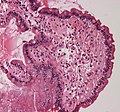 Small bowel duodenum with amyloid deposition 20X
 -
 Amyloidosis, Node, Congo Red
 -
 Amyloidosis, blood vessels, H&E
 -
 Amyloidosis, lymph node, H&E
 -
 Amyloidosis, lymph node, polarizer
 -

-
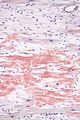 Micrographshowing amyloid deposition (red fluffy material) in theheart(cardiac amyloidosis).Congo red stain.


Classification
editHistorical classification systems were based on clinical factors. Until the early 1970s, the idea of a single amyloid substance predominated. Various descriptive classification systems were proposed based on the organ distribution of amyloid deposits and clinical findings. Most classification systems included primary (i.e.,idiopathic) amyloidosis, in which no associated clinical condition was identified, and secondary amyloidosis (i.e.,secondaryto chronic inflammatory conditions). Some classification systems included myeloma-associated, familial, and localized amyloidosis.[citation needed]
The modern era of amyloidosis classification began in the late 1960s with the development of methods to make amyloid fibrils soluble. These methods permitted scientists to study the chemical properties of amyloids.[medical citation needed]Descriptive terms such as primary amyloidosis, secondary amyloidosis, and others (e.g., senile amyloidosis), which are not based on cause, provide little useful information and are no longer recommended.
The modern classification of amyloid disease tends to use an abbreviation of the protein that makes the majority of deposits, prefixed with the letter A. For example, amyloidosis caused bytransthyretinis termed "ATTR".[medical citation needed]Deposition patterns vary between people but are almost always composed of just one amyloidogenic protein. Deposition can besystemic(affecting many different organ systems) or organ-specific. Many amyloidoses areinherited,due tomutationsin the precursor protein.[medical citation needed]
Other forms are due to different diseases causing overabundant or abnormal protein production – such as with overproduction ofimmunoglobulin light chains(termedAL amyloidosis), or with continuous overproduction ofacute phase proteinsinchronic inflammation(which can lead toAA amyloidosis).[medical citation needed]
About 60 amyloid proteins have been identified so far.[27]Of those, at least 36 have been associated with a human disease.[28]
All amyloid fibril proteins start with the letter "A" followed by the protein suffix (and any applicable specification). See below for a list of amyloid fibril proteins which have been found in humans:[29]
| Fibril protein | Precursor protein | Target Organs | Systemic and/or localized | Acquired or hereditary |
|---|---|---|---|---|
| AL | Immunoglobulin light chain | All organs, usually exceptCNS | S, L | A, H |
| AH | Immunoglobulin heavy chain | All organs exceptCNS | S, L | A |
| AA | (Apo) serum amyloid A | All organs exceptCNS | S | A |
| ATTR | Transthyretin,wild type
Transthyretin,variants |
Heart mainly in males, lung, ligaments, tenosynovium
PNS, ANS, heart, eye, leptomeninges |
S
S |
A
H |
| Aβ2M | β2-microglobulin,wild type
β2-microglobulin,variants |
Musculoskeletal system
ANS |
S
S |
A
H |
| AApoAI | Apolipoprotein A I,variants | Heart, liver, kidney, PNS, testis, larynx (C
terminal variants), skin (C terminal variants) |
S | H |
| AApoAII | Apolipoprotein A II,variants | Kidney | S | H |
| AApoAIV | Apolipoprotein A IV, wild type | Kidney medulla and systemic | S | A |
| AApoCII | Apolipoprotein C II, variants | Kidney | S | H |
| AApoCIII | Apolipoprotein C III, variants | Kidney | S | H |
| AGel | Gelsolin, variants | Kidney, PNS, cornea | S | H |
| ALys | Lysozyme, variants | Kidney | S | H |
| ALECT2 | Leukocyte chemotactic factor-2 | Kidney, primarily | S | A |
| AFib | Fibrinogen a, variants | Kidney, primarily | S | H |
| ACys | Cystatin C, variants | CNS,PNS, skin | S | H |
| ABri | ABriPP, variants | CNS | S | H |
| ADanb | ADanPP, variants | CNS | L | H |
| Aβ | Aβ protein precursor, wild type
Aβ protein precursor, variant |
CNS | L
L |
A
H |
| AαSyn | α-Synuclein | CNS | L | A |
| ATau | Tau | CNS | L | A |
| APrP | Prion protein, wild type
Prion protein variants Prion protein variant |
CJD, fatal insomnia
CJD, GSS syndrome, fatal insomnia PNS |
L
L S |
A
H H |
| ACal | (Pro)calcitonin | C-cell thyroid tumours
Kidney |
L
S |
A
A |
| AIAPP | Islet amyloid polypeptidec | Islets of Langerhans, insulinomas | L | A |
| AANF | Atrial natriuretic factor | Cardiac atria | L | A |
| APro | Prolactin | Pituitary prolactinomas, aging pituitary | L | A |
| AIns | Insulin | Iatrogenic, local injection | L | A |
| ASPCd | Lung surfactant protein | Lung | L | A |
| ACor | Corneodesmosin | Cornified epithelia, hair follicles | L | A |
| AMed | Lactadherin | Senile aortic, media | L | A |
| AKer | Kerato-epithelin | Cornea, hereditary | L | A |
| ALac | Lactoferrin | Cornea | L | A |
| AOAAP | Odontogenic ameloblast-associated protein | Odontogenic tumours | L | A |
| ASem1 | Semenogelin 1 | Vesicula seminalis | L | A |
| AEnf | Enfurvitide | Iatrogenic | L | A |
| ACatKe | Cathepsin K | Tumour associated | L | A |
| AEFEMP1e | EGF-containing fibulin-like extracellular
matrix protein 1 (EFEMP1) |
Portal veins, Aging associated | L | A |
Alternative
editAn older clinical method of classification refers to amyloidoses as systemic or localised:
- Systemic amyloidoses affect more than one body organ or system. Examples are AL, AA and Aβ2m.[30]
- Localised amyloidoses affect only one body organ or tissue type. Examples areAβ,IAPP,Atrial natriuretic factor(inisolated atrial amyloidosis), andCalcitonin(inmedullary carcinoma of the thyroid)[30]
Another classification is primary or secondary.[medical citation needed]
- Primary amyloidosesarise from a disease with disordered immune cell function, such asmultiple myelomaor other immunocyte dyscrasias.
- Secondary (reactive) amyloidoses occur as a complication of some other chronic inflammatory or tissue-destroying disease. Examples arereactive systemic amyloidosisandsecondary cutaneous amyloidosis.[30]
Additionally, based on the tissues in which it is deposited, it is divided into mesenchymal (organs derived frommesoderm) or parenchymal (organs derived fromectodermorendoderm).[medical citation needed]
Treatment
editTreatment depends on the type of amyloidosis that is present. Treatment with high dosemelphalan,achemotherapyagent, followed bystem celltransplantation has shown promise in early studies and is recommended for stage I and II AL amyloidosis.[26]However, only 20–25% of people are eligible for stem cell transplant. Chemotherapy treatment includingcyclophosphamide-bortezomib-dexamethasone-daratumumab(Dara-Cybord) is currently the recommended treatment option for people with AL Amyloidosis not eligible for transplant.[5][31]
In AA, symptoms may improve if the underlying condition is treated. In people who have inflammation caused by AA amyloidosis,tumour necrosis factor (TNF)-alpha inhibitorssuch asinfliximabandetanerceptare used for an average duration of 20 months. If TNF-alpha inhibitors are not effective,Interleukin-1inhibitors (e.g.,anakinra,canakinumab,rilonacept) andinterleukin-6inhibitors (e.g., tocilizumab) may be considered.[32]
Management of ATTR amyloidosis will depend on its classification as wild type or variant.[5]Both may be treated withtafamidis,a low toxicity oral agent that prevents destabilization of correctly folded protein.[5]Studies showed tafamidis reduced mortality and hospitalization due toheart failure.[5]Previously, for variant ATTR amyloidosis,liver transplantwas the only effective treatment.[5]New therapies includediflunisal,inotersen,andpatisiran.
Diflunisal binds to misfolded mutant TTR protein to prevent its buildup, like how tafamidis works. Low-certainty evidence indicates that it mitigates worsening ofperipheral neuropathyand disability from disease progression.[33]
Inotersen blocks gene expression of both wild-type and mutant TTR, reducing amyloid precursor. Moderate-certainty evidence suggests that it mitigates worsening of peripheral neuropathy. Long-term efficacy and safety of inotersen use in people with mutant TTR-related amyloidosis is still be evaluated in a phase-III clinical trial as of 2021. Both diflunisal and inotersen may also mitigate declines in quality-of-life, though the evidence for this effect is unclear.[33]For people with cardiac ATTR the effect of inotersen use is inconclusive and requires further investigation.[34]In 2018,inotersenwas approved by the European Medicines Agency to treat polyneuropathy in adults with hereditary transthyretin amyloidosis.[35]It has since been approved for use in Canada, the European Union and in the USA.[36]
Patisiranfunctions similarly to inotersen. Moderate-certainty evidence suggests that patisiran mitigates worsening of peripheral neuropathy and disability from disease progression. Additionally, low-certainty evidence suggests that patisiran mitigates decreases in quality-of-life and slightly reduces the rate of adverse events versus placebo. There is no evidence of an effect on mortality rate.[33]A review of early data from use of patisiran in people with variant cardiac ATTR suggests that it may reduce mortality and hospitalization, however this is still being investigated and requires further investigation.[34]In 2018, patisiran was not recommended by NICE in the UK for hereditary transthyretin-related amyloidosis.[37]As of July 2019 further review however is occurring.[38]It was approved for this use in the United States, however.[39]
The roles of inotersen and patisiran in cardiac ATTR amyloidosis are still being investigated.[5]
In 2021, in a clinical trial using theCRISPRgene-editing technique, several participants had an "80% to 96% drop in TTR levels, on par or better than the average of 81%" who were given patisiran.[40]
Vutrisiranwas approved by the U.S.Food and Drug Administration(FDA) in June 2022, for the treatment of the polyneuropathy of hereditary transthyretin-mediated (hATTR) amyloidosis in adults.[41]
Support groups
editPeople affected by amyloidosis are supported by organizations, including the Amyloidosis Research Consortium, Amyloidosis Foundation, Amyloidosis Support Groups, and Australian Amyloidosis Network.[42][43]
Prognosis
editPrognosis varies with the type of amyloidosis and the affected organ system. Prognosis for untreated AL cardiac amyloidosis is poor, with a median survival of six months.[44]More specifically, AL amyloidosis can be classified as stage I, II or III based on cardiac biomarkers like Nt-proBNP and cardiac troponin.[45]Survival diminishes with increasing stage, but recent advancements in treatments have improved median survival rates for stages I, II, and III, to 91.2, 60, and 7 months respectively.[45]
Outcomes in a person with AA amyloidosis depend on the underlying disease, organ(s) affected, and correlate with the concentration of serum amyloid A protein.[5]
People with ATTR, mutant ATTR and wild-type ATTR have a better prognosis when compared to people with AL and may survive for over a decade.[10][46]Survival time is not associated with gender or age, however, some measures of reduced heart function are associated with a shorter survival time.[46]
Senile systemic amyloidosis was determined to be the primary cause of death for 70% ofpeople over 110who have beenautopsied.[47][48]
Epidemiology
editAmyloidosis has a combined estimated prevalence of 30 per 100,000 persons with the three most common forms being AL, ATTR, and AA.[49]The median age at diagnosis is 64.[11]
AL has the highest incidence at approximately 12 cases per million persons per year and an estimated prevalence of 30,000 to 45,000 cases in the US and European Union.[49][5]
AA amyloidoses is the most common form in developing countries and can complicate longstanding infections withtuberculosis,osteomyelitis,andbronchiectasis.AA amyloidosis is caused by an increase in extracellular deposition of serum amyloid A (SAA) protein. SAA protein levels can rise in both direct and indirect manners, through infection, inflammation, and malignancies.[50]The most common causes of AA amyloidosis in the West are rheumatoid arthritis, inflammatory bowel disease, psoriasis, andfamilial Mediterranean fever.[10]
People undergoing long-term hemodialysis (14–15 years) can develop amyloidosis from accumulation of light chains of the HLA 1 complex which is normally filtered out by the kidneys.[11]
Wild-type transthyretin (ATTR) amyloidosis is found in a quarter of elderly at postmortem.[51]ATTR is found in 13–19% of people experiencingheart failurewith preserved ejection fraction, making it a very common form of systemic amyloidosis.[52]
Research
editTreatments for ATTR-relatedneuropathyinclude TTR-specificoligonucleotidesin the form ofsmall interfering RNA(patisiran) orantisenseinotersen,[53]the former having recently received FDA approval.[54]Research into treatments for ATTR amyloidosis have compared liver transplantation, oral drugs that stabilize the misfolding protein (including tafamidis and diflunisal), and newer therapeutic agents still being investigated (including patisiran).[55]
Based on available research, liver transplant remains the most effective treatment option for advanced ATTR amyloidosis, protein stabilizing drugs may slow disease progression but were insufficient to justify delay of liver transplant, and newer agents such as patisiran require additional studies.[55]
See also
editReferences
edit- ^"Amyloidosis Awareness"(PDF).amyloidaware.com.Amyloidosis Support Groups. 1 March 2022.Retrieved15 June2024.
- ^abcdefghijklmnHazenberg BP (May 2013)."Amyloidosis: a clinical overview"(PDF).Rheumatic Disease Clinics of North America.39(2):323–345.doi:10.1016/j.rdc.2013.02.012.PMID23597967.S2CID215069282.
- ^abcdefPepys MB (2006). "Amyloidosis".Annual Review of Medicine.57:223–241.doi:10.1146/annurev.med.57.121304.131243.PMID16409147.
- ^"AL amyloidosis".rarediseases.info.nih.gov.Genetic and Rare Diseases Information Center (GARD).Archivedfrom the original on 24 April 2017.Retrieved22 April2017.
- ^abcdefghijklmnGertz MA, Dispenzieri A (July 2020). "Systemic Amyloidosis Recognition, Prognosis, and Therapy: A Systematic Review".JAMA.324(1):79–89.doi:10.1001/jama.2020.5493.PMID32633805.S2CID220385853.
- ^abcPicken MM (2020)."The Pathology of Amyloidosis in Classification: A Review".Acta Haematologica.143(4):322–334.doi:10.1159/000506696.PMID32392555.S2CID218600304.
- ^Ando Y, Coelho T, Berk JL, Cruz MW, Ericzon BG, Ikeda S, et al. (February 2013)."Guideline of transthyretin-related hereditary amyloidosis for clinicians".Orphanet Journal of Rare Diseases.8:31.doi:10.1186/1750-1172-8-31.PMC3584981.PMID23425518.
- ^abc"Amyloidosis & Kidney Disease".National Institute of Diabetes and Digestive and Kidney Diseases.U.S. Department of Health and Human Services.Archivedfrom the original on 19 November 2021.Retrieved19 November2021.
- ^abLewis JB, Neilson EG (2018)."Glomerular Diseases".In Jameson J, Fauci AS, Kasper DL, Hauser SL, Longo DL, Loscalzo J (eds.).Harrison's Principles of Internal Medicine(20 ed.). McGraw Hill.Archivedfrom the original on 29 November 2021.Retrieved29 November2021.
- ^abcdefghijklmnFalk RH, Comenzo RL, Skinner M (September 1997). "The systemic amyloidoses".The New England Journal of Medicine.337(13):898–909.doi:10.1056/NEJM199709253371306.PMID9302305.
- ^abcdefgEbert EC, Nagar M (March 2008). "Gastrointestinal manifestations of amyloidosis".The American Journal of Gastroenterology.103(3):776–787.doi:10.1111/j.1572-0241.2007.01669.x.PMID18076735.S2CID25431033.
- ^abKyriakou P, Mouselimis D, Tsarouchas A, Rigopoulos A, Bakogiannis C, Noutsias M, Vassilikos V (December 2018)."Diagnosis of cardiac amyloidosis: a systematic review on the role of imaging and biomarkers".BMC Cardiovascular Disorders.18(1): 221.doi:10.1186/s12872-018-0952-8.PMC6278059.PMID30509186.
- ^Soprano DR, Herbert J, Soprano KJ, Schon EA, Goodman DS. Demonstration of transthyretin mRNA in the brain and other extrahepatic tissues in the rat. J Biol Chem 1985; 260 (21) 11793-11798
- ^abcKaku M, Berk JL (October 2019). "Neuropathy Associated with Systemic Amyloidosis".Seminars in Neurology.39(5):578–588.doi:10.1055/s-0039-1688994.PMID31639841.S2CID204850185.
- ^abcRowe K, Pankow J, Nehme F, Salyers W (May 2017)."Gastrointestinal Amyloidosis: Review of the Literature".Cureus.9(5): e1228.doi:10.7759/cureus.1228.PMC5464793.PMID28611935.
- ^abTakahashi N, Glockner J, Howe BM, Hartman RP, Kawashima A (May 2016). "Taxonomy and Imaging Manifestations of Systemic Amyloidosis".Radiologic Clinics of North America.54(3):597–612.doi:10.1016/j.rcl.2015.12.012.PMID27153791.
- ^Eldhagen P, Berg S, Lund LH, Sörensson P, Suhr OB, Westermark P (June 2021)."Transthyretin amyloid deposits in lumbar spinal stenosis and assessment of signs of systemic amyloidosis".Journal of Internal Medicine.289(6):895–905.doi:10.1111/joim.13222.ISSN1365-2796.PMC8248398.PMID33274477.
- ^abcdefNguyen TX, Naqvi A, Thompson TL, Wilson RH (Spring 2018). "Musculoskeletal Manifestations of Amyloidosis: A Focused Review".Journal of Surgical Orthopaedic Advances.27(1):1–5.PMID29762107.
- ^abcdefgChiti F, Dobson CM (June 2017). "Protein Misfolding, Amyloid Formation, and Human Disease: A Summary of Progress Over the Last Decade".Annual Review of Biochemistry.86:27–68.doi:10.1146/annurev-biochem-061516-045115.hdl:2158/1117236.PMID28498720.
- ^abcMerlini G, Seldin DC, Gertz MA (May 2011)."Amyloidosis: pathogenesis and new therapeutic options".Journal of Clinical Oncology.29(14):1924–1933.doi:10.1200/JCO.2010.32.2271.PMC3138545.PMID21483018.
- ^Gertz MA, Rajkumar SV (2010).Amyloidosis.Totowa, N.J.: Humana.ISBN978-1-60761-631-3.OCLC654382006.
- ^abDember LM (December 2006)."Amyloidosis-associated kidney disease".Journal of the American Society of Nephrology.17(12):3458–3471.doi:10.1681/ASN.2006050460.PMID17093068.Archivedfrom the original on 5 December 2011.
- ^Sachchithanantham S, Wechalekar AD (2013)."Imaging in systemic amyloidosis".British Medical Bulletin.107:41–56.doi:10.1093/bmb/ldt021.PMID23896486.
- ^abcMerlini G, Dispenzieri A, Sanchorawala V, Schönland SO, Palladini G, Hawkins PN, Gertz MA (October 2018)."Systemic immunoglobulin light chain amyloidosis".Nature Reviews. Disease Primers.4(1): 38.doi:10.1038/s41572-018-0034-3.PMID30361521.S2CID53023121.Archivedfrom the original on 14 June 2022.Retrieved25 December2020.
- ^abWechalekar AD, Gillmore JD, Hawkins PN (June 2016). "Systemic amyloidosis".Lancet.387(10038):2641–2654.doi:10.1016/S0140-6736(15)01274-X.PMID26719234.S2CID4762107.
- ^abRosenzweig M, Landau H (November 2011)."Light chain (AL) amyloidosis: update on diagnosis and management".Journal of Hematology & Oncology.4(1): 47.doi:10.1186/1756-8722-4-47.PMC3228694.PMID22100031.
- ^Mok KH, Pettersson J, Orrenius S, Svanborg C (March 2007). "HAMLET, protein folding, and tumor cell death".Biochemical and Biophysical Research Communications.354(1):1–7.doi:10.1016/j.bbrc.2006.12.167.PMID17223074.
- ^Pettersson-Kastberg J, Aits S, Gustafsson L, Mossberg A, Storm P, Trulsson M, et al. (November 2008). "Can misfolded proteins be beneficial? The HAMLET case".Annals of Medicine.41(3):162–176.doi:10.1080/07853890802502614.PMID18985467.S2CID31198109.
- ^Benson MD, Buxbaum JN, Eisenberg DS, Merlini G, Saraiva MJ, Sekijima Y, et al. (December 2020)."Amyloid nomenclature 2020: update and recommendations by the International Society of Amyloidosis (ISA) nomenclature committee".Amyloid.27(4):217–222.doi:10.1080/13506129.2020.1835263.PMID33100054.S2CID225073269.
- ^abcTable 5-12 in:Mitchell RS, Kumar V, Abbas AK, Fausto N (2007).Robbins Basic Pathology.Philadelphia: Saunders.ISBN978-1-4160-2973-1.8th edition.
- ^"FDA grants accelerated approval to Darzalex Faspro for newly diagnosed light chain amyloidosis".U.S. Food and Drug Administration.11 June 2021.
- ^ter Haar NM, Oswald M, Jeyaratnam J, Anton J, Barron KS, Brogan PA, et al. (September 2015). "Recommendations for the management of autoinflammatory diseases".Annals of the Rheumatic Diseases.74(9):1636–1644.doi:10.1136/annrheumdis-2015-207546.hdl:2445/108897.PMID26109736.S2CID18876892.
- ^abcMagrinelli F, Fabrizi GM, Santoro L, Manganelli F, Zanette G, Cavallaro T, Tamburin S, et al. (Cochrane Neuromuscular Group) (April 2020)."Pharmacological treatment for familial amyloid polyneuropathy".The Cochrane Database of Systematic Reviews.4(4): CD012395.doi:10.1002/14651858.CD012395.pub2.PMC7170468.PMID32311072.
- ^abMarques N, Azevedo O, Almeida AR, Bento D, Cruz I, Correia E, et al. (October 2020)."Specific Therapy for Transthyretin Cardiac Amyloidosis: A Systematic Literature Review and Evidence-Based Recommendations".Journal of the American Heart Association.9(19): e016614.doi:10.1161/JAHA.120.016614.PMC7792401.PMID32969287.
- ^"Tegsedi".Europeans Medicines Agency.Archivedfrom the original on 8 October 2020.Retrieved12 March2021.
- ^Mathew V, Wang AK (6 May 2019)."Inotersen: new promise for the treatment of hereditary transthyretin amyloidosis".Drug Design, Development and Therapy.13:1515–1525.doi:10.2147/DDDT.S162913.PMC6507904.PMID31118583.
- ^"Patisiran for treating hereditary transthyretinrelated amyloidosis".Archivedfrom the original on 4 July 2019.Retrieved20 July2019.
- ^"Patisiran for treating hereditary transthyretin-related amyloidosis [ID1279] | Guidance | NICE".National Institute for Health and Care Excellence (Nice).Archivedfrom the original on 20 July 2019.Retrieved20 July2019.
- ^Hoy SM (October 2018). "Patisiran: First Global Approval".Drugs.78(15):1625–1631.doi:10.1007/s40265-018-0983-6.PMID30251172.S2CID52813638.
- ^Gillmore JD, Gane E, Taubel J, Kao J, Fontana M, Maitland ML, et al. (August 2021)."CRISPR-Cas9 In Vivo Gene Editing for Transthyretin Amyloidosis".The New England Journal of Medicine.385(6):493–502.doi:10.1056/NEJMoa2107454.PMID34215024.S2CID235722446.
- ^"Alnylam Announces FDA Approval of Amvuttra (vutrisiran), an RNAi Therapeutic for the Treatment of the Polyneuropathy of Hereditary Transthyretin-Mediated Amyloidosis in Adults".Alnylam. 13 June 2022.Archivedfrom the original on 14 June 2022.Retrieved14 June2022– via Business Wire.
- ^"Amyloidosis - NORD (National Organization for Rare Disorders)".NORD (National Organization for Rare Disorders).Archivedfrom the original on 16 March 2016.Retrieved15 March2016.
- ^"Amyloidosis primary cutaneous – Disease – Organizations – Genetic and Rare Diseases Information Center (GARD) – NCATS Program".rarediseases.info.nih.gov.Archivedfrom the original on 15 March 2016.Retrieved15 March2016.
- ^Merlini G (December 2017)."AL amyloidosis: from molecular mechanisms to targeted therapies".Hematology. American Society of Hematology. Education Program.2017(1):1–12.doi:10.1182/asheducation-2017.1.1.PMC6142527.PMID29222231.
- ^abFalk RH, Alexander KM, Liao R, Dorbala S (September 2016)."AL (Light-Chain) Cardiac Amyloidosis: A Review of Diagnosis and Therapy".Journal of the American College of Cardiology.68(12):1323–1341.doi:10.1016/j.jacc.2016.06.053.PMID27634125.
- ^abXin Y, Hu W, Chen X, Hu J, Sun Y, Zhao Y (November 2019)."Prognostic impact of light-chain and transthyretin-related categories in cardiac amyloidosis: A systematic review and meta-analysis".Hellenic Journal of Cardiology.60(6):375–383.doi:10.1016/j.hjc.2019.01.015.PMID30742933.S2CID73419672.
- ^Coles LS, Young RD (May 2012). "Supercentenarians and transthyretin amyloidosis: the next frontier of human life extension".Preventive Medicine.54(Suppl): S9-11.doi:10.1016/j.ypmed.2012.03.003.PMID22579241.
- ^"Searching for the Secrets of the Super Old".Science. 26 September 2008. pp.1764–1765.Archivedfrom the original on 9 March 2013.Retrieved22 February2013.
- ^abLin HM, Gao X, Cooke CE, Berg D, Labotka R, Faller DV, et al. (June 2017). "Disease burden of systemic light-chain amyloidosis: a systematic literature review".Current Medical Research and Opinion.33(6):1017–1031.doi:10.1080/03007995.2017.1297930.PMID28277869.S2CID205541963.
- ^Brunger AF, Nienhuis HL, Bijzet J, Hazenberg BP (March 2020)."Causes of AA amyloidosis: a systematic review".Amyloid.27(1):1–12.doi:10.1080/13506129.2019.1693359.PMID31766892.S2CID208299494.
- ^Tanskanen M, Peuralinna T, Polvikoski T, Notkola IL, Sulkava R, Hardy J, Singleton A, Kiuru-Enari S, Paetau A, Tienari PJ, Myllykangas L (1 January 2008)."Senile systemic amyloidosis affects 25% of the very aged and associates with genetic variation in alpha2-macroglobulin and tau: A population-based autopsy study".Annals of Medicine.40(3):232–239.doi:10.1080/07853890701842988.ISSN0785-3890.PMID18382889.S2CID23446885.Archivedfrom the original on 14 June 2022.Retrieved18 March2022.
- ^Hasib Sidiqi M, Gertz MA (May 2021)."Immunoglobulin light chain amyloidosis diagnosis and treatment algorithm 2021".Blood Cancer Journal.11(5): 90.doi:10.1038/s41408-021-00483-7.PMC8124067.PMID33993188.
- ^Buxbaum JN (July 2018). "Oligonucleotide Drugs for Transthyretin Amyloidosis".The New England Journal of Medicine.379(1):82–85.doi:10.1056/nejme1805499.PMID29972750.S2CID49658028.
- ^Office of the Commissioner."Press Announcements - FDA approves first-of-its kind targeted RNA-based therapy to treat a rare disease".www.fda.gov.Archivedfrom the original on 7 September 2018.Retrieved11 August2018.
- ^abCristóbal Gutiérrez H, Pelayo-Negro AL, Gómez Gómez D, Martín Vega MÁ, Valero Domínguez M (July 2020)."Overview of treatments used in transthyretin-related hereditary amyloidosis: a systematic review".European Journal of Hospital Pharmacy.27(4):194–201.doi:10.1136/ejhpharm-2018-001823.PMC7335620.PMID32587078.