Afish scaleis a small rigid plate that grows out of theskinof a fish. The skin of mostjawed fishesis covered with these protectivescales,which can also provide effectivecamouflagethrough the use ofreflectionandcolouration,as well as possible hydrodynamic advantages. The termscalederives from theOld Frenchescale,meaning a shell pod or husk.[1]

Scales vary enormously in size, shape, structure, and extent, ranging from strong and rigid armour plates in fishes such asshrimpfishesandboxfishes,to microscopic or absent in fishes such aseelsandanglerfishes.Themorphologyof a scale can be used to identify the species of fish it came from. Scales originated within the jawlessostracoderms,ancestors to all jawed fishes today. Mostbony fishesare covered with the cycloid scales ofsalmonandcarp,or the ctenoid scales ofperch,or the ganoid scales ofsturgeonsandgars.Cartilaginous fishes(sharksandrays) are covered with placoid scales. Some species are covered instead byscutes,and others have no outer covering on part or all of the skin.
Fish scales are part of the fish'sintegumentary system,and are produced from themesodermlayer of thedermis,which distinguishes them fromreptile scales.[2][3]The samegenesinvolved in tooth and hair development inmammalsare also involved in scale development. The placoid scales of cartilaginous fishes are also called dermal denticles and are structurallyhomologouswith vertebrate teeth. Most fish are also covered in a layer ofmucusor slime which can protect against pathogens such as bacteria, fungi, and viruses, and reduce surface resistance when the fish swims.
Thelodont scales
edit
The bony scales ofthelodonts,the most abundant form offossil fish,are well understood. The scales were formed and shed throughout the organisms' lifetimes, and quickly separated after their death.[4]
Bone, a tissue that is both resistant to mechanical damage and relatively prone to fossilization, often preserves internal detail, which allows thehistologyand growth of the scales to be studied in detail. The scales comprise a non-growing "crown" composed ofdentine,with a sometimes-ornamentedenameloidupper surface and an aspidine base.[5]Its growing base is made of cell-free bone, which sometimes developed anchorage structures to fix it in the side of the fish.[6]Beyond that, there appear to be five types of bone growth, which may represent five natural groupings within the thelodonts—or a spectrum ranging between the end members meta- (or ortho-) dentine and mesodentine tissues.[7]Each of the five scale morphs appears to resemble the scales of more derived groupings of fish, suggesting that thelodont groups may have been stem groups to succeeding clades of fish.[6]
However, using scale morphology alone to distinguish species has some pitfalls. Within each organism, scale shape varies hugely according to body area,[8]with intermediate forms appearing between different areas—and to make matters worse, scale morphology may not even be constant within one area. To confuse things further, scale morphologies are not unique to taxa, and may be indistinguishable on the same area of two different species.[9]
The morphology and histology of thelodonts provides the main tool for quantifying their diversity and distinguishing between species, although ultimately using suchconvergenttraits is prone to errors. Nonetheless, a framework comprising three groups has been proposed based upon scale morphology and histology.[7]Comparisons to modern shark species have shown that thelodont scales were functionally similar to those of modern cartilaginous fish, and likewise has allowed an extensive comparison between ecological niches.[10]
Cosmoid scales
edit
Cosmoid scales are found only on ancientlobe-finned fishes,including some of the earliestlungfishes(subclassDipnoi), and inCrossopterygii,including the livingcoelacanthin a modified form (see elasmoid scales, below). They were probably derived from a fusion of placoid-ganoid scales. The inner part of the scales is made of denselamellarbone called isopedine. On top of this lies a layer of spongy orvascularbone supplied with blood vessels, followed by a complexdentine-like layer calledcosminewith a superficial outer coating ofvitrodentine.The upper surface iskeratin.Cosmoid scales increase in size through the growth of the lamellar bone layer.[11]
Elasmoid scales
edit
Elasmoid scales are thin,imbricatedscales composed of a layer of dense, lamellar collagen bone called isopedine, above which is a layer of tubercles usually composed of bone, as inEusthenopteron.The layer of dentine that was present in the first lobe-finned fish is usually reduced, as in the extantcoelacanth,or entirely absent, as in extantlungfishand in the DevonianEusthenopteron.[12]Elasmoid scales have appeared several times over the course of fish evolution. They are present in somelobe-finned fishes,such as all extant and some extinctlungfishes,as well as thecoelacanthswhich have modified cosmoid scales that lack cosmine and are thinner than true cosmoid scales. They are also present in some tetrapodomorphs likeEusthenopteron,amiids, and teleosts, whose cycloid and ctenoid scales represent the least mineralized elasmoid scales.
Thezebrafishelasmoid scales are used in the lab to study bone mineralization process, and can be cultured (kept) outside of the organism.[13][14]
Ganoid scales
edit

Ganoid scales are found in thesturgeons,paddlefishes,gars,bowfin,andbichirs.They are derived from cosmoid scales and often have serrated edges. They are covered with a layer of hard enamel-likedentinein the place ofcosmine,and a layer of inorganic bone salt calledganoinein place ofvitrodentine.
Ganoine is a characteristic component of ganoid scales. It is a glassy, often multi-layered mineralizedtissuethat covers the scales, as well as thecranialbones andfin raysin some non-teleostray-finned fishes,[15]such asgars,bichirs,andcoelacanths.[16][17]It is composed of rod-likeapatitecrystallites.[18]Ganoine is an ancient feature of ray-finned fishes, being found for example on the scales ofstem groupactinopteryigianCheirolepis.[17]While often considered asynapomorphic characterof ray-finned fishes, ganoine or ganoine-like tissues are also found on the extinctacanthodii.[17]It has been suggested ganoine ishomologoustotooth enamelin vertebrates[15]or even considered a type of enamel.[18]
 Amblypterus striatus
|
Ganoid scales of the extinctCarboniferousfish,Amblypterus striatus.(a) shows the outer surface of four of the scales, and (b) shows the inner surface of two of the scales. Each of the rhomboidal-shaped ganoid scales of Amblypterus has a ridge on the inner surface which is produced at one end into a projecting peg which fits into a notch in the next scale, similar to the manner in which tiles are pegged together on the roof of a house. | 
|
Most ganoid scales arerhomboidal(diamond-shaped) and connected by peg-and-socket joints. They are usually thick and fit together more like a jigsaw rather than overlapping like other scales.[19]In this way, ganoid scales are nearly impenetrable and are excellent protection against predation.
-
![The alligator gar has a tough armouring of rhomboidal-shaped ganoid scales.[19]](https://upload.wikimedia.org/wikipedia/commons/thumb/b/bd/Alligator_gar_fish.jpg/395px-Alligator_gar_fish.jpg)
-
 Thesturgeonhas rows of ganoid scales enlarged intoscute-likearmour plates.
 -
 The ganoid scales on abowfinare reduced in size and resemblecycloid scales.
![The alligator gar has a tough armouring of rhomboidal-shaped ganoid scales.[19]](/translate/en.m.wikipedia.org?u=https%3A%2F%2Fen.m.wikipedia.org%2Fwiki%2FFile%3AAlligator_gar_fish.jpg&t=hv)


In sturgeons, the scales are greatly enlarged into armour plates along the sides and back, while in the bowfin the scales are greatly reduced in thickness to resemblecycloid scales.
-
 Earrings made from the ganoid scales of an alligator gar
 -
 Fossil of a primitive rayfin with ganoid scales
 -


Native Americansandpeople of the Caribbeanused the tough ganoid scales of thealligator garfor arrow heads, breastplates, and as shielding to cover plows. In current times jewellery is made from these scales.[20]
Leptoid scales
editLeptoid (bony-ridge) scales are found on higher-order bony fish, theteleosts(the morederivedcladeof ray-finned fishes). The outer part of these scales fan out with bony ridges while the inner part is criss-crossed with fibrous connective tissue. Leptoid scales are thinner and more translucent than other types of scales, and lack the hardened enamel-like or dentine layers. Unlike ganoid scales, further scales are added in concentric layers as the fish grows.[21]
Leptoid scales overlap in a head-to-tail configuration, like roof tiles, making them more flexible than cosmoid and ganoid scales. This arrangement allows a smoother flow of water over the body, and reducesdrag.[22]The scales of some species exhibit bands of uneven seasonal growth calledannuli(singularannulus). These bands can be used toage the fish.
Leptoid scales come in two forms: cycloid (smooth) and ctenoid (comb-like).[23]
Cycloid scales
editCycloid (circular) scales have a smooth texture and are uniform, with a smooth outer edge or margin. They are most common on fish with soft fin rays, such assalmonandcarp.

|

|
Asian arowanahave large cycloid scales arranged on the fish in amosaicof raised ribs (left). The scales themselves are covered with a delicate net pattern (right).[24][25]
| |


Cycloid (circular) scales are usually found on carp-like or salmon-like fishes.
|


Ctenoid scales
editCtenoid (toothed) scales are like cycloid scales, except they have small teeth orspinulescalledcteniialong their outer or posterior edges. Because of these teeth, the scales have a rough texture. They are usually found on fishes with spiny fin rays, such as theperch-likefishes. These scales contain almost no bone, being composed of a surface layer containinghydroxyapatiteandcalcium carbonateand a deeper layer composed mostly ofcollagen.The enamel of the other scale types is reduced to superficial ridges and ctenii.


|

|
The size of the teeth on ctenoid scales can vary with position, as these scales from therattailCetonurus crassicepsshow.
| |


Ctenoid (toothed) scales are usually found on perch-like fishes.
|


Ctenoid scales, similar to other epidermal structures, originate fromplacodesand distinctive cellular differentiation makes them exclusive from other structures that arise from theintegument.[27]Development starts near thecaudal fin,along thelateral lineof the fish.[28]The development process begins with an accumulation offibroblastsbetween theepidermisanddermis.[27]Collagen fibrilsbegin to organize themselves in the dermal layer, which leads to the initiation ofmineralization.[27]The circumference of the scales grows first, followed by thickness when overlapping layers mineralize together.[27]
Ctenoid scales can be further subdivided into three types:
- Crenatescales, where the margin of the scale bears indentations and projections.
- Spinoidscales, where the scale bears spines that are continuous with the scale itself.
- True ctenoidscales, where the spines on the scale are distinct structures.
Most ray-finned fishes have ctenoid scales. Some species offlatfisheshave ctenoid scales on the eyed side and cycloid scales on the blind side, while other species have ctenoid scales in males and cycloid scales in females.
Reflection
edit


Many teleost fish are covered with highly reflective scales which function as small mirrors and give the appearance of silvered glass. Reflection through silvering is widespread or dominant in fish of the open sea, especially those that live in the top 100 metres. Atransparencyeffect can be achieved by silvering to make an animal's body highly reflective. At medium depths at sea, light comes from above, so a mirror oriented vertically makes animals such as fish invisible from the side.[29]
Themarine hatchetfishis extremely flattened laterally (side to side), leaving the body just millimetres thick, and the body is so silvery as to resemblealuminium foil.The mirrors consist of microscopic structures similar to those used to providestructural coloration:stacks of between 5 and 10 crystals ofguaninespaced about ¼ of a wavelength apart to interfere constructively and achieve nearly 100 per cent reflection. In the deep waters that the hatchetfish lives in, only blue light with a wavelength of 500 nanometres percolates down and needs to be reflected, so mirrors 125 nanometres apart provide good camouflage.[29]
Most fish in the upper ocean are camouflaged by silvering. In fish such as theherring,which lives in shallower water, the mirrors must reflect a mixture of wavelengths, and the fish accordingly has crystal stacks with a range of different spacings. A further complication for fish with bodies that are rounded in cross-section is that the mirrors would be ineffective if laid flat on the skin, as they would fail to reflect horizontally. The overall mirror effect is achieved with many small reflectors, all oriented vertically.[29]
Fish scales with these properties are used in some cosmetics, since they can give a shimmering effect to makeup and lipstick.[30]
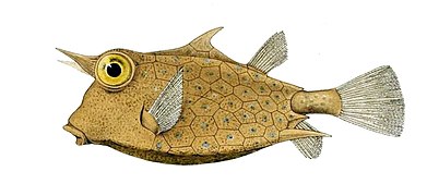
Placoid scales
edit
Placoid(pointed, tooth-shaped) scales are found in thecartilaginous fishes:sharks,rays.They are also calleddermal denticles.Placoid scales are structurallyhomologouswithvertebrateteeth( "denticle" translates to "small tooth" ), having a centralpulp cavitysupplied withblood vessels,surrounded by a conical layer ofdentine,all of which sits on top of a rectangular basal plate that rests on thedermis.The outermost layer is composed ofvitrodentine,a largely inorganicenamel-like substance. Placoid scales cannot grow in size, but rather more scales are added as the fish increases in size.
Similar scales can also be found under the head of thedenticle herring.The amount of scale coverage is much less in rays.
Rhomboidal scales with the properties of both placoid and ganoid scales are suspected to exist in modern jawed fish ancestors: jawlessostracodermsand then jawedplacoderms.
Shark skin
edit
Shark skin is almost entirely covered by small placoid scales. The scales are supported by spines, which feel rough when stroked in a backward direction, but when flattened by the forward movement of water, create tinyvorticesthat reducehydrodynamicdragand reduceturbulence,making swimming both more efficient and quieter compared to that of bony fishes.[31]It also serves a role inanti-foulingby exhibiting thelotus effect.[32]
All denticles are composed of an interior pulp cavity with a nervous and arterial supply rooted in thedermisto supply the denticle with mucus.[33]Denticles contain riblet structures that protrude from the surface of the scale; under a microscope this riblet can look like a hook or ridges coming out of the scale. The overall shape of the protrusion from the denticle is dependent on the type of shark and can be generally described with two appearances.[34]The first is a scale in which ridges are placed laterally down the shark and parallel with the flow of the water. The second form is a smooth scale with what looks like a hooked riblet curling out of the surface aiming towards theposterior sideof the shark.[34]Both riblet shapes assist in creating a turbulentboundary layerforcing thelaminar flowfarther away from the sharks skin.[35]
Unlike bony fish, sharks have a complicated dermal corset made of flexiblecollagenousfibersarranged as ahelicalnetwork surrounding their body. The corset works as an outer skeleton, providing attachment for their swimming muscles and thus saving energy.[36]Depending on the position of these placoid scales on the body, they can be flexible and can be passively erected, allowing them to change their angle of attack. These scales also have riblets which are aligned in the direction of flow, these riblets reduce the drag force acting on the shark skin by pushing the vortex further away from the skin surface, inhibiting any high-velocity cross-stream flow.[37]
Scale morphology
editThe general anatomy of the scales varies, but all of them can be divided into three parts: the crown, the neck and the base. The scale pliability is related to the size of the base of the scale. The scales with higher flexibility have a smaller base, and thus are less rigidly attached to thestratum laxum.On the crown of the fast-swimming sharks there are a series of parallel riblets or ridges which run from an anterior to posterior direction.[38]
Analyzing the three components of the scale it can be concluded that the base of the denticle does not come into contact with any portion of the fluid flow.[39]The crown and the neck of the denticles however play a key role and are responsible for creating the turbulent vortices andeddiesfound near the skin's surface.[39]Because denticles come in so many different shapes and sizes, it can be expected that not all shapes will produce the same type ofturbulent flow.During a recent research experimentbiomimeticsamples of shark denticles with a crescent like microstructure were tested in a water tank using a traction table as a slide. The experiment showed that the surface with denticles experienced a 10% drag reduction overall versus the smooth sample. The reason for this drag reduction was that the turbulent vortices became trapped between the denticles, creating a ‘cushion like’ barrier against the laminar flow.[40]This same type of experiment was performed by another research group which implemented more variation in their biomimetic sample. The second group arrived at the same conclusion as the first. However, because their experiment contained more variation within the samples they were able to achieve a high degree of experimental accuracy. In conclusion, they stated that more practical shapes were more durable than ones with intricate ridge-lines. The practical shapes were low profile and contained trapezoidal or semi-circular trough-like cross sections, and were less effective but nonetheless reduced drag by 6 or 7%.[41]
Drag reduction
edit

Sharks decrease drag and overallcost of transport(COT) through multiple different avenues.Pressure dragis created from the pressure difference between the anterior and posterior sides of the shark due to the amount of volume that is pushed past the shark to propel itself forward.[42]This type of drag is also directly proportional to thelaminar flow.When the laminar flow increases around the fish the pressure drag does as well.[43]Frictional drag is a result of the interaction between the fluid against the shark's skin and can vary depending on how the boundary layer changes against the surface of the fish.[42]
The riblets impede the cross-stream translation of the streamwise vortices in the viscous sublayer. The mechanism is complex and not yet understood fully. Basically, the riblets inhibit the vortex formation near the surface because the vortex cannot fit in the valleys formed by the riblets. This pushes the vortex further up from the surface, interacting only with the riblet tips, not causing any high-velocity flow in the valleys. Since this high-velocity flow now only interacts with the riblet-tip, which is a very small surface area, the momentum transfer which causes drag is now much lower than before, thereby effectively reducing drag. Also, this reduces the cross-stream velocity fluctuations, which aids in momentum transfer too.[38]
Recent research has shown that there is a pre and post-breakdown regime in the near-wall boundary layer where thesublayerthickens at a declining rate and then abruptly undergoes a breakdown into turbulent vortices before finally collapsing. This system is completely self-regulating and mediates the growth and decay cycle; the vortices accumulate during the growth period and are abruptly liquidated intoStrouhal arraysof hairpin vortices lifting off the wall. Lifting vortices are what push the boundary layer out and away from the surface of the shark which results in reducing the overall drag experienced by the fish.[44]
Technical application
editThe rough,sandpaper-like texture of shark and ray skin, coupled with its toughness, has led it to be valued as a source of rawhideleather,calledshagreen.One of the many historical applications of shark shagreen was in making hand-grips forswords.The rough texture of the skin is also used inJapanese cuisineto makegraterscalledoroshiki,by attaching pieces of shark skin to wooden boards. The small size of the scales grates the food very finely.

In the marine industry, there is an extremely large market and need foranti-fouling surfaces.In laymen's terms, fouling is known as the process by which something becomes encrusted with material from the surrounding environment such asbarnacles,algae,andgreen sludge.Dermal denticles are an extremely promising area of research for this type of application due to the fact that sharks are among the only fish without build up or growth on their scales. Studies by theU.S. Navyhave shown that if a biomimetic material can be engineered, it could potentially lead to fuel cost savings for military vessels of up to 45%.[45]
There are many examples ofbiomimetic materialsand surfaces based on the structure of aquatic organisms, including sharks. Such applications intend to enable more efficient movement through fluid mediums such as air, water, and oil.
Surfaces that mimic the skin of sharks have also been used in order to keep microorganisms andalgaefrom coating the hulls of submarines and ships. One variety is traded as "sharklet".[46][47]
A lot of the new methods for replicating shark skin involve the use ofpolydimethylsiloxane(PDMS) for creating a mold. Usually the process involves taking a flat piece of shark skin, covering it with the PDMS to form a mold and pouring PDMS into that mold again to get a shark skin replica. This method has been used to create a biomimetic surface which hassuperhydrophobicproperties, exhibiting thelotus effect.[46]One study found that these biomimetic surfaces reduced drag by up to 9%,[37]while with flapping motion drag reduction reached 12.3%.[48]
Denticles also provide drag reduction on objects where the main form of drag is caused by turbulent flow at the surface. A large portion of the total drag on long objects with relatively flat sides usually comes from turbulence at the wall, so riblets will have an appreciable effect. Along with marine applications, the aerospace industry can benefit greatly from these biomimetic designs. Other applications include pipes, where they score the insides to a riblet-like roughness and have discovered a 5% drag reduction, and a few percent reduction is claimed with competitive swimwear.[49]
Parametric modelinghas been done on shark denticles with a wide range of design variations such as low and high-profile vortex generators.[50]Through this method, the most thorough characterization has been completed for symmetrical two-dimensional riblets with sawtooth, scalloped and blade cross sections.[49]These biomimetic models were designed and analyzed to see the effects of applying the denticle-like structures to the wings of various airplanes. During the simulation, it was noted that the sample altered how the low and highangles of attackreacted. Both the geometry of the denticles and their arrangement have a profound effect on the aerodynamic response of the aerofoils. Out of both the low and high-profile samples tested, the low-profile vortex generators outperformed the current smooth wing structures by 323%. This increase in performance is due to a separation bubble in the denticle's wake and stream-wise vortices that replenish momentum lost in the boundary layer due to skin friction.[50]
Scutes
edit
Scutesare similar to scales and serve the same function. Unlike the scales of fish, which are formed from the epidermis, scutes are formed in the lower vascular layer of the skin and the epidermal element is only the top surface. Forming in the living dermis, the scutes produce a horny outer layer, that is superficially similar to that of scales.
Scute comes from Latin forshield,and can take the form of:
- an external shield-like bony plate, or
- a modified, thickened scale that often is keeled or spiny, or
- a projecting, modified (rough and strongly ridged) scale, usually associated with the lateral line, or on the caudal peduncle forming caudal keels, or along the ventral profile.
Some fish, such aspineconefish,are completely or partially covered in scutes.River herringsandthreadfinshave an abdominal row of scutes, which are scales with raised, sharp points that are used for protection. Somejackshave a row of scutes following thelateral lineon either side.
Scale development
editScales typically appear late in the development of fish. In the case ofzebrafish,it takes 30 days after fertilization before the different layers needed to start forming the scales have differentiated and become organized. For this it is necessary that consolidation of themesenchymeoccurs, thenmorphogenesisis induced, and finally the process of differentiation or latemetamorphosisoccurs.[51][52]
- Mesenchyme consolidation: The consolidation or structuring of the mesenchyme originates during the development of thedermis.This process depends on whether the fish is cartilaginous or bony. For cartilaginous fish the structuring originates through the formation of two layers. The first is superficial and wide and the second is thin and compact. These two layers are separated bymesenchymal cells.Bony fish generate anacellularsubstrate organized by perpendicularly bycollagen fibers.Subsequently, for both fish thefibroblastselongate. These penetrate the compact layer of the mesenchyme, which consolidates prior to the formation of the scale, in order to initiate the dermal plate.[51][52][53]
- Morphogenesis induction: The morphogenesis is due to the formation of the epidermalpapilla,which is generated by joining theepidermisanddermisthrough a process ofinvagination.Morphogenesis begins at the time when fibroblasts are relocated to the upper part of the compact mesenchyme. Throughout this process, thebasal cellsof theepitheliumform a delimiting layer, which is located in the upper part of the mesenchyme. Subsequently, these cells will differentiate in the area where the scaleprimordiumwill arise.[51][52][53]
- Differentiation or late metamorphosis: This differentiation is generated by two different forms according to the type of scale being formed. The formation of elasmoid scales (cycloids and ctenoids) occurs through the formation of a space between the matrix of the epidermal papilla. This space contains collagen fibers. Around this space elasmoblasts differentiate and are responsible for generating the necessary material for the formation of the scale. Subsequently,matrix mineralizationoccurs, allowing the scale to acquire the rigid characteristic that identifies them.[51][52][53]
Unlike elasmoid scales,ganoid scalesare composed of mineralized and non-mineralized collagen in different regions. The formation of these occurs through the entry of the surface cells of the mesenchyme into the matrix, the latter is composed of collagen fibers and is located around the vascular capillaries, thus giving rise to vascular cavities. At this point, elasmoblasts are replaced byosteoblasts,thus forming bone. The patches of the matrix of the scale that are notossifiedare composed of compacted collagen that allow it to maintain the union with the mesenchyme. This are known asSharpey fibers.[51][52][53]
One of the genes that regulate the development of scale formation in fish is thesonic hedgehog(shh) gene, which by means of the (shh) protein, involved inorganogenesisand in the process ofcellular communication,enable the formation of the scales.[54][55]Theapolipoprotein E(ApoE), that allows the transport and metabolism oftriglyceridesandcholesterol,has an interaction with shh, because ApoE provides cholesterol to theshh signaling pathway.It has been shown that during the process ofcell differentiationandinteraction,the level of ApoE transcription is high, which has led to the conclusion that this protein is important for the late development of scales.[54][55]
Modified scales
edit


Different groups of fish haveevolveda number of modified scales to serve various functions.
- Almost all fishes have alateral line,a system ofmechanoreceptorsthat detect water movements. In bony fishes, the scales along the lateral line have central pores that allow water to contact the sensory cells.
- The dorsal fin spines ofdogfish sharksand chimaeras, the stinging tail spines ofstingrays,and the "saw" teeth ofsawfishesandsawsharksare fused and modified placoid scales.
- Surgeonfishhave a scalpel-like blade, which is a modified scale, on either side of thecaudal peduncle.[56]
- Someherrings,anchovies,andhalfbeakshavedeciduous scales,which are easily shed and aid in escaping predators.
- MalePercinadarters have a row of enlargedcaducous scalesbetween thepelvic finsand theanus.
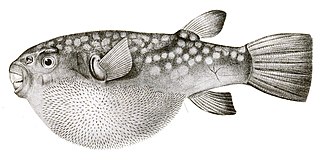
- Porcupine fisheshave scales modified into large externalspines.
- By contrast,pufferfishhave thinner, more hidden spines than porcupine fish, which become visible only when the fish puffs up. Unlike the porcupine fish, these spines are not modified scales, but develop under the control of the same network of genes that produce feathers and hairs in other vertebrates.[57][58]
-
 Porcupine fishhave scales modified intospines.
 -
 Pufferfishspines are not modified scales but are developed by an independent gene network.


Fish without scales
edit-
 Mandarinfishlack scales and protect themselves with a layer of smelly and bitter slime.

Fish without scales usually evolve alternatives to the protection scales can provide, such as tough leathery skin or bony plates.
- Jawless fish(lampreysandhagfishes) have smooth skin without scales and without dermal bone.[59]Lampreys get some protection from a tough leathery skin. Hagfish exude copious quantities slime ormucusif they are threatened.[60]They can tie themselves in anoverhand knot,scraping off the slime as they go and freeing themselves from a predator.[61]
- Mosteelsare scaleless, though some species are covered with tiny smooth cycloid scales.
- Mostcatfishlack scales, though several families have body armour in the form of dermal plates or some sort of scute.[62]
- Mandarinfishlack scales and have a layer of smelly and bitter slime which blocks out disease and probably discourages predators, implying their bright coloration isaposematic.[63]
- Anglerfishhave loose, thin skin often covered with fine forked dermal prickles ortubercles,but they do not have regular scales. They rely on camouflage to avoid the attention of predators, while their loose skin makes it difficult for predators to grab them.
Many groups of bony fishes, includingpipefish,seahorses,boxfish,poachers,and several families ofsticklebacks,have developed external bony plates, structurally resembling placoid scales, as protective armour against predators.
- Seahorses lack scales but have thin skin stretched over a bony plate armour arranged in rings through the length of their bodies.
- In boxfish, the plates fuse together to form a rigid shell orexoskeletonenclosing the entire body. These bony plates are not modified scales but skin that has beenossified.Because of this heavy armour boxfish are limited to slow movements, but few other fish are able to eat the adults.

| |
Eelsseem scaleless, but some species are covered with tiny smooth cycloid scales.
| |
-
 Boxfishhave plates of ossified skin fused together to form a rigid shell.
 -
 Seahorseshave thin skin stretched over bony plates arranged in rings.


Some fish, such as hoki and swordfish, are born with scales but shed them as they grow.
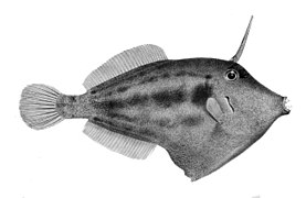
Filefish have rough non-overlapping scales with small spikes, which is why they are called filefish. Some filefish appear scaleless because their scales are so small.
Prominent scaling appears ontunaonly along the lateral line and in thecorselet,a protective band of thickened and enlarged scales in the shoulder region. Over most of their body tuna have scales so small that to casual inspection they seems scaleless.[64]
-
 Somefilefishappear scaleless because their scales are so small.
 -
 To casual examinationtunaseem largely free of scales, but they are not.


Leviticus
editA passage inLeviticusdeclares that, "of all that are in the waters... in the seas, and in the rivers" those that do not have both fins and scales "shall be an abomination unto you" and may not be eaten.[65]This eliminates all aquaticinvertebratesasabominationsandunclean,as well as any fish that lack scales (there do not seem to be fish that lack fins).
According to thechokor divine decrees of theTorahand theTalmud,for a fish to be declaredkosher,it must have scales and fins.[66]The definition of "scale" differs from the definitions presented in biology, in that the scales of a kosher fish must be visible to the eye, and can be easily removed from the skin either by hand or scaling knife.[66]According to thekosher certification agencyof theOrthodox Union,a fish is kosher if the scales can be removed without tearing its skin.[67]Thus carp and salmon are kosher, whereas a shark, whose scales are microscopic, a sturgeon, whose scutes can not be easily removed without cutting them out of the body, are all not kosher. Other non-kosher fish include catfish, eels,freshwater cod,snake mackerels, and puffer fish.[66]
Lepidophagy
edit
Lepidophagy(Ancient Greek forscale-eating) is a specialised feeding behaviour in fish that involves eating the scales of other fish.[69]Lepidophagy has independentlyevolvedin at least five freshwater families and seven marine families.[70]
Fish scales can be nutritious, containing a dermal portion and a layer of protein-rich mucus apart from the layers ofkeratinandenamel.They are a rich source ofcalcium phosphate.[70]However, the energy expended to make a strike versus the amount of scales consumed per strike puts a limit on the size of lepidophagous fish, and they are usuallymuch smaller than their prey.[70]Scale eating behaviour usually evolves because of lack of food and extreme environmental conditions. The eating of scales and the skin surrounding the scales provides protein rich nutrients that may not be available elsewhere in the niche.[71]
Fish jawsnormally showbilateral symmetry.An exception occurs with the scale-eatingcichlidPerissodus microlepis.The jaws of this fish occur in two distinctmorphologicalforms. One morph has its jaw twisted to the left, allowing it to eat scales more readily on its victim's right flank. The other morph has its jaw twisted to the right, which makes it easier to eat scales on its victim's left flank. The relative abundance of the two morphs in populations is regulated byfrequency-dependent selection.[68][72][73]
See also
editReferences
edit- ^ScaleEtymonline.Retrieved 28 April 2019.
- ^Mongera, A.; Nüsslein-Volhard, C. (2013)."Scales of fish arise from mesoderm".Current Biology.23(9): R338–R339.doi:10.1016/j.cub.2013.02.056.PMID23660349.
- ^Sharpe, P. T. (2001)."Fish scale development: Hair today, teeth and scales yesterday?".Current Biology.11(18): R751–R752.doi:10.1016/S0960-9822(01)00438-9.PMID11566120.S2CID18868124.
- ^Turner, S.; Tarling, D. H. (1982). "Thelodont and other agnathan distributions as tests of Lower Paleozoic continental reconstructions".Palaeogeography, Palaeoclimatology, Palaeoecology.39(3–4): 295–311.Bibcode:1982PPP....39..295T.doi:10.1016/0031-0182(82)90027-X.
- ^Märss, T. (2006). "Exoskeletal ultrasculpture of early vertebrates".Journal of Vertebrate Paleontology.26(2): 235–252.doi:10.1671/0272-4634(2006)26[235:EUOEV]2.0.CO;2.S2CID85993241.
- ^abJanvier, Philippe (1998). "Early vertebrates and their extant relatives".Early Vertebrates.Oxford University Press.pp. 123–127.ISBN978-0-19-854047-2.
- ^abTurner, S. (1991). "Monophyly and interrelationships of the Thelodonti". In M. M. Chang; Y. H. Liu; G. R. Zhang (eds.).Early Vertebrates and Related Problems of Evolutionary Biology.Science Press, Beijing. pp. 87–119.
- ^Märss, T. (1986). "Squamation of the thelodont agnathanPhlebolepis".Journal of Vertebrate Paleontology.6(1): 1–11.doi:10.1080/02724634.1986.10011593.
- ^Botella, H.; J. I. Valenzuela-Rios; P. Carls (2006)."A New Early Devonian thelodont from Celtiberia (Spain), with a revision of Spanish thelodonts".Palaeontology.49(1): 141–154.doi:10.1111/j.1475-4983.2005.00534.x.S2CID128939911.
- ^Ferrón, Humberto G.; Botella, Héctor (2017)."Squamation and ecology of thelodonts".PLOS ONE.12(2): e0172781.Bibcode:2017PLoSO..1272781F.doi:10.1371/journal.pone.0172781.PMC5328365.PMID28241029.
- ^MICHAEL ALLABY "cosmoid scale." A Dictionary of Zoology.. Encyclopedia.com. 29 Oct. 2019 <https://www.encyclopedia.com>
- ^Zylberberg, L., Meunier, F.J., Laurin, M. (2010).A microanatomical and histological study of the postcranial dermal skeleton in the Devonian sarcopterygianEusthenopteron foordi,Acta Palaeontologica Polonica55: 459–470.
- ^Bergen, Dylan J. M.; Kague, Erika; Hammond, Chrissy L. (2019)."Zebrafish as an Emerging Model for Osteoporosis: A Primary Testing Platform for Screening New Osteo-Active Compounds".Frontiers in Endocrinology.10:6.doi:10.3389/fendo.2019.00006.ISSN1664-2392.PMC6361756.PMID30761080.
- ^de Vrieze, E.; van Kessel, M. A. H. J.; Peters, H. M.; Spanings, F. A. T.; Flik, G.; Metz, J. R. (1 February 2014). "Prednisolone induces osteoporosis-like phenotype in regenerating zebrafish scales".Osteoporosis International.25(2): 567–578.doi:10.1007/s00198-013-2441-3.ISSN1433-2965.PMID23903952.S2CID21829206.
- ^abZylberberg, L.; Sire, J. -Y.; Nanci, A. (1997)."Immunodetection of amelogenin-like proteins in the ganoine of experimentally regenerating scales of Calamoichthys calabaricus, a primitive actinopterygian fish".The Anatomical Record.249(1): 86–95.doi:10.1002/(SICI)1097-0185(199709)249:1<86::AID-AR11>3.0.CO;2-X.PMID9294653.
- ^Sire, Jean-Yves; Donoghue, Philip C. J.; Vickaryous, Matthews K. (2009)."Origin and evolution of the integumentary skeleton in non-tetrapod vertebrates".Journal of Anatomy.214(4): 409–440.doi:10.1111/j.1469-7580.2009.01046.x.ISSN0021-8782.PMC2736117.PMID19422423.
- ^abcRichter, M. (1995). "A microstructural study of the ganoine tissue of selected lower vertebrates".Zoological Journal of the Linnean Society.114(2): 173–212.doi:10.1006/zjls.1995.0023.
- ^abBruet, B. J. F.; Song, J.; Boyce, M. C.; Ortiz, C. (2008). "Materials design principles of ancient fish armour".Nature Materials.7(9): 748–756.Bibcode:2008NatMa...7..748B.doi:10.1038/nmat2231.PMID18660814.
- ^abSherman, Vincent R.; Yaraghi, Nicholas A.; Kisailus, David; Meyers, Marc A. (1 December 2016)."Microstructural and geometric influences in the protective scales of Atractosteus spatula".Journal of the Royal Society Interface.13(125): 20160595.doi:10.1098/rsif.2016.0595.ISSN1742-5689.PMC5221522.PMID27974575.
- ^"Missouri Alligator Gar Management and Restoration Plan"(PDF).Missouri Department of Conservation Fisheries Division. January 22, 2013. Archived fromthe original(PDF)on May 6, 2016.RetrievedApril 12,2019.
- ^Lagler, K. F., J. E. Bardach, and R. R. Miller (1962)Ichthyology.New York: John Wiley & Sons.
- ^Ballard, Bonnie; Cheek, Ryan (2 July 2016).Exotic Animal Medicine for the Veterinary Technician.John Wiley & Sons.ISBN978-1-118-92421-1.
- ^McGrouther, Mark (2 December 2019)."Cycloid and Ctenoid Scales".The Australian Museum.Retrieved29 December2021.
- ^Pouyaud, L.; Sudarto, Guy G. Teugels (2003). "The different colour varieties of the Asian arowana Scleropages formosus (Osteoglossidae) are distinct species: morphologic and genetic evidences".Cybium.27(4): 287–305.
- ^Ismail, M. (1989).Systematics, Zoogeography, and Conservation of the Freshwater Fishes of Peninsular Malaysia(Doctoral Dissertation ed.). Colorado State University.
- ^E.J. Brill (1953).The Fishes of the Indo-Australian Archipelago.E.J. Brill. pp. 306–307.
- ^abcdKawasaki, Kenta C., "A Genetic Analysis of Cichlid Scale Morphology" (2016). Masters Theses May 2014 - current. 425.http://scholarworks.umass.edu/masters_theses_2/425
- ^Helfman, Gene (2009).The Diversity of Fishes Biology, Evolution, and Ecology.Wiley-Blackwell.
- ^abcHerring, Peter(2002).The Biology of the Deep Ocean.Oxford: Oxford University Press. pp. 193–195.ISBN9780198549567.
- ^"There Are Probably Fish Scales In Your Lipstick".HuffPost India.23 April 2015.Retrieved6 May2019.
- ^Martin, R. Aidan."Skin of the Teeth".Retrieved28 August2007.
- ^Fürstner, Reiner; Barthlott, Wilhelm; Neinhuis, Christoph; Walzel, Peter (1 February 2005). "Wetting and Self-Cleaning Properties of Artificial Superhydrophobic Surfaces".Langmuir.21(3): 956–961.doi:10.1021/la0401011.ISSN0743-7463.PMID15667174.
- ^Lauder, George V.; Wainwright, Dylan K.; Domel, August G.; Weaver, James C.; Wen, Li; Bertoldi, Katia (2016)."Structure, biomimetics, and fluid dynamics of fish skin surfaces".Physical Review Fluids.1(6): 060502.Bibcode:2016PhRvF...1f0502L.doi:10.1103/PhysRevFluids.1.060502.S2CID18118663.
- ^abFeld, Katrine; Kolborg, Anne Noer; Nyborg, Camilla Marie; Salewski, Mirko; Steffensen, John Fleng; Berg-Sørensen, Kirstine (24 May 2019)."Dermal Denticles of Three Slowly Swimming Shark Species: Microscopy and Flow Visualization".Biomimetics.4(2): 38.doi:10.3390/biomimetics4020038.ISSN2313-7673.PMC6631580.PMID31137624.
- ^Fletcher, Thomas; Altringham, John; Peakall, Jeffrey; Wignall, Paul; Dorrell, Robert (7 August 2014)."Hydrodynamics of fossil fishes".Proceedings of the Royal Society B: Biological Sciences.281(1788): 20140703.doi:10.1098/rspb.2014.0703.ISSN0962-8452.PMC4083790.PMID24943377.
- ^Martin, R. Aidan."The Importance of Being Cartilaginous".ReefQuest Centre for Shark Research.Retrieved29 August2009.
- ^abHage, W.; Bruse, M.; Bechert, D. W. (1 May 2000). "Experiments with three-dimensional riblets as an idealized model of shark skin".Experiments in Fluids.28(5): 403–412.Bibcode:2000ExFl...28..403B.doi:10.1007/s003480050400.ISSN1432-1114.S2CID122574419.
- ^abMotta, Philip; Habegger, Maria Laura; Lang, Amy; Hueter, Robert; Davis, Jessica (1 October 2012). "Scale morphology and flexibility in the shortfin mako Isurus oxyrinchus and the blacktip shark Carcharhinus limbatus".Journal of Morphology.273(10): 1096–1110.doi:10.1002/jmor.20047.ISSN1097-4687.PMID22730019.S2CID23881820.
- ^abDou, Zhaoliang; Wang, Jiadao; Chen, Darong (1 December 2012)."Bionic Research on Fish Scales for Drag Reduction".Journal of Bionic Engineering.9(4): 457–464.doi:10.1016/S1672-6529(11)60140-6.ISSN1672-6529.S2CID137143652.
- ^"Experimental investigations on drag-reduction characteristics of bionic surface with water-trapping microstructures of fish scales"(PDF).
- ^Palmer, Colin; Young, Mark T. (14 January 2015)."Surface drag reduction and flow separation control in pelagic vertebrates, with implications for interpreting scale morphologies in fossil taxa".Royal Society Open Science.2(1): 140163.Bibcode:2015RSOS....240163P.doi:10.1098/rsos.140163.ISSN2054-5703.PMC4448786.PMID26064576.
- ^abLauder, George V.; Wainwright, Dylan K.; Domel, August G.; Weaver, James C.; Wen, Li; Bertoldi, Katia (18 October 2016)."Structure, biomimetics, and fluid dynamics of fish skin surfaces".Physical Review Fluids.1(6): 060502.Bibcode:2016PhRvF...1f0502L.doi:10.1103/PhysRevFluids.1.060502.
- ^Muthuramalingam, Muthukumar; Villemin, Leo S.; Bruecker, Christoph (29 April 2019). "Streak formation in flow over Biomimetic Fish Scale Arrays".The Journal of Experimental Biology.222(Pt 16): jeb205963.arXiv:1904.12752.Bibcode:2019arXiv190412752M.doi:10.1242/jeb.205963.PMID31375542.S2CID139103148.
- ^Bandyopadhyay, Promode R.; Hellum, Aren M. (23 October 2014)."Modeling how shark and dolphin skin patterns control transitional wall-turbulence vorticity patterns using spatiotemporal phase reset mechanisms".Scientific Reports.4:6650.Bibcode:2014NatSR...4E6650B.doi:10.1038/srep06650.ISSN2045-2322.PMC4206846.PMID25338940.
- ^Magin, Chelsea M.; Cooper, Scott P.; Brennan, Anthony B. (1 April 2010)."Non-toxic antifouling strategies".Materials Today.13(4): 36–44.doi:10.1016/S1369-7021(10)70058-4.ISSN1369-7021.
- ^abLiu, Yunhong; Li, Guangji (15 December 2012). "A new method for producing" Lotus Effect "on a biomimetic shark skin".Journal of Colloid and Interface Science.388(1): 235–242.Bibcode:2012JCIS..388..235L.doi:10.1016/j.jcis.2012.08.033.ISSN0021-9797.PMID22995249.
- ^"Sharklet Discovery | Sharklet Technologies, Inc".www.sharklet.com.Retrieved26 September2018.
- ^Lauder, George V.; Oeffner, Johannes (1 March 2012)."The hydrodynamic function of shark skin and two biomimetic applications".Journal of Experimental Biology.215(5): 785–795.doi:10.1242/jeb.063040.ISSN1477-9145.PMID22323201.
- ^abDean, Brian & Bhushan, Bharat. (2010). Shark-Skin Surfaces for Fluid-Drag Reduction in Turbulent Flow: A Review. Philosophical transactions. Series A, Mathematical, physical, and engineering sciences. 368. 4775-806. 10.1098/rsta.2010.0201.
- ^abDomel, August G.; Saadat, Mehdi; Weaver, James C.; Haj-Hariri, Hossein; Bertoldi, Katia; Lauder, George V. (28 February 2018)."Shark skin-inspired designs that improve aerodynamic performance".Journal of the Royal Society Interface.15(139): 20170828.doi:10.1098/rsif.2017.0828.PMC5832729.PMID29436512.
- ^abcdeSire, J.Y.; Huysseune, A.N.N. (2003). "Formation of dermal skeletal and dental tissues in fish: a comparative and evolutionary approach".Biological Reviews.78(2): 219–249.doi:10.1017/S1464793102006073.PMID12803422.S2CID19556201.
- ^abcdeLe Guellec, D.; Morvan-Dubois, G.; Sire, J.Y. (2004)."Skin development in bony fish with particular emphasis on collagen deposition in the dermis of the zebrafish (Danio rerio) ".International Journal of Developmental Biology.48(2–3): 217–231.doi:10.1387/ijdb.15272388.PMID15272388.
- ^abcdSire, J.Y. (2001). "Teeth outside the mouth in teleost fishes: how to benefit from a developmental accident".Evolution & Development.3(2): 104–108.doi:10.1046/j.1525-142x.2001.003002104.x.PMID11341672.S2CID13353402.
- ^abSire, J.Y.; Akimenko, M.A. (2003)."Scale development in fish: a review, with description of sonic hedgehog (shh) expression in the zebrafish (Danio rerio) ".International Journal of Developmental Biology.48(2–3): 233–247.doi:10.1387/ijdb.15272389.PMID15272389.
- ^abMonnot, M.J.; Babin, P.J.; Poleo, G.; Andre, M.; Laforest, L.; Ballagny, C.; Akimenko, M.A. (1999)."Epidermal expression of apolipoprotein E gene during fin and scale development and fin regeneration in zebrafish".Developmental Dynamics.214(3): 207–215.doi:10.1002/(SICI)1097-0177(199903)214:3<207::AID-AJA4>3.0.CO;2-5.PMID10090147.
- ^Sorenson, L.; Santini, F.; Carnevale, G.; Alfaro, M.E. (2013). "A multi-locus timetree of surgeonfishes (Acanthuridae, Percomorpha), with revised family taxonomy".Molecular Phylogenetics & Evolution.68(1): 150–160.doi:10.1016/j.ympev.2013.03.014.PMID23542000.
- ^How the pufferfish got its wacky spinesPhys.org,25 July 2019.
- ^Shono, T.; Thiery, A.P.; Cooper, R.L.; Kurokawa, D.; Britz, R.; Okabe, M.; Fraser, G.J. (2019)."Evolution and Developmental Diversity of Skin Spines in Pufferfishes".iScience.19:1248–1259.Bibcode:2019iSci...19.1248S.doi:10.1016/j.isci.2019.06.003.PMC6831732.PMID31353167.
- ^Coolidge E, Hedrick MS and Milsom WK (2011)"Ventilatory Systems".In: McKenzie DJ, Farrell AP and Brauner CJ (Eds)Fish Physiology: Primitive Fishes,Elsevier, Page 182–213.ISBN9780080549521
- ^Rothschild, Anna (1 April 2013)."Hagfish slime: The clothing of the future?".BBC News.Retrieved2 April2013.
- ^Yong, Ed (23 January 2019)."No One Is Prepared for Hagfish Slime".The Atlantic.Retrieved26 January2019.
- ^Friel, J P; Lundberg, J G (1996). "Micromyzon akamai,gen. et sp. nov., a small and eyeless banjo catfish (Siluriformes: Aspredinidae) from the river channels of the lower Amazon basin ".Copeia.1996(3): 641–648.doi:10.2307/1447528.JSTOR1447528.
- ^Sadovy, Y.; Randall, J. E.; Rasotto, Maria B. (May 2005). "Skin structure in six dragonet species (Gobiesociformes; Callionymidae): Interspecific differences in glandular cell types and mucus secretion".Journal of Fish Biology.66(5): 1411–1418.doi:10.1111/j.0022-1112.2005.00692.x.
- ^Do tunas have scales?Northeast Fisheries Science Center, NOAA Fisheries. Accessed 4 August 2019.
- ^Leviticus 11:9–10
- ^abcAryeh Citron,"All About Kosher Fish"
- ^Verifying Kosher FishOU Kosher Certification.Retrieved 9 August 2019.
- ^abLee, H. J.; Kusche, H.; Meyer, A. (2012)."Handed Foraging Behavior in Scale-Eating Cichlid Fish: Its Potential Role in Shaping Morphological Asymmetry".PLOS ONE.7(9): e44670.Bibcode:2012PLoSO...744670L.doi:10.1371/journal.pone.0044670.PMC3435272.PMID22970282.
- ^Froese, R. and D. Pauly. Editors."Glossary: Lepidophagy".FishBase.Retrieved12 April2007.
{{cite web}}:|author=has generic name (help) - ^abcJanovetz, Jeff (2005)."Functional morphology of feeding in the scale-eating specialistCatoprion mento"(PDF).The Journal of Experimental Biology.208(Pt 24): 4757–4768.doi:10.1242/jeb.01938.PMID16326957.S2CID15566769.
- ^Martin, C.; P.C. Wainwright (2011)."Trophic novelty is linked to exceptional rates of morphological diversification in two adaptive radiations of Cyprinodon pupfish".Evolution.65(8): 2197–2212.doi:10.1111/j.1558-5646.2011.01294.x.PMID21790569.S2CID23695342.
- ^Hori, M. (1993). "Frequency-dependent natural selection in the handedness of scale-eating cichlid fish".Science.260(5105): 216–219.Bibcode:1993Sci...260..216H.doi:10.1126/science.260.5105.216.PMID17807183.S2CID33113282.
- ^Stewart, T. A.; Albertson, R. C. (2010)."Evolution of a unique predatory feeding apparatus: functional anatomy, development and a genetic locus for jaw laterality in Lake Tanganyika scale-eating cichlids".BMC Biology.8(1): 8.doi:10.1186/1741-7007-8-8.PMC2828976.PMID20102595.
Further reading
edit- Helfman, G.S., B.B. Collette and D.E. Facey (1997).The Diversity of Fishes.Blackwell Science. pp. 33–36.ISBN978-0-86542-256-8.
{{cite book}}:CS1 maint: multiple names: authors list (link) - Schultze, H.P. (2016)."Scales, enamel, cosmine, ganoine, and early osteichthyans".Comptes Rendus Palevol.15(1–2): 83–102.doi:10.1016/j.crpv.2015.04.001.