This articleneeds additional citations forverification.(December 2018) |
Esophagogastroduodenoscopy(EGD) oroesophagogastroduodenoscopy(OGD), also called byvarious other names,is adiagnosticendoscopicprocedure that visualizes the upper part of thegastrointestinal tractdown to theduodenum.It is considered aminimally invasive proceduresince it does not require anincisioninto one of the major body cavities and does not require any significant recovery after the procedure (unlesssedationoranesthesiahas been used). However, asore throatis common.[1][2][3]
| Esophagogastroduodenoscopy | |
|---|---|
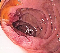
 Endoscopic still of esophageal ulcers seen after banding ofesophageal varices,at time of esophagogastroduodenoscopy | |
| Other names | EGD OGD Upper endoscopy |
| ICD-9-CM | 45.13 |
| MeSH | D016145 |
| OPS-301 code | 1-631,1-632 |
Alternative names
editThe wordsesophagogastroduodenoscopy(EGD;American English) andoesophagogastroduodenoscopy(OGD;British English;seespelling differences) are both pronounced/ɪˌsɒfəɡoʊˌɡæstroʊˌduːoʊdɪˈnɒskoʊpi/.It is also calledpanendoscopy(PES) andupper GI endoscopy.It is also often called justupper endoscopy,upper GI,or even justendoscopy;because EGD is the most commonly performed type of endoscopy, the ambiguous termendoscopyis sometimes informally used to refer to EGD by default. The termgastroscopyliterally focuses on the stomach alone, but in practice, the usage overlaps.
Medical uses
edit
Diagnostic
edit- Unexplainedanemia(usually along with acolonoscopy)
- Upper gastrointestinal bleedingas evidenced byhematemesisormelena
- Persistentdyspepsiain patients over the age of 45 years
- Heartburn and chronic acid reflux – this can lead to a precancerous lesion calledBarrett's esophagus
- Persistent emesis
- Dysphagia– difficulty in swallowing
- Odynophagia– painful swallowing
- Persistentnausea
- IBD (inflammatory bowel diseases)
Surveillance
edit- Surveillance ofBarrett's esophagus
- Surveillance ofgastric ulcerorduodenal ulcer
- Occasionally after gastric surgery
Confirmation of diagnosis/biopsy
edit- Abnormalbarium swalloworbarium meal
- Confirmation ofceliac disease(via biopsy)
Therapeutic
edit- Treatment (banding/sclerotherapy) of esophageal varices
- Injection therapy (e.g.,epinephrinein bleeding lesions)
- Cutting off of larger pieces of tissue with asnare device(e.g.,polyps,endoscopic mucosal resection)
- Application ofcauteryto tissues
- Removal offoreign bodies(e.g., food) that have been ingested
- Tamponade of bleedingesophageal variceswith aballoon
- Application of photodynamic therapy for treatment of esophageal malignancies
- Endoscopic drainage ofpancreatic pseudocyst
- Tightening thelower esophageal sphincter
- Dilating or stenting ofstenosisorachalasia
- Percutaneous endoscopic gastrostomy(feeding tube placement)
- Endoscopic retrograde cholangiopancreatography(ERCP) combines EGD withfluoroscopy
- Endoscopic ultrasound(EUS) combines EGD with 5–12 MHzultrasoundimaging
Newer interventions
edit- Endoscopic trans-gastric laparoscopy
- Placement ofgastric balloonsinbariatric surgery
Complications
editThe complication rate is about 1 in 1000.[4]They include:
- aspiration, causingaspiration pneumonia
- bleeding
- perforation
- cardiopulmonary problems
When used ininfants,the esophagogastroduodenoscope may compress thetrachealis muscle,which narrows thetrachea.[5]This can result in reduced airflow to thelungs.[5]Infants may beintubatedto make sure that the trachea is fixed open.[5]
Limitations
editProblems of gastrointestinalfunctionare usually not well diagnosed by endoscopy sincemotionorsecretionof the gastrointestinal tract is not easily inspected by EGD. Nonetheless, findings such as excess fluid or poor motion of the gut during endoscopy can be suggestive of disorders of function.Irritable bowel syndromeandfunctional dyspepsiaare not diagnosed with EGD, but EGD may be helpful in excluding other diseases that mimic these common disorders.[citation needed]
Procedure
editThis sectionneeds additional citations forverification.(May 2024) |
The tip of the endoscope should be lubricated and checked for critical functions including tip angulations, air and water suction, and image quality.
The patient is keptNPO (nil per os) or NBM (nothing by mouth)for at least 4 hours before the procedure. Most patients tolerate the procedure with onlytopical anesthesiaof theoropharynxusinglidocainespray. However, some patients may need sedation and the very anxious/agitated patient may even need a general anesthetic.Informed consentis obtained before the procedure. The main risks are bleeding and perforation. The risk is increased when a biopsy or other intervention is performed.
The patient lies on their left side with the head resting comfortably on a pillow. A mouth-guard is placed between the teeth to prevent the patient from biting on the endoscope. The endoscope is then passed over the tongue and into the oropharynx. This is the most uncomfortable stage for the patient. Quick and gentle manipulation under vision guides the endoscope into the esophagus. The endoscope is gradually advanced down the esophagus making note of any pathology. Excessiveinsufflationof the stomach is avoided at this stage. The endoscope is quickly passed through the stomach and through thepylorusto examine the first and second parts of theduodenum.Once this has been completed, the endoscope is withdrawn into the stomach and a more thorough examination is performed including a J-maneuver. This involves retroflexing the tip of the scope so it resembles a 'J' shape in order to examine thefundusand gastroesophageal junction. Any additional procedures are performed at this stage. The air in the stomach is aspirated before removing the endoscope. Stillphotographscan be made during the procedure and later shown to the patient to help explain any findings.
In its most basic use, the endoscope is used to inspect the internal anatomy of the digestive tract. Often inspection alone is sufficient, butbiopsyis a valuable adjunct to endoscopy. Small biopsies can be made with a pincer (biopsyforceps) which is passed through the scope and allows sampling of 1 to 3 mm pieces of tissue under direct vision. The intestinal mucosa heals quickly from such biopsies.
Clinical practice varies with respect to routine biopsy for histological analysis of the examined upper gastrointestinal system. A rapid urease test is quick, easy, and cost-effective screening for Helicobacter pylori infection.
Equipment
edit
- Endoscope
- Non-coaxialoptic fibersystem to carry light to the tip of the endoscope
- A chip camera at the tip of the endoscope – this has now replaced the coaxial optic fibers of older scopes that were prone to damage and consequent loss of picture quality
- Air/water channel to clean the lens using the water and air channel for drying the lens itself and to insufflate the esophagus and the stomach during the operation to prevent from collapsing the track to better vision in the procedure
- Suction/Working channels – these may be in the form of one or more channels
- Control handle – this houses the controls
- Umbilical Cords that connect to the light source and video processor to supply the endoscope with suction and air pressure and water for (suction and irrigation process) and light to transmit in the body to deliver the video signal to the processor to show the live image on the monitor
- Stack
- Light source
- Suction
- Electrosurgicalunit
- Video recorder/photo printer
- Instruments
- Biopsy forceps
- Snares
- Injecting needles
- Chemical agents
-
 Endoscopic image ofadenocarcinoma of duodenumseen in the post-bulbar duodenum.
 -
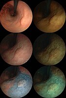
 Endoscopic image ofgastric antral vascular ectasiaseen as a radial pattern around thepylorusbefore (top) and after (bottom) treatment withargon plasma coagulation
 -
 Endoscopic image ofBarrett's esophagus,which is the area of red mucosa projecting like a tongue.
 -
 Deepgastric ulcer
 -
 Endoscopic still ofduodenumof patient withceliac diseaseshowing scalloping of folds.
 -

-
 Endoscopic image of a posterior wallduodenal ulcerwith a clean base, which is a common cause ofupper GI hemorrhage.
 -


See also
editReferences
edit- ^"Gastroscopy – examination of oesophagus and stomach by endoscope".BUPA.December 2006. Archived fromthe originalon 2007-10-06.Retrieved2007-10-07.
- ^National Digestive Diseases Information Clearinghouse(November 2004)."Upper Endoscopy".National Institutes of Health.Retrieved2007-10-07.
- ^"What is Upper GI Endoscopy?".Patient Center -- Procedures.American Gastroenterological Association.Archived fromthe originalon 2007-09-28.Retrieved2007-10-07.
- ^"EGD – esophagogastroduodenoscopy".
- ^abcCravero, Joseph P.; Landrigan-Ossar, Mary (2019-01-01), Coté, Charles J.; Lerman, Jerrold; Anderson, Brian J. (eds.),"46 - Anesthesia Outside the Operating Room",A Practice of Anesthesia for Infants and Children (Sixth Edition),Philadelphia: Elsevier, pp. 1077–1094.e4,ISBN978-0-323-42974-0,retrieved2021-01-23