Glioblastoma,previously known asglioblastoma multiforme(GBM), is the most aggressive and most common type ofcancerthat originates in thebrain,and has a very poor prognosis for survival.[6][7][8]Initial signs and symptoms of glioblastoma are nonspecific.[1]They may includeheadaches,personality changes,nausea,and symptoms similar to those of astroke.[1]Symptoms often worsen rapidly and may progress tounconsciousness.[2]
| Glioblastoma | |
|---|---|
| Other names | Glioblastoma multiforme |
 | |
| CoronalMRI(viewed from the back of the head) with contrast of a glioblastoma in a 38-year-old woman | |
| Specialty | Neuro-oncology,neurosurgery |
| Symptoms | Initially nonspecific,headaches,personality changes,nausea,symptoms similar to astroke[1] |
| Usual onset | ~64 years old[2][3] |
| Causes | Usually unclear[2] |
| Risk factors | Genetic disorders(neurofibromatosis,Li–Fraumeni syndrome), previousradiation therapy[2][3] |
| Diagnostic method | CT scan,MRI scan,tissue biopsy[1] |
| Prevention | Unknown[3] |
| Treatment | Surgery,chemotherapy,radiation[3] |
| Medication | Temozolomide,steroids[1][4] |
| Prognosis | Life expectancy~12 months with treatment (5 year survival <10%)[2][5] |
| Frequency | 3 per 100,000 per year[3] |
The cause of most cases of glioblastoma is not known.[2]Uncommon risk factors includegenetic disorders,such asneurofibromatosisandLi–Fraumeni syndrome,and previousradiation therapy.[2][3]Glioblastomas represent 15% of allbrain tumors.[1]They are thought to arise fromastrocytes.[9]The diagnosis typically is made by a combination of aCT scan,MRI scan,andtissue biopsy.[1]
There is no known method of preventing the cancer.[3]Treatment usually involvessurgery,after whichchemotherapyand radiation therapy are used.[3]The medicationtemozolomideis frequently used as part of chemotherapy.[3][4][10]High-dosesteroidsmay be used to help reduce swelling and decrease symptoms.[1]Surgical removal (decompression) of the tumor is linked to increased survival, but only by some months.[11]
Despite maximum treatment, thecancer almost always recurs.[3]The typical duration of survival following diagnosis is 10–13 months, with fewer than 5–10% of people surviving longer than five years.[12][13][5]Without treatment, survival is typically three months.[14]It is the most common cancer that begins within the brain and the second-most common brain tumor, aftermeningioma,which is benign in most cases.[6][15]About 3 in 100,000 people develop the disease per year.[3]The average age at diagnosis is 64, and the disease occurs more commonly in males than females.[2][3]
Tumors of the central nervous system are the 10th leading cause of death worldwide, with up to 90% being brain tumors.[16]Glioblastoma multiforme (GBM) is derived from astrocytes and accounts for 49% of all malignant central nervous system tumors, making it the most common form of central nervous system cancer. Despite countless efforts to develop new therapies for GBM over the years, the median survival rate of GBM patients worldwide is 8 months; radiation and chemotherapy standard-of-care treatment beginning shortly after diagnosis improve the median survival length to around 14 months and a five-year survival rate of 5–10%. The five-year survival rate for individuals with any form of primary malignant brain tumor is 20%.[17]Even when all detectable traces of the tumor are removed through surgery, most patients with GBM experience recurrence of their cancer.
Signs and symptoms
editCommon symptoms includeseizures,headaches, nausea andvomiting,memory loss,changes to personality, mood or concentration, and localized neurological problems.[18]The kinds of symptoms produced depend more on the location of the tumor than on its pathological properties. The tumor can start producing symptoms quickly, but occasionally is anasymptomatic conditionuntil it reaches an enormous size.[19]
Risk factors
editThe cause of most cases is unclear.[2]The best known risk factor is exposure to ionizing radiation, and CT scan radiation is an important cause.[20][21]About 5% develop from certain hereditary syndromes.[18]
Genetics
editUncommon risk factors include genetic disorders such as neurofibromatosis, Li–Fraumeni syndrome,tuberous sclerosis,orTurcot syndrome.[18]Previous radiation therapy is also a risk.[2][3]For unknown reasons, it occurs more commonly in males.[22]
Environmental
editOther associations include exposure tosmoking,pesticides,and working inpetroleum refiningorrubber manufacturing.[18]
Glioblastoma has been associated with the virusesSV40,[23]HHV-6,[24][25]andcytomegalovirus(CMV).[26]Infection with an oncogenic CMV may even be necessary for the development of glioblastoma.[27][28]
Other
editResearch has been done to see if consumption ofcured meatis a risk factor. No risk had been confirmed as of 2003.[29]Similarly, exposure toformaldehyde,and residentialelectromagnetic fields,such as from cell phones and electrical wiring within homes, have been studied as risk factors. As of 2015, they had not been shown to cause GBM.[18][30][31]
Pathogenesis
editThe cellular origin of glioblastoma is unknown. Because of the similarities inimmunostainingofglial cellsand glioblastoma, gliomas such as glioblastoma have long been assumed to originate from glial-type stem cells found in thesubventricular zone.More recent studies suggest thatastrocytes,oligodendrocyte progenitor cells,andneural stem cellscould all serve as the cell of origin.[32][33]
GBMs usually form in the cerebral white matter, grow quickly, and can become very large before producing symptoms. Since the function of glial cells in the brain is to support neurons, they have the ability to divide, to enlarge, and to extend cellular projections along neurons and blood vessels. Once cancerous, these cells are predisposed to spread along existing paths in the brain, typically along white-matter tracts, blood vessels and theperivascular space.[34]The tumor may extend into the meninges orventricularwall, leading to high protein content in thecerebrospinal fluid(CSF) (> 100 mg/dl), as well as an occasionalpleocytosisof 10 to 100 cells, mostlylymphocytes.Malignantcells carried in the CSF may spread (rarely) to thespinal cordor cause meningeal gliomatosis. However,metastasisof GBM beyond thecentral nervous systemis extremely unusual. About 50% of GBMs occupy more than one lobe of a hemisphere or are bilateral. Tumors of this type usually arise from thecerebrumand may exhibit the classic infiltration across thecorpus callosum,producing a butterfly (bilateral)glioma.[35]
Glioblastoma classification
editBrain tumorclassification has been traditionally based on histopathology at macroscopic level, measured inhematoxylin-eosinsections. TheWorld Health Organizationpublished the first standard classification in 1979[36]and has been doing so since. The 2007 WHO Classification of Tumors of the Central Nervous System[37]was the last classification mainly based on microscopy features. The new 2016 WHO Classification of Tumors of the Central Nervous System[38]was a paradigm shift: some of the tumors were defined also by their genetic composition as well as their cell morphology.
In 2021, the fifth edition of the WHO Classification of Tumors of the Central Nervous System was released. This update eliminated the classification of secondary glioblastoma and reclassified those tumors as Astrocytoma, IDH mutant, grade 4. Only tumors that are IDH wild type are now classified as glioblastoma.[39]
| Synonyms | Glioblastoma, GBM |
| Cell of origin | Astrocyte |
| Median age at diagnosis | ~62 years |
| Male:Female ratio | 1.42:1 |
| Median length of clinical history at diagnosis | 4 months |
| Median overall survival | |
| Surgery + radiotherapy | 9.9 months |
| Surgery + radiotherapy + chemotherapy | 15 months |
| Location | Usually supratentorial, rarely cerebellum or spine |
| Necrosis and microvascular proliferation | Extensive |
| Associated molecular/genetic mutations | TERT promoter mutation, combined gain of chromosome 7 and loss of chromosome 10; EGFR amplification |
Molecular alterations
editThere are currently three molecular subtypes of glioblastoma that were identified based on gene expression:[41]
- Classical: Around 97% of tumors in this subtype carry extra copies of theepidermal growth factor receptor(EGFR) gene, and most have higher than normal expression ofEGFR,whereas the geneTP53(p53), which is often mutated in glioblastoma, is rarely mutated in this subtype.[42]Loss of heterozygosityin chromosome 10 is also frequently seen in the classical subtype alongside chromosome 7amplification.[43]
- The proneural subtype often has high rates of alterations inTP53(p53), and inPDGFRAthe gene encoding a-typeplatelet-derived growth factor receptor.[44]
- The mesenchymal subtype is characterized by high rates of mutations or other alterations inNF1,the gene encodingneurofibromin 1and fewer alterations in theEGFRgene and less expression ofEGFRthan other types.[45]
Initial analyses of gene expression had revealed a fourth neural subtype.[44]However, further analyses revealed that this subtype is non-tumor specific and is potential contamination caused by the normal cells.[41]
Many other genetic alterations have been described in glioblastoma, and the majority of them are clustered in two pathways, theRBand thePI3K/AKT.[46]68–78% and 88% of Glioblastomas have alterations in these pathways, respectively.[6]
Another important alteration is methylation ofMGMT,a "suicide" DNA repair enzyme. Methylation impairs DNA transcription and expression of the MGMT gene. Since the MGMT enzyme can repair only one DNA alkylation due to its suicide repair mechanism, reserve capacity is low and methylation of theMGMT gene promotergreatly affects DNA-repair capacity.[47][48]MGMT methylation is associated with an improved response to treatment with DNA-damaging chemotherapeutics, such as temozolomide.[49]
Studies using genome-wide profiling have revealed glioblastomas to have a remarkable genetic variety.[50]
At least three distinct paths in the development of Glioblastomas have been identified with the aid of molecular investigations.
- The first pathway involves the amplification and mutational activation ofreceptor tyrosine kinase(RTK) genes, leading to the dysregulation of growth factor signaling.Epithelial growth factor(EGF), vascular endothelial growth factor (VEGF), and platelet-derived growth factor (PDGF) are all recognized by transmembrane proteins called RTKs. Additionally, they can function as receptors for hormones, cytokines, and other signaling pathways.
- The second method involves activating the intracellular signaling system known as phosphatidylinositol-3-OH kinase (PI3K)/AKT/mTOR,which is crucial for controlling cell survival.
- The third pathway is defined by p53 and retinoblastoma (Rb) tumor suppressor pathway inactivation.[51]
Cancer stem cells
editGlioblastoma cells with properties similar to progenitor cells (glioblastomacancer stem cells) have been found in glioblastomas. Their presence, coupled with the glioblastoma's diffuse nature results in difficulty in removing them completely by surgery, and is therefore believed to be the possible cause behind resistance to conventional treatments, and the high recurrence rate.[52]Glioblastoma cancer stem cells share some resemblance with neural progenitor cells, both expressing the surface receptorCD133.[53]CD44can also be used as a cancer stem cell marker in a subset of glioblastoma tumour cells.[54]Glioblastoma cancer stem cells appear to exhibit enhanced resistance to radiotherapy and chemotherapy mediated, at least in part, by up-regulation of theDNA damage response.[55]
Metabolism
editTheIDH1gene encodes for the enzymeisocitrate dehydrogenase1 and is not mutated in glioblastoma. As such, these tumors behave more aggressively compared to IDH1-mutated astrocytomas.[48]
Ion channels
editFurthermore, GBM exhibits numerous alterations in genes that encode forion channels,including upregulation ofgBK potassium channelsandClC-3 chloride channels.By upregulating these ion channels, glioblastoma tumor cells are hypothesized to facilitate increased ion movement over the cell membrane, thereby increasing H2O movement through osmosis, which aids glioblastoma cells in changing cellular volume very rapidly. This is helpful in their extremely aggressive invasive behavior because quick adaptations in cellular volume can facilitate movement through the sinuous extracellular matrix of the brain.[56]
MicroRNA
editAs of 2012,RNA interference,usually microRNA, was under investigation in tissue culture, pathology specimens, and preclinical animal models of glioblastoma.[57]Additionally, experimental observations suggest thatmicroRNA-451is a key regulator ofLKB1/AMPKsignaling in cultured glioma cells[58]and that miRNA clustering controls epigenetic pathways in the disease.[59]
Tumor vasculature
editGBM is characterized by abnormal vessels that present disrupted morphology and functionality.[60]The high permeability and poor perfusion of the vasculature result in a disorganized blood flow within the tumor and can lead to increased hypoxia, which in turn facilitates cancer progression by promoting processes such as immunosuppression.[60][61]
Diagnosis
edit


When viewed with MRI, glioblastomas often appear as ring-enhancing lesions. The appearance is not specific, however, as other lesions such asabscess,metastasis,tumefactive multiple sclerosis,and other entities may have a similar appearance.[63]Definitive diagnosis of a suspected GBM on CT or MRI requires astereotactic biopsyor acraniotomywith tumor resection and pathologic confirmation. Because the tumor grade is based upon the most malignant portion of the tumor, biopsy or subtotal tumor resection can result in undergrading of the lesion. Imaging of tumor blood flow using perfusion MRI and measuring tumor metabolite concentration withMR spectroscopymay add diagnostic value to standard MRI in select cases by showing increased relative cerebral blood volume and increased choline peak, respectively, but pathology remains the gold standard for diagnosis and molecular characterization.[citation needed]
Distinguishing glioblastoma from high-grade astrocytoma is important. These tumors occur spontaneously (de novo) and have not progressed from a lower-grade glioma, as in high-grade astrocytomas[6]Glioblastomas have a worse prognosis and different tumor biology, and may have a different response to therapy, which makes this a critical evaluation to determine patient prognosis and therapy.[47][64]Astrocytomas carry a mutation inIDH1orIDH2,whereas this mutation is not present in glioblastoma. Thus,IDH1andIDH2mutations are a useful tool to distinguish glioblastomas from astrocytomas, since histopathologically they are similar and the distinction without molecular biomarkers is unreliable.[48]IDH-wildtype glioblastomas usually have lowerOLIG2expression compared with IDH-mutant lower grade astrocytomas.[65]In patients aged over 55 years with a histologically typical glioblastoma, without a pre-existing lower grade glioma, with a non-midline tumor location and with retained nuclearATRXexpression, immunohistochemical negativity forIDH1R132H suffices for the classification as IDH-wild-type glioblastoma.[62]In all other instances of diffuse gliomas, a lack of IDH1 R132H immunopositivity should be followed by IDH1 and IDH2 DNA sequencing to detect or exclude the presence of non-canonical mutations.[62]IDH-wild-type diffuse astrocytic gliomas without microvascular proliferation or necrosis should be tested for EGFR amplification, TERT promoter mutation and a +7/–10 cytogenetic signature as molecular characteristics of IDH-wild-type glioblastomas.[62]
-
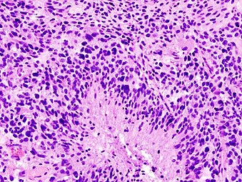
 Histopathologyof glioblastoma, showing high grade astrocytoma features of marked nuclear pleomorphism, multiple mitoses (one at white arrow) and multinucleated cells (one at black arrow), with cells having a patternless arrangement in a pink fibrillary background on H&E stain.
 -
 Lower magnification histopathology, showing necrosis surrounded bypseudopalisadesof tumor cells, conferring a diagnosis of glioblastoma rather thananaplastic astrocytoma


Prevention
editThere are no known methods to prevent glioblastoma.[3]It is the case for most gliomas, unlike for some other forms of cancer, that they happen without previous warning and there are no known ways to prevent them.[66]
Treatment
edit
Treating glioblastoma is difficult due to several complicating factors:[67]
- The tumor cells are resistant to conventional therapies.
- The brain is susceptible to damage from conventional therapy.
- The brain has a limited capacity to repair itself.
- Many drugs cannot cross theblood–brain barrierto act on the tumor.
Treatment of primary brain tumors consists of palliative (symptomatic) care and therapies intended to improve survival.
Symptomatic therapy
editSupportive treatment focuses on relieving symptoms and improving the patient's neurologic function. The primary supportive agents areanticonvulsantsandcorticosteroids.
- Historically, around 90% of patients with glioblastoma underwent anticonvulsant treatment, although only an estimated 40% of patients required this treatment. Neurosurgeons have recommended that anticonvulsants not be administered prophylactically, and should wait until a seizure occurs before prescribing this medication.[68]Those receivingphenytoinconcurrent with radiation may have serious skin reactions such aserythema multiformeandStevens–Johnson syndrome.
- Corticosteroids, usuallydexamethasone,can reduce peritumoral edema (through rearrangement of the blood–brain barrier), diminishing mass effect and lowering intracranial pressure, with a decrease in headache or drowsiness.
Surgery
edit
Surgery is the first stage of treatment of glioblastoma. An average GBM tumor contains 1011cells, which is on average reduced to 109cells after surgery (a reduction of 99%). Benefits of surgery include resection for a pathological diagnosis, alleviation of symptoms related to mass effect, and potentially removing disease before secondary resistance to radiotherapy and chemotherapy occurs.[69]
The greater the extent of tumor removal, the better. In retrospective analyses, removal of 98% or more of the tumor has been associated with a significantly longer healthier time than if less than 98% of the tumor is removed.[70]The chances of near-complete initial removal of the tumor may be increased if the surgery is guided by a fluorescent dye known as5-aminolevulinic acid.[71][72]GBM cells are widely infiltrative through the brain at diagnosis, and despite a "total resection" of all obvious tumor, most people with GBM later develop recurrent tumors either near the original site or at more distant locations within the brain. Other modalities, typically radiation and chemotherapy, are used after surgery in an effort to suppress and slow recurrent disease through damaging the DNA of rapidly proliferative GBM cells.[73]
Between 60-85% of glioblastoma patients report cancer-related cognitive impairments following surgery, which refers to problems with executive functioning, verbal fluency, attention, and speed of processing.[74][75][76]These symptoms may be managed with cognitive behavioral therapy,[77][75]physical exercise, yoga and meditation.[77][75][78]
Radiotherapy
edit
Subsequent to surgery, radiotherapy becomes the mainstay of treatment for people with glioblastoma. It is typically performed along with givingtemozolomide.[10]A pivotal clinical trial carried out in the early 1970s showed that among 303 GBM patients randomized to radiation or best medical therapy, those who received radiation had a median survival more than double those who did not.[79]Subsequent clinical research has attempted to build on the backbone of surgery followed by radiation. Whole-brain radiotherapy does not improve when compared to the more precise and targeted three-dimensional conformal radiotherapy.[80]A total radiation dose of 60–65 Gy has been found to be optimal for treatment.[81]
GBM tumors are well known to contain zones of tissue exhibitinghypoxia,which are highly resistant to radiotherapy. Various approaches to chemotherapy radiosensitizers have been pursued, with limited success as of 2016[update].As of 2010[update],newer research approaches included preclinical and clinical investigations into the use of anoxygen diffusion-enhancing compoundsuch astrans sodium crocetinateasradiosensitizers,[82]and as of 2015[update]a clinical trial was underway.[83]Boron neutron capture therapyhas been tested as an alternative treatment for glioblastoma, but is not in common use.
Chemotherapy
editMost studies show no benefit from the addition of chemotherapy. However, a large clinical trial of 575 participants randomized to standard radiation versus radiation plus temozolomide chemotherapy showed that the group receiving temozolomide survived a median of 14.6 months as opposed to 12.1 months for the group receiving radiation alone.[10][84]This treatment regimen is now standard for most cases of glioblastoma where the person is not enrolled in aclinical trial.[85][86]Temozolomide seems to work by sensitizing the tumor cells to radiation, and appears more effective for tumors withMGMTpromoter methylation.[87]High doses of temozolomide in high-grade gliomas yield low toxicity, but the results are comparable to the standard doses.[88]Antiangiogenic therapywith medications such asbevacizumabcontrol symptoms, but do not appear to affect overall survival in those with glioblastoma. A 2018systematic reviewfound that the overall benefit of anti-angiogenic therapies was unclear.[89]In elderly people with newly diagnosed glioblastoma who are reasonably fit, concurrent and adjuvant chemoradiotherapy gives the best overall survival but is associated with a greater risk of haematological adverse events than radiotherapy alone.[90]
Immunotherapy
editPhase 3 clinical trials of immunotherapy treatments for glioblastoma have largely failed.[91]
Other procedures
editAlternating electric field therapyis an FDA-approved therapy for newly diagnosed[92]and recurrent glioblastoma.[93]In 2015, initial results from a phase-III randomized clinical trial of alternating electric field therapy plus temozolomide in newly diagnosed glioblastoma reported a three-month improvement in progression-free survival, and a five-month improvement in overall survival compared to temozolomide therapy alone,[94][95]representing the first large trial in a decade to show a survival improvement in this setting.[95]Despite these results, the efficacy of this approach remains controversial among medical experts.[96]However, increasing understanding of the mechanistic basis through which alternating electric field therapy exerts anti-cancer effects and results from ongoing phase-III clinical trials in extracranial cancers may help facilitate increased clinical acceptance to treat glioblastoma in the future.[97]
Prognosis
editThe most common length of survival following diagnosis is 10 to 13 months (although recent research points to a median survival rate of 15 months),[98][99][8]with fewer than 1–3% of people surviving longer than five years.[2][5][100]In the United States between 2012 and 2016 five-year survival was 6.8%.[5]Without treatment, survival is typically three months.[14]Complete cures are extremely rare, but have been reported.[101][102]
Increasing age (> 60 years) carries a worse prognostic risk. Death is usually due to widespread tumor infiltration withcerebral edemaand increasedintracranial pressure.[103]
A good initialKarnofsky performance score(KPS) andMGMTmethylationare associated with longer survival.[103]A DNA test can be conducted on glioblastomas to determine whether or not thepromoterof theMGMTgeneismethylated.Patients with a methylated MGMT promoter have longer survival than those with an unmethylated MGMT promoter, due in part to increased sensitivity to temozolomide.[104]
Long-term benefits have also been associated with those patients who receive surgery, radiotherapy, and temozolomide chemotherapy.[103]However, much remains unknown about why some patients survive longer with glioblastoma. Age under 50 is linked to longer survival in GBM, as is 98%+ resection and use of temozolomide chemotherapy and better KPSs. A recent study confirms that younger age is associated with a much better prognosis, with a small fraction of patients under 40 years of age achieving a population-based cure. Cure is thought to occur when a person's risk of death returns to that of the normal population, and in GBM, this is thought to occur after 10 years.[102]
UCLA Neuro-oncology publishes real-time survival data for patients with this diagnosis.[105]
According to a 2003 study, GBM prognosis can be divided into three subgroups dependent on KPS, the age of the patient, and treatment.[106]
| Recursive partitioning analysis (RPA) class |
Definition | Historical Median Survival Time | Historical 1-Year Survival | Historical 3-Year Survival | Historical 5-Year Survival |
|---|---|---|---|---|---|
| III | Age < 50,KPS≥ 90 | 17.1 months | 70% | 20% | 14% |
| IV | Age < 50, KPS < 90 | 11.2 months | 46% | 7% | 4% |
| Age ≥ 50, KPS ≥ 70, surgical removal with good neurologic function | |||||
| V + VI | Age ≥ 50, KPS ≥ 70, no surgical removal | 7.5 months | 28% | 1% | 0% |
| Age ≥ 50, KPS < 70 |
Epidemiology
editAbout three per 100,000 people develop the disease a year,[3]although regional frequency may be much higher.[107]The frequency in England doubled between 1995 and 2015.[108]
It is the second-most commoncentral nervous systemtumor aftermeningioma.[15]It occurs more commonly in males than females.[2][3]Although the median age at diagnosis is 64,[2][3]in 2014, the broad category of brain cancers was second only to leukemia in people in the United States under 20 years of age.[109]
History
editThe termglioblastoma multiformewas introduced in 1926 byPercival BaileyandHarvey Cushing,based on the idea that the tumor originates from primitive precursors ofglial cells(glioblasts), and the highly variable appearance due to the presence of necrosis, hemorrhage, and cysts (multiform).[110]
Research
editGene therapy
editGene therapyhas been explored as a method to treat glioblastoma, and while animal models and early-phase clinical trials have been successful, as of 2017, all gene-therapy drugs that had been tested in phase-III clinical trials for glioblastoma had failed.[111][112][113]Scientists have developed the core–shell nanostructured LPLNP-PPT (long persistent luminescencenanoparticles.PPT refers to polyetherimide, PEG and trans-activator of transcription, and TRAIL is the human tumor necrosis factor-related apoptosis-induced ligand[114]) for effective gene delivery and tracking, with positive results. This is a TRAIL ligand that has been encoded to induce apoptosis of cancer cells, more specifically glioblastomas. Although this study was still in clinical trials in 2017, it has shown diagnostic and therapeutic functionalities, and will open great interest for clinical applications in stem-cell-based therapy.[115]
Other gene therapy approaches have also been explored in the context of glioblastoma, including suicide gene therapy.Suicide genetherapy is a two-step approach that includes the delivery of a foreign enzyme-gene to the cancer cells followed by activation with a pro-drug causing toxicities in the cancer-cells, which induces cell death. This approach has succeeded in animal models and small clinical studies but has not shown survival benefit in larger clinical studies. Using new, more efficient delivery vectors and suicide gene-prodrug systems could improve the clinical benefit from these types of therapies.[116]
Oncolytic virotherapy
editOncolytic virotherapyis an emerging novel treatment that is under investigation both at preclinical and clinical stages. Several viruses including herpes simplex virus, adenovirus, poliovirus, and reovirus are currently being tested in phases I and II of clinical trials for glioblastoma therapy and have shown to improve overall survival.[117]
Intranasal drug delivery
editDirect nose-to-brain drug delivery is being explored as a means to achieve higher, and hopefully more effective, drug concentrations in the brain.[118][119]A clinical phase-I/II study with glioblastoma patients in Brazil investigated the natural compoundperillyl alcoholfor intranasal delivery as anaerosol.The results were encouraging[118][120][121]and, as of 2016, a similar trial has been initiated in the United States.[122]
See also
editReferences
edit- ^abcdefghYoung RM, Jamshidi A, Davis G, Sherman JH (June 2015)."Current trends in the surgical management and treatment of adult glioblastoma".Annals of Translational Medicine.3(9): 121.doi:10.3978/j.issn.2305-5839.2015.05.10.PMC4481356.PMID26207249.
- ^abcdefghijklm"Chapter 5.16".World Cancer Report 2014.World Health Organization. 2014.ISBN978-92-832-0429-9.
- ^abcdefghijklmnopqGallego O (August 2015)."Nonsurgical treatment of recurrent glioblastoma".Current Oncology.22(4):e273 –e281.doi:10.3747/co.22.2436.PMC4530825.PMID26300678.
- ^abHart MG, Garside R, Rogers G, Stein K, Grant R (April 2013)."Temozolomide for high grade glioma".The Cochrane Database of Systematic Reviews.2013(4): CD007415.doi:10.1002/14651858.CD007415.pub2.PMC6457743.PMID23633341.
- ^abcdOstrom QT, Cioffi G, Gittleman H, Patil N, Waite K, Kruchko C, et al. (November 2019)."CBTRUS Statistical Report: Primary Brain and Other Central Nervous System Tumors Diagnosed in the United States in 2012–2016".Neuro-Oncology.21(Suppl 5):v1 –v100.doi:10.1093/neuonc/noz150.PMC6823730.PMID31675094.
- ^abcdBleeker FE, Molenaar RJ, Leenstra S (May 2012)."Recent advances in the molecular understanding of glioblastoma".Journal of Neuro-Oncology.108(1):11–27.doi:10.1007/s11060-011-0793-0.PMC3337398.PMID22270850.
- ^Tan AC, Ashley DM, López GY, Malinzak M, Friedman HS, Khasraw M (July 2020). "Management of glioblastoma: State of the art and future directions".CA.70(4). Wiley:299–312.doi:10.3322/caac.21613.hdl:10536/DRO/DU:30138185.PMID32478924.S2CID219170898.
- ^abTran B, Rosenthal MA (April 2010). "Survival comparison between glioblastoma multiforme and other incurable cancers".Journal of Clinical Neuroscience.17(4):417–421.doi:10.1016/j.jocn.2009.09.004.PMID20167494.S2CID5492993.
- ^"Chapter 3.8".World Cancer Report 2014.World Health Organization. 2014.ISBN978-92-832-0429-9.
- ^abcKhosla D (February 2016)."Concurrent therapy to enhance radiotherapeutic outcomes in glioblastoma".Annals of Translational Medicine.4(3): 54.doi:10.3978/j.issn.2305-5839.2016.01.25.PMC4740000.PMID26904576.
- ^Van Meir EG, Hadjipanayis CG, Norden AD, Shu HK, Wen PY, Olson JJ (2010)."Exciting new advances in neuro-oncology: the avenue to a cure for malignant glioma".CA.60(3):166–193.doi:10.3322/caac.20069.PMC2888474.PMID20445000.
- ^McKenney AS, Weg E, Bale TA, Wild AT, Um H, Fox MJ, et al. (6 February 2022)."Radiomic Analysis to Predict Histopathologically Confirmed Pseudoprogression in Glioblastoma Patients".Advances in Radiation Oncology.8(1): 100916.doi:10.1016/j.adro.2022.100916.PMC9873493.PMID36711062.S2CID246647975.
- ^Stupp R, Hegi ME, Mason WP, van den Bent MJ, Taphoorn MJ, Janzer RC, et al. (May 2009). "Effects of radiotherapy with concomitant and adjuvant temozolomide versus radiotherapy alone on survival in glioblastoma in a randomised phase III study: 5-year analysis of the EORTC-NCIC trial".The Lancet. Oncology.10(5):459–466.doi:10.1016/S1470-2045(09)70025-7.PMID19269895.S2CID25150249.
- ^abSchapira AH (2007).Neurology and clinical neuroscience.Philadelphia: Mosby Elsevier. p. 1336.ISBN978-0-323-07053-9.Archivedfrom the original on 29 July 2017.
- ^abMcNeill KA (November 2016). "Epidemiology of Brain Tumors".Neurologic Clinics.34(4):981–998.doi:10.1016/j.ncl.2016.06.014.PMID27720005.
- ^Ostrom QT, Patil N, Cioffi G, Waite K, Kruchko C, Barnholtz-Sloan JS (October 2020)."CBTRUS Statistical Report: Primary Brain and Other Central Nervous System Tumors Diagnosed in the United States in 2013–2017".Neuro-Oncology.22(12 Suppl 2):iv1 –iv96.doi:10.1093/neuonc/noaa200.PMC7596247.PMID33123732.
- ^Visser O, Ardanaz E, Botta L, Sant M, Tavilla A, Minicozzi P (October 2015). "Survival of adults with primary malignant brain tumours in Europe; Results of the EUROCARE-5 study".European Journal of Cancer.51(15):2231–2241.doi:10.1016/j.ejca.2015.07.032.PMID26421825.
- ^abcdeAlifieris C, Trafalis DT (August 2015). "Glioblastoma multiforme: Pathogenesis and treatment".Pharmacology & Therapeutics.152:63–82.doi:10.1016/j.pharmthera.2015.05.005.PMID25944528.
- ^Iacob G, Dinca EB (25 November 2009)."Current data and strategy in glioblastoma multiforme".Journal of Medicine and Life.2(4):386–393.PMC3019011.PMID20108752.
- ^Smoll NR, Brady Z, Scurrah KJ, Lee C, Berrington de González A, Mathews JD (6 July 2023)."Computed tomography scan radiation and brain cancer incidence".Neuro-Oncology.25(7):1368–1376.doi:10.1093/neuonc/noad012.PMC10326490.PMID36638155.
- ^Smoll NR, Brady Z, Scurrah K, Mathews JD (June 2016). "Exposure to ionizing radiation and brain cancer incidence: The Life Span Study cohort".Cancer Epidemiology.42:60–65.doi:10.1016/j.canep.2016.03.006.PMID27038588.ProQuest1797583676.
- ^Ohgaki H, Kleihues P (June 2005)."Population-based studies on incidence, survival rates, and genetic alterations in astrocytic and oligodendroglial gliomas".Journal of Neuropathology and Experimental Neurology.64(6):479–489.doi:10.1093/jnen/64.6.479.PMID15977639.
- ^Vilchez RA, Kozinetz CA, Arrington AS, Madden CR, Butel JS (June 2003). "Simian virus 40 in human cancers".The American Journal of Medicine.114(8):675–684.doi:10.1016/S0002-9343(03)00087-1.PMID12798456.
- ^Crawford JR, Santi MR, Thorarinsdottir HK, Cornelison R, Rushing EJ, Zhang H, et al. (September 2009)."Detection of human herpesvirus-6 variants in pediatric brain tumors: association of viral antigen in low grade gliomas".Journal of Clinical Virology.46(1):37–42.doi:10.1016/j.jcv.2009.05.011.PMC2749001.PMID19505845.
- ^Chi J, Gu B, Zhang C, Peng G, Zhou F, Chen Y, et al. (November 2012)."Human herpesvirus 6 latent infection in patients with glioma".The Journal of Infectious Diseases.206(9):1394–1398.doi:10.1093/infdis/jis513.PMID22962688.
- ^McFaline-Figueroa JR, Wen PY (February 2017). "The Viral Connection to Glioblastoma".Current Infectious Disease Reports.19(2): 5.doi:10.1007/s11908-017-0563-z.PMID28233187.S2CID30446699.
- ^El Baba R, Pasquereau S, Haidar Ahmad S, Monnien F, Abad M, Bibeau F, et al. (9 June 2023)."EZH2-Myc driven glioblastoma elicited by cytomegalovirus infection of human astrocytes".Oncogene.42(24):2031–2045.doi:10.1038/s41388-023-02709-3.PMC10256614.PMID37147437.
- ^Guyon J, Haidar Ahmad S, El Baba R, Le Quang M, Bikfalvi A, Daubon T, et al. (29 March 2024)."Generation of glioblastoma in mice engrafted with human cytomegalovirus-infected astrocytes".Cancer Gene Therapy.31(7):1070–1080.doi:10.1038/s41417-024-00767-7.PMC11257955.PMID38553638.
- ^Huncharek M, Kupelnick B, Wheeler L (2003). "Dietary cured meat and the risk of adult glioma: a meta-analysis of nine observational studies".Journal of Environmental Pathology, Toxicology and Oncology.22(2):129–137.doi:10.1615/JEnvPathToxOncol.v22.i2.60.PMID14533876.
- ^Kan P, Simonsen SE, Lyon JL, Kestle JR (January 2008). "Cellular phone use and brain tumor: a meta-analysis".Journal of Neuro-Oncology.86(1):71–78.doi:10.1007/s11060-007-9432-1.PMID17619826.S2CID23460254.
- ^Hardell L, Carlberg M, Hansson Mild K (August 2009). "Epidemiological evidence for an association between use of wireless phones and tumor diseases".Pathophysiology.16(2–3):113–122.doi:10.1016/j.pathophys.2009.01.003.PMID19268551.
- ^Zong H, Verhaak RG, Canoll P (May 2012)."The cellular origin for malignant glioma and prospects for clinical advancements".Expert Review of Molecular Diagnostics.12(4):383–394.doi:10.1586/erm.12.30.PMC3368274.PMID22616703.
- ^Zong H, Parada LF, Baker SJ (January 2015)."Cell of origin for malignant gliomas and its implication in therapeutic development".Cold Spring Harbor Perspectives in Biology.7(5): a020610.doi:10.1101/cshperspect.a020610.PMC4448618.PMID25635044.
- ^Seker-Polat F, Pinarbasi Degirmenci N, Solaroglu I, Bagci-Onder T (2022)."Tumor Cell Infiltration into the Brain in Glioblastoma: From Mechanisms to Clinical Perspectives".Cancers.14(2): 443.doi:10.3390/cancers14020443.PMC8773542.PMID35053605.
- ^Kazi AZ, Joshi PC, Kelkar AB, Mahajan MS, Ghawate AS (October 2013)."MRI evaluation of pathologies affecting the corpus callosum: A pictorial essay".The Indian Journal of Radiology & Imaging.23(4):321–332.doi:10.4103/0971-3026.125604.PMC3932574.PMID24604936.
- ^Kleihues P, Burger PC, Scheithauer BW (1993). "Introduction".Histological Typing of Tumours of the Central Nervous System.pp.1–3.doi:10.1007/978-3-642-84988-6_1.ISBN978-3-540-56971-8.
- ^Louis DN, Ohgaki H, Wiestler OD, Cavenee WK, Burger PC, Jouvet A, et al. (August 2007)."The 2007 WHO classification of tumours of the central nervous system".Acta Neuropathologica.114(2):97–109.doi:10.1007/s00401-007-0243-4.PMC1929165.PMID17618441.
- ^Louis DN, Perry A, Reifenberger G, von Deimling A, Figarella-Branger D, Cavenee WK, et al. (June 2016)."The 2016 World Health Organization Classification of Tumors of the Central Nervous System: a summary".Acta Neuropathologica.131(6):803–820.doi:10.1007/s00401-016-1545-1.PMID27157931.
- ^Louis DN, Perry A, Wesseling P, Brat DJ, Cree IA, Figarella-Branger D, et al. (August 2021)."The 2021 WHO Classification of Tumors of the Central Nervous System: a summary".Neuro-Oncology.23(8):1231–1251.doi:10.1093/neuonc/noab106.PMC8328013.PMID34185076.
- ^Louis DN, Perry A, Wesseling P, Brat DJ, Cree IA, Figarella-Branger D, et al. (2 August 2021)."The 2021 WHO Classification of Tumors of the Central Nervous System: a summary".Neuro-Oncology.23(8):1231–1251.doi:10.1093/neuonc/noab106.PMC8328013.PMID34185076.
- ^abWang Q, Hu B, Hu X, Kim H, Squatrito M, Scarpace L, et al. (July 2017)."Tumor Evolution of Glioma-Intrinsic Gene Expression Subtypes Associates with Immunological Changes in the Microenvironment".Cancer Cell.32(1): 42–56.e6.doi:10.1016/j.ccell.2017.06.003.PMC5599156.PMID28697342.
- ^Hayden EC (January 2010)."Genomics boosts brain-cancer work".Nature.463(7279): 278.doi:10.1038/463278a.PMID20090720.
- ^Sasmita AO, Wong YP, Ling AP (February 2018)."Biomarkers and therapeutic advances in glioblastoma multiforme".Asia-Pacific Journal of Clinical Oncology.14(1):40–51.doi:10.1111/ajco.12756.PMID28840962.
- ^abVerhaak RG, Hoadley KA, Purdom E, Wang V, Qi Y, Wilkerson MD, et al. (January 2010)."Integrated genomic analysis identifies clinically relevant subtypes of glioblastoma characterized by abnormalities in PDGFRA, IDH1, EGFR, and NF1".Cancer Cell.17(1):98–110.doi:10.1016/j.ccr.2009.12.020.PMC2818769.PMID20129251.
- ^Kuehn BM (March 2010). "Genomics illuminates a deadly brain cancer".JAMA.303(10):925–927.doi:10.1001/jama.2010.236.PMID20215599.
- ^Bleeker FE, Lamba S, Zanon C, Molenaar RJ, Hulsebos TJ, Troost D, et al. (September 2014)."Mutational profiling of kinases in glioblastoma".BMC Cancer.14(1): 718.doi:10.1186/1471-2407-14-718.PMC4192443.PMID25256166.
- ^abMolenaar RJ, Verbaan D, Lamba S, Zanon C, Jeuken JW, Boots-Sprenger SH, et al. (September 2014)."The combination of IDH1 mutations and MGMT methylation status predicts survival in glioblastoma better than either IDH1 or MGMT alone".Neuro-Oncology.16(9):1263–1273.doi:10.1093/neuonc/nou005.PMC4136888.PMID24510240.
- ^abcMolenaar RJ, Radivoyevitch T, Maciejewski JP, van Noorden CJ, Bleeker FE (December 2014). "The driver and passenger effects of isocitrate dehydrogenase 1 and 2 mutations in oncogenesis and survival prolongation".Biochimica et Biophysica Acta (BBA) - Reviews on Cancer.1846(2):326–341.doi:10.1016/j.bbcan.2014.05.004.PMID24880135.
- ^Hegi ME, Diserens AC, Gorlia T, Hamou MF, de Tribolet N, Weller M, et al. (March 2005)."MGMT gene silencing and benefit from temozolomide in glioblastoma".The New England Journal of Medicine.352(10):997–1003.doi:10.1056/NEJMoa043331.PMID15758010.
- ^Furnari FB, Fenton T, Bachoo RM, Mukasa A, Stommel JM, Stegh A, et al. (November 2007). "Malignant astrocytic glioma: genetics, biology, and paths to treatment".Genes & Development.21(21):2683–2710.doi:10.1101/gad.1596707.PMID17974913.
- ^Greenberg MS (2016).Handbook of Neurosurgery.doi:10.1055/b-006-149702.ISBN978-1-62623-242-6.[page needed]
- ^Murat A, Migliavacca E, Gorlia T, Lambiv WL, Shay T, Hamou MF, et al. (June 2008)."Stem cell-related" self-renewal "signature and high epidermal growth factor receptor expression associated with resistance to concomitant chemoradiotherapy in glioblastoma".Journal of Clinical Oncology.26(18):3015–3024.doi:10.1200/JCO.2007.15.7164.PMID18565887.
- ^Gilbertson RJ, Rich JN (October 2007). "Making a tumour's bed: glioblastoma stem cells and the vascular niche".Nature Reviews. Cancer.7(10):733–736.doi:10.1038/nrc2246.PMID17882276.S2CID2634215.
- ^Brown DV, Stylli SS, Kaye AH, Mantamadiotis T (2019). "Multilayered Heterogeneity of Glioblastoma Stem Cells: Biological and Clinical Significance".Stem Cells Heterogeneity in Cancer.Advances in Experimental Medicine and Biology. Vol. 1139. pp.1–21.doi:10.1007/978-3-030-14366-4_1.ISBN978-3-030-14365-7.PMID31134492.S2CID167220115.
- ^Annovazzi L, Mellai M, Schiffer D (May 2017)."Chemotherapeutic Drugs: DNA Damage and Repair in Glioblastoma".Cancers.9(6): 57.doi:10.3390/cancers9060057.PMC5483876.PMID28587121.
- ^Molenaar RJ (2011)."Ion channels in glioblastoma".ISRN Neurology.2011:590249.doi:10.5402/2011/590249.PMC3263536.PMID22389824.
- ^Møller HG, Rasmussen AP, Andersen HH, Johnsen KB, Henriksen M, Duroux M (February 2013)."A systematic review of microRNA in glioblastoma multiforme: micro-modulators in the mesenchymal mode of migration and invasion".Molecular Neurobiology.47(1):131–144.doi:10.1007/s12035-012-8349-7.PMC3538124.PMID23054677.
- ^Godlewski J, Nowicki MO, Bronisz A, Nuovo G, Palatini J, De Lay M, et al. (March 2010)."MicroRNA-451 regulates LKB1/AMPK signaling and allows adaptation to metabolic stress in glioma cells".Molecular Cell.37(5):620–632.doi:10.1016/j.molcel.2010.02.018.PMC3125113.PMID20227367.
- ^Bhaskaran V, Nowicki MO, Idriss M, Jimenez MA, Lugli G, Hayes JL, et al. (January 2019)."The functional synergism of microRNA clustering provides therapeutically relevant epigenetic interference in glioblastoma".Nature Communications.10(1): 442.Bibcode:2019NatCo..10..442B.doi:10.1038/s41467-019-08390-z.PMC6347618.PMID30683859.
- ^abDimberg A (December 2014)."The glioblastoma vasculature as a target for cancer therapy"(PDF).Biochemical Society Transactions.42(6):1647–1652.doi:10.1042/BST20140278.PMID25399584.
- ^Jain RK (June 2013)."Normalizing tumor microenvironment to treat cancer: bench to bedside to biomarkers".Journal of Clinical Oncology.31(17):2205–2218.doi:10.1200/JCO.2012.46.3653.PMC3731977.PMID23669226.
- ^abcdeWeller M, van den Bent M, Preusser M, Le Rhun E, Tonn JC, Minniti G, et al. (March 2021)."EANO guidelines on the diagnosis and treatment of diffuse gliomas of adulthood".Nature Reviews. Clinical Oncology.18(3):170–186.doi:10.1038/s41571-020-00447-z.PMC7904519.PMID33293629.
- ^Smirniotopoulos JG, Murphy FM, Rushing EJ, Rees JH, Schroeder JW (2007). "Patterns of contrast enhancement in the brain and meninges".Radiographics.27(2):525–551.doi:10.1148/rg.272065155.PMID17374867.
- ^Eibl RH, Schneemann M (February 2023)."Liquid biopsy and glioblastoma".Exploration of Targeted Anti-Tumor Therapy.4(1):28–41.doi:10.37349/etat.2023.00121.PMC10017188.PMID36937320.
- ^Mo H, Magaki S, Deisch JK, Raghavan R (August 2022)."Isocitrate Dehydrogenase Mutations Are Associated with Different Expression and DNA Methylation Patterns of OLIG2 in Adult Gliomas".Journal of Neuropathology and Experimental Neurology.81(9):707–716.doi:10.1093/jnen/nlac059.PMC9614687.PMID35856894.
- ^"Gliomas Prevention".The James Cancer Hospital.Ohio State University.Archivedfrom the original on 9 October 2021.Retrieved9 October2021.
- ^Lawson HC, Sampath P, Bohan E, Park MC, Hussain N, Olivi A, et al. (May 2007)."Interstitial chemotherapy for malignant gliomas: the Johns Hopkins experience".Journal of Neuro-Oncology.83(1):61–70.doi:10.1007/s11060-006-9303-1.PMC4086528.PMID17171441.
- ^Stevens GH (July 2006). "Antiepileptic therapy in patients with central nervous system malignancies".Current Neurology and Neuroscience Reports.6(4):311–318.doi:10.1007/s11910-006-0024-9.PMID16822352.S2CID37712742.
- ^Das S, Marsden PA (October 2013). Phimister EG (ed.)."Angiogenesis in glioblastoma".The New England Journal of Medicine.369(16):1561–1563.doi:10.1056/NEJMcibr1309402.PMC5378489.PMID24131182.
- ^Lacroix M, Abi-Said D, Fourney DR, Gokaslan ZL, Shi W, DeMonte F, et al. (August 2001). "A multivariate analysis of 416 patients with glioblastoma multiforme: prognosis, extent of resection, and survival".Journal of Neurosurgery.95(2):190–198.doi:10.3171/jns.2001.95.2.0190.PMID11780887.
- ^Stummer W, Pichlmeier U, Meinel T, Wiestler OD, Zanella F, Reulen HJ (May 2006). "Fluorescence-guided surgery with 5-aminolevulinic acid for resection of malignant glioma: a randomised controlled multicentre phase III trial".The Lancet. Oncology.7(5):392–401.doi:10.1016/S1470-2045(06)70665-9.PMID16648043.
- ^Schatlo B, Fandino J, Smoll NR, Wetzel O, Remonda L, Marbacher S, et al. (December 2015)."Outcomes after combined use of intraoperative MRI and 5-aminolevulinic acid in high-grade glioma surgery".Neuro-Oncology.17(12):1560–1567.doi:10.1093/neuonc/nov049.PMC4633924.PMID25858636.
- ^Rominiyi O, Collis SJ (January 2022)."DDRugging glioblastoma: understanding and targeting the DNA damage response to improve future therapies".Molecular Oncology.16(1):11–41.doi:10.1002/1878-0261.13020.PMC8732357.PMID34036721.
- ^Sinha R, Stephenson JM, Price SJ (March 2020)."A systematic review of cognitive function in patients with glioblastoma undergoing surgery".Neuro-Oncology Practice.7(2):131–142.doi:10.1093/nop/npz018.PMC7318858.PMID32626582.
- ^abcPendergrass JC, Targum SD, Harrison JE (February 2018)."Cognitive Impairment Associated with Cancer: A Brief Review".Innovations in Clinical Neuroscience.15(1–2):36–44.PMC5819720.PMID29497579.
- ^Janelsins MC, Kesler SR, Ahles TA, Morrow GR (February 2014)."Prevalence, mechanisms, and management of cancer-related cognitive impairment".International Review of Psychiatry.26(1):102–113.doi:10.3109/09540261.2013.864260.PMC4084673.PMID24716504.
- ^abLange M, Joly F, Vardy J, Ahles T, Dubois M, Tron L, et al. (December 2019)."Cancer-related cognitive impairment: an update on state of the art, detection, and management strategies in cancer survivors".Annals of Oncology.30(12):1925–1940.doi:10.1093/annonc/mdz410.PMC8109411.PMID31617564.
- ^Biegler KA, Chaoul MA, Cohen L (2009). "Cancer, cognitive impairment, and meditation".Acta Oncologica.48(1):18–26.doi:10.1080/02841860802415535.PMID19031161.
- ^Walker MD, Alexander E, Hunt WE, MacCarty CS, Mahaley MS, Mealey J, et al. (September 1978). "Evaluation of BCNU and/or radiotherapy in the treatment of anaplastic gliomas. A cooperative clinical trial".Journal of Neurosurgery.49(3):333–343.doi:10.3171/jns.1978.49.3.0333.PMID355604.
- ^Showalter TN, Andrel J, Andrews DW, Curran WJ, Daskalakis C, Werner-Wasik M (November 2007). "Multifocal glioblastoma multiforme: prognostic factors and patterns of progression".International Journal of Radiation Oncology, Biology, Physics.69(3):820–824.doi:10.1016/j.ijrobp.2007.03.045.PMID17499453.
- ^Fulton DS, Urtasun RC, Scott-Brown I, Johnson ES, Mielke B, Curry B, et al. (September 1992). "Increasing radiation dose intensity using hyperfractionation in patients with malignant glioma. Final report of a prospective phase I-II dose response study".Journal of Neuro-Oncology.14(1):63–72.doi:10.1007/BF00170946.PMID1335044.S2CID24245934.
- ^Sheehan JP, Shaffrey ME, Gupta B, Larner J, Rich JN, Park DM (October 2010). "Improving the radiosensitivity of radioresistant and hypoxic glioblastoma".Future Oncology.6(10):1591–1601.doi:10.2217/fon.10.123.PMID21062158.
- ^ Clinical trial numberNCT01465347for "Safety and Efficacy Study of Trans Sodium Crocetinate (TSC) With Concomitant Radiation Therapy and Temozolomide in Newly Diagnosed Glioblastoma (GBM)" atClinicalTrials.gov,accessed 1 February 2016
- ^Stupp R, Mason WP, van den Bent MJ, Weller M, Fisher B, Taphoorn MJ, et al. (European Organisation for Research Treatment of Cancer Brain Tumor Radiotherapy Groups, National Cancer Institute of Canada Clinical Trials Group) (March 2005)."Radiotherapy plus concomitant and adjuvant temozolomide for glioblastoma".The New England Journal of Medicine.352(10):987–996.doi:10.1056/NEJMoa043330.PMID15758009.S2CID41340212.
- ^Mason WP, Mirimanoff RO, Stupp R (2006). "Radiotherapy with Concurrent and Adjuvant Temozolomide: A New Standard of Care for Glioblastoma Multiforme".Progress in Neurotherapeutics and Neuropsychopharmacology.1:37–52.doi:10.1017/S1748232105000054.
- ^"Temozolomide Plus Radiation Helps Brain Cancer – National Cancer Institute".Archived fromthe originalon 15 August 2007.Retrieved15 September2007.
- ^Chamberlain MC, Glantz MJ, Chalmers L, Van Horn A, Sloan AE (March 2007). "Early necrosis following concurrent Temodar and radiotherapy in patients with glioblastoma".Journal of Neuro-Oncology.82(1):81–83.doi:10.1007/s11060-006-9241-y.PMID16944309.S2CID6262668.
- ^Dall'oglio S, D'Amico A, Pioli F, Gabbani M, Pasini F, Passarin MG, et al. (December 2008). "Dose-intensity temozolomide after concurrent chemoradiotherapy in operated high-grade gliomas".Journal of Neuro-Oncology.90(3):315–319.doi:10.1007/s11060-008-9663-9.PMID18688571.S2CID21517366.
- ^Ameratunga M, Pavlakis N, Wheeler H, Grant R, Simes J, Khasraw M (November 2018)."Anti-angiogenic therapy for high-grade glioma".The Cochrane Database of Systematic Reviews.2018(11): CD008218.doi:10.1002/14651858.CD008218.pub4.PMC6516839.PMID30480778.
The use of anti-angiogenic therapy does not significantly improve overall survival in newly diagnosed people with glioblastoma. Thus, there is insufficient evidence to support the use of anti-angiogenic therapy for people with newly diagnosed glioblastoma at this time.
- ^Hanna C, Lawrie TA, Rogozińska E, Kernohan A, Jefferies S, Bulbeck H, et al. (March 2020)."Treatment of newly diagnosed glioblastoma in the elderly: a network meta-analysis".The Cochrane Database of Systematic Reviews.2020(3): CD013261.doi:10.1002/14651858.cd013261.pub2.PMC7086476.PMID32202316.
- ^Habashy KJ, Mansour R, Moussalem C, Sawaya R, Massaad MJ (October 2022)."Challenges in glioblastoma immunotherapy: mechanisms of resistance and therapeutic approaches to overcome them".British Journal of Cancer.127(6):976–987.doi:10.1038/s41416-022-01864-w.PMC9470562.PMID35662275.
- ^"FDA approves expanded indication for medical device to treat a form of brain cancer".Food and Drug Administration.Archivedfrom the original on 23 March 2016.Retrieved19 March2016.
- ^"FDA approval letter – NovoTTF-100A System"(PDF).www.fda.gov.Archived(PDF)from the original on 22 September 2015.Retrieved26 December2014.
- ^Stupp R, Taillibert S, Kanner AA, Kesari S, Steinberg DM, Toms SA, et al. (December 2015)."Maintenance Therapy With Tumor-Treating Fields Plus Temozolomide vs Temozolomide Alone for Glioblastoma: A Randomized Clinical Trial".JAMA.314(23):2535–2543.doi:10.1001/jama.2015.16669.PMID26670971.
- ^abSampson JH (December 2015). "Alternating Electric Fields for the Treatment of Glioblastoma".JAMA.314(23):2511–2513.doi:10.1001/jama.2015.16701.PMID26670969.
- ^Wick W (March 2016)."TTFields: where does all the skepticism come from?".Neuro-Oncology.18(3):303–305.doi:10.1093/neuonc/now012.PMC4767251.PMID26917587.
- ^Rominiyi O, Vanderlinden A, Clenton SJ, Bridgewater C, Al-Tamimi Y, Collis SJ (February 2021)."Tumour treating fields therapy for glioblastoma: current advances and future directions".British Journal of Cancer.124(4):697–709.doi:10.1038/s41416-020-01136-5.PMC7884384.PMID33144698.
- ^Grochans S, Cybulska AM, Simińska D, Korbecki J, Kojder K, Chlubek D, et al. (May 2022)."Epidemiology of Glioblastoma Multiforme-Literature Review".Cancers.14(10): 2412.doi:10.3390/cancers14102412.PMC9139611.PMID35626018.
- ^Koshy M, Villano JL, Dolecek TA, Howard A, Mahmood U, Chmura SJ, et al. (March 2012)."Improved survival time trends for glioblastoma using the SEER 17 population-based registries".Journal of Neuro-Oncology.107(1):207–212.doi:10.1007/s11060-011-0738-7.PMC4077033.PMID21984115.
- ^Smoll NR, Schaller K, Gautschi OP (May 2013). "Long-term survival of patients with glioblastoma multiforme (GBM)".Journal of Clinical Neuroscience.20(5):670–675.doi:10.1016/j.jocn.2012.05.040.PMID23352352.S2CID5088829.
- ^Caruso R, Pesce A, Wierzbicki V (20 February 2017)."A very rare case report of long-term survival: A patient operated on in 1994 of glioblastoma multiforme and currently in perfect health".International Journal of Surgery Case Reports.33:41–43.doi:10.1016/j.ijscr.2017.02.025.PMC5338899.PMID28273605.
- ^abSmoll NR, Schaller K, Gautschi OP (2012)."The cure fraction of glioblastoma multiforme".Neuroepidemiology.39(1):63–69.doi:10.1159/000339319.PMID22776797.
- ^abcKrex D, Klink B, Hartmann C, von Deimling A, Pietsch T, Simon M, et al. (October 2007)."Long-term survival with glioblastoma multiforme".Brain.130(Pt 10):2596–2606.doi:10.1093/brain/awm204.PMID17785346.
- ^Martinez R, Schackert G, Yaya-Tur R, Rojas-Marcos I, Herman JG, Esteller M (May 2007). "Frequent hypermethylation of the DNA repair gene MGMT in long-term survivors of glioblastoma multiforme".Journal of Neuro-Oncology.83(1):91–93.doi:10.1007/s11060-006-9292-0.PMID17164975.S2CID34370292.
- ^"University of California, Los Angeles Neuro-Oncology: How Our Patients Perform: Glioblastoma Multiforme [GBM]".Archived fromthe originalon 9 June 2012..Neurooncology.ucla.edu. Retrieved on 19 October 2010.
- ^Shaw EG, Seiferheld W, Scott C, Coughlin C, Leibel S, Curran W, et al. (2003)."Reexamining the radiation therapy oncology group (RTOG) recursive partitioning analysis (RPA) for glioblastoma multiforme (GBM) patients".International Journal of Radiation Oncology, Biology, Physics.57(2): S135–36.doi:10.1016/S0360-3016(03)00843-5.
- ^Xu H, Chen J, Xu H, Qin Z (2017)."Geographic Variations in the Incidence of Glioblastoma and Prognostic Factors Predictive of Overall Survival in US Adults from 2004–2013".Frontiers in Aging Neuroscience.9:352.doi:10.3389/fnagi.2017.00352.PMC5681990.PMID29163134.
- ^Philips A, Henshaw DL, Lamburn G, O'Carroll MJ (2018)."Brain Tumours: Rise in Glioblastoma Multiforme Incidence in England 1995–2015 Suggests an Adverse Environmental or Lifestyle Factor".Journal of Environmental and Public Health.2018:7910754.doi:10.1155/2018/7910754.PMC6035820.PMID30034480.
- ^Siegel DA, Li J, Henley SJ, Wilson RJ, Lunsford NB, Tai E, et al. (June 2018)."Geographic Variation in Pediatric Cancer Incidence – United States, 2003–2014".MMWR. Morbidity and Mortality Weekly Report.67(25):707–713.doi:10.15585/mmwr.mm6725a2.PMC6023185.PMID29953430.
- ^Bailey & Cushing:Tumors of the Glioma Group.JB Lippincott, Philadelphia, 1926.[page needed]
- ^Rajesh Y, Pal I, Banik P, Chakraborty S, Borkar SA, Dey G, et al. (May 2017)."Insights into molecular therapy of glioma: current challenges and next generation blueprint".Acta Pharmacologica Sinica.38(5):591–613.doi:10.1038/aps.2016.167.PMC5457688.PMID28317871.
- ^Tobias A, Ahmed A, Moon KS, Lesniak MS (February 2013)."The art of gene therapy for glioma: a review of the challenging road to the bedside".Journal of Neurology, Neurosurgery, and Psychiatry.84(2):213–222.doi:10.1136/jnnp-2012-302946.PMC3543505.PMID22993449.
- ^Fulci G, Chiocca EA (February 2007)."The status of gene therapy for brain tumors".Expert Opinion on Biological Therapy.7(2):197–208.doi:10.1517/14712598.7.2.197.PMC2819130.PMID17250458.
- ^Borchers A, Pieler T (November 2010)."Programming pluripotent precursor cells derived from Xenopus embryos to generate specific tissues and organs".Genes.1(3). MDPI AG:413–426.doi:10.3390/chemosensors8040117.PMC3966229.PMID24710095.
- ^Wu SQ, Yang CX, Yan XP (March 2017). "A Dual-Functional Persistently Luminescent Nanocomposite Enables Engineering of Mesenchymal Stem Cells for Homing and Gene Therapy of Glioblastoma".Advanced Functional Materials.27(11): 1604992.doi:10.1002/adfm.201604992.S2CID99147218.
- ^Hossain JA, Marchini A, Fehse B, Bjerkvig R, Miletic H (1 January 2020)."Suicide gene therapy for the treatment of high-grade glioma: past lessons, present trends, and future prospects".Neuro-Oncology Advances.2(1): vdaa013.doi:10.1093/noajnl/vdaa013.PMC7212909.PMID32642680.
- ^Suryawanshi YR, Schulze AJ (July 2021)."Oncolytic Viruses for Malignant Glioma: On the Verge of Success?".Viruses.13(7): 1294.doi:10.3390/v13071294.PMC8310195.PMID34372501.
- ^abvan Woensel M, Wauthoz N, Rosière R, Amighi K, Mathieu V, Lefranc F, et al. (August 2013)."Formulations for Intranasal Delivery of Pharmacological Agents to Combat Brain Disease: A New Opportunity to Tackle GBM?".Cancers.5(3):1020–1048.doi:10.3390/cancers5031020.PMC3795377.PMID24202332.
- ^Pardeshi CV, Belgamwar VS (July 2013). "Direct nose to brain drug delivery via integrated nerve pathways bypassing the blood-brain barrier: an excellent platform for brain targeting".Expert Opinion on Drug Delivery.10(7):957–972.doi:10.1517/17425247.2013.790887.PMID23586809.S2CID8020921.
- ^Peterson A, Bansal A, Hofman F, Chen TC, Zada G (February 2014). "A systematic review of inhaled intranasal therapy for central nervous system neoplasms: an emerging therapeutic option".Journal of Neuro-Oncology.116(3):437–446.doi:10.1007/s11060-013-1346-5.PMID24398618.S2CID2414770.
- ^Chen TC, Fonseca CO, Schönthal AH (2015)."Preclinical development and clinical use of perillyl alcohol for chemoprevention and cancer therapy".American Journal of Cancer Research.5(5):1580–1593.PMC4497427.PMID26175929.
- ^Clinical trial numberNCT02704858for "Safety and Efficacy Study in Recurrent Grade IV Glioma" atClinicalTrials.gov
External links
edit- Information about glioblastomaArchived5 February 2024 at theWayback Machinefrom theAmerican Brain Tumor Association