Grey matter,orgray matterinAmerican English,is a major component of thecentral nervous system,consisting ofneuronalcell bodies,neuropil(dendritesand unmyelinatedaxons),glial cells(astrocytesandoligodendrocytes),synapses,andcapillaries.Grey matter is distinguished fromwhite matterin that it contains numerous cell bodies and relatively few myelinated axons, while white matter contains relatively few cell bodies and is composed chiefly of long-range myelinated axons.[1]The colour difference arises mainly from the whiteness ofmyelin.In living tissue, grey matter actually has a very light grey colour with yellowish or pinkish hues, which come from capillary blood vessels and neuronal cell bodies.[2]
| Grey matter | |
|---|---|
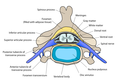
 The formation of the spinal nerve from the dorsal and ventral roots (with grey matter labelled at centre right). | |
 Micrographshowing grey matter, with the characteristicneuronal cell bodies(dark shade of pink), andwhite matterwith its characteristic fine meshwork-like appearance (left of image; lighter shade of pink).HPS stain. | |
| Details | |
| Identifiers | |
| Latin | substantia grisea |
| MeSH | D066128 |
| TA98 | A14.1.00.002 A14.1.02.020 A14.1.04.201 A14.1.05.201 A14.1.05.401 A14.1.06.301 |
| TA2 | 5365 |
| FMA | 67242 |
| Anatomical terminology | |
Structure
editGrey matter refers to unmyelinatedneuronsand other cells of thecentral nervous system.It is present in thebrain,brainstemandcerebellum,and present throughout thespinal cord.
Grey matter is distributed at the surface of thecerebral hemispheres(cerebral cortex) and of the cerebellum (cerebellar cortex), as well as in the depths of the cerebrum (thethalamus;hypothalamus;subthalamus,basal ganglia–putamen,globus pallidusandnucleus accumbens;as well as theseptal nuclei), cerebellum (deep cerebellar nuclei – thedentate nuclei,globose nucleus,emboliform nucleus,andfastigial nucleus), andbrainstem(thesubstantia nigra,red nucleus,olivary nuclei,andcranial nerve nuclei).
Grey matter in the spinal cord is known as thegrey columnwhich travels down the spinal cord distributed in three grey columns that are presented in an "H" shape. The forward-facing column is theanterior grey column,the rear-facing one is theposterior grey columnand the interlinking one is thelateral grey column.The grey matter on the left and right side is connected by thegrey commissure.The grey matter in the spinal cord consists ofinterneurons,as well as thecell bodiesofprojection neurons.
-
 Cross-section of a spinalvertebrawith the spinal cord in the centre (and grey matter labelled).
 -
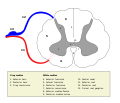 Cross-section ofspinal cordwith the grey matter labelled.


Grey matter undergoes development and growth throughout childhood and adolescence.[3]Recent studies using cross-sectional neuroimaging have shown that by around the age of 8 the volume of grey matter begins to decrease.[4]However, the density of grey matter appears to increase as a child develops into early adulthood.[4]Males tend to exhibit grey matter of increased volume but lower density than that of females.[5]
Function
editGrey matter contains most of the brain's neuronal cell bodies.[6]The grey matter includes regions of the brain involved in muscle control, and sensory perception such as seeing and hearing, memory, emotions, speech, decision-making, and self-control.
The grey matter in thespinal cordis split into three grey columns:
- Theanterior grey columncontainsmotor neurons.Thesesynapsewith interneurons and theaxonsof cells that have travelled down thepyramidal tract.These cells are responsible for the movement of muscles.
- Theposterior grey columncontains the points wheresensory neuronssynapse. These receive sensory information from the body, includingfine touch,proprioception,andvibration.This information is sent from receptors of the skin, bones, and joints through sensory neurons whose cell bodies lie in the dorsal root ganglion. This information is then transmitted in axons up the spinal cord in spinal tracts, including thedorsal column-medial lemniscus tractand thespinothalamic tract.
- Thelateral grey columnis the third column of the spinal cord.
The grey matter of the spinal cord can be divided into different layers, calledRexed laminae.These describe, in general, the purpose of the cells within the grey matter of the spinal cord at a particular location.
-
 Interneuronspresent in the grey matter of the spinal cord
 -
 Rexed laminaegroups the grey matter in the spinal cord according to its function.


Clinical significance
editHighalcohol consumptionhas been correlated with significant reductions in grey matter volume.[7][8]Short-termcannabisuse (30 days) is not correlated with changes inwhiteor grey matter.[9]However, several cross-sectional studies have shown that repeated long-term cannabis use is associated with smaller grey matter volumes in thehippocampus,amygdala,medialtemporal cortex,andprefrontal cortex,with increased grey matter volume in the cerebellum.[10][11][12]Long-term cannabis use is also associated with alterations in white matter integrity in an age-dependent manner,[13]with heavy cannabis use during adolescence and early adulthood associated with the greatest amount of change.[14]
Meditation has been shown to change grey matter structure.[15][16][17][18][19]
Habitual playing of action video games has been reported to promote a reduction of grey matter in the hippocampus while 3D platformer games have been reported to increase grey matter in the hippocampus.[20][21][22]
Women and men with equivalent IQ scores have differing proportions of grey to white matter in cortical brain regions associated with intelligence.[23]
Pregnancy renders substantial changes in brain structure, primarily reductions in grey matter volume in regions subserving social cognition. Grey matter reductions endure for at least 2 years post-pregnancy.[24]The profile of brain changes is comparable to that taking place during adolescence, a hormonally similar transitional period of life.[25]
History
editEtymology
editIn the current edition[26]of the official Latin nomenclature,Terminologia Anatomica,substantia griseais used for Englishgrey matter.The adjectivegriseaforgreyis however not attested inclassical Latin.[27]The adjectivegriseais derived from theFrenchword for grey,gris.[27]Alternative designations likesubstantia cana[28]andsubstantia cinerea[29]are being used alternatively. The adjectivecana,attested in classical Latin,[30]can meangrey,[27]orgreyish white.[31]The classical Latincinereameansash-coloured.[30]
Additional images
edit-
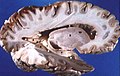 Human brain right dissected lateral view
 -
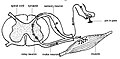
 Schematic representation of the chief ganglionic categories (I to V).


See also
editReferences
edit- ^Purves D, Augustine GJ, Fitzpatrick D, Hall WC, LaMantia AS, McNamara JO, White LE (2008).Neuroscience(4th ed.). Sinauer Associates. pp.15–16.ISBN978-0-87893-697-7.
- ^Kolb B, Whishaw IQ (2003).Fundamentals of human neuropsychology(5th ed.). New York: Worth Publishing. p. 49.ISBN978-0-7167-5300-1.
- ^Sowell ER, Thompson PM, Tessner KD, Toga AW (November 2001)."Mapping continued brain growth and gray matter density reduction in dorsal frontal cortex: Inverse relationships during postadolescent brain maturation".The Journal of Neuroscience.21(22):8819–29.doi:10.1523/JNEUROSCI.21-22-08819.2001.PMC6762261.PMID11698594.
- ^abGennatas ED, Avants BB, Wolf DH, Satterthwaite TD, Ruparel K, Ciric R, Hakonarson H, Gur RE, Gur RC (May 2017)."Age-Related Effects and Sex Differences in Gray Matter Density, Volume, Mass, and Cortical Thickness from Childhood to Young Adulthood".The Journal of Neuroscience.37(20):5065–5073.doi:10.1523/JNEUROSCI.3550-16.2017.PMC5444192.PMID28432144.
- ^Luders, Eileen; Gaser, Christian; Narr, Katherine L.; Toga, Arthur W. (11 November 2009)."Why Sex Matters: Brain Size Independent Differences in Gray Matter Distributions between Men and Women".The Journal of Neuroscience.29(45):14265–14270.doi:10.1523/JNEUROSCI.2261-09.2009.PMC3110817.PMID19906974.
- ^Miller AK, Alston RL, Corsellis JA (1980). "Variation with age in the volumes of grey and white matter in the cerebral hemispheres of man: measurements with an image analyser".Neuropathology and Applied Neurobiology.6(2):119–32.doi:10.1111/j.1365-2990.1980.tb00283.x.PMID7374914.S2CID23201991.
- ^Yang X, Tian F, Zhang H, Zeng J, Chen T, Wang S, Jia Z, Gong Q (July 2016). "Cortical and subcortical gray matter shrinkage in alcohol-use disorders: a voxel-based meta-analysis".Neuroscience and Biobehavioral Reviews.66:92–103.doi:10.1016/j.neubiorev.2016.03.034.PMID27108216.S2CID19928689.
- ^Xiao P, Dai Z, Zhong J, Zhu Y, Shi H, Pan P (August 2015). "Regional gray matter deficits in alcohol dependence: A meta-analysis of voxel-based morphometry studies".Drug and Alcohol Dependence.153:22–8.doi:10.1016/j.drugalcdep.2015.05.030.PMID26072220.
- ^Thayer RE, YorkWilliams S, Karoly HC, Sabbineni A, Ewing SF, Bryan AD, Hutchison KE (December 2017)."Structural neuroimaging correlates of alcohol and cannabis use in adolescents and adults".Addiction.112(12):2144–2154.doi:10.1111/add.13923.PMC5673530.PMID28646566.
- ^Lorenzetti V, Lubman DI, Whittle S, Solowij N, Yücel M (September 2010). "Structural MRI findings in long-term cannabis users: what do we know?".Substance Use & Misuse.45(11):1787–808.doi:10.3109/10826084.2010.482443.PMID20590400.S2CID22127231.
- ^Matochik JA, Eldreth DA, Cadet JL, Bolla KI (January 2005). "Altered brain tissue composition in heavy marijuana users".Drug and Alcohol Dependence.77(1):23–30.doi:10.1016/j.drugalcdep.2004.06.011.PMID15607838.
- ^Yücel M, Solowij N, Respondek C, Whittle S, Fornito A, Pantelis C, Lubman DI (June 2008)."Regional brain abnormalities associated with long-term heavy cannabis use".Archives of General Psychiatry.65(6):694–701.doi:10.1001/archpsyc.65.6.694.PMID18519827.
- ^Jakabek D, Yücel M, Lorenzetti V, Solowij N (October 2016)."An MRI study of white matter tract integrity in regular cannabis users: effects of cannabis use and age".Psychopharmacology.233(19–20):3627–37.doi:10.1007/s00213-016-4398-3.PMID27503373.S2CID5968884.
- ^Becker MP, Collins PF, Lim KO, Muetzel RL, Luciana M (December 2015)."Longitudinal changes in white matter microstructure after heavy cannabis use".Developmental Cognitive Neuroscience.16:23–35.doi:10.1016/j.dcn.2015.10.004.PMC4691379.PMID26602958.
- ^Kurth F, Luders E, Wu B, Black DS (2014)."Brain Gray Matter Changes Associated with Mindfulness Meditation in Older Adults: An Exploratory Pilot Study using Voxel-based Morphometry".Neuro.1(1):23–26.doi:10.17140/NOJ-1-106.PMC4306280.PMID25632405.
- ^Hölzel BK, Carmody J, Vangel M, Congleton C, Yerramsetti SM, Gard T, Lazar SW (January 2011)."Mindfulness practice leads to increases in regional brain gray matter density".Psychiatry Research.191(1):36–43.doi:10.1016/j.pscychresns.2010.08.006.PMC3004979.PMID21071182.
- ^Kurth F, MacKenzie-Graham A, Toga AW, Luders E (January 2015)."Shifting brain asymmetry: the link between meditation and structural lateralization".Social Cognitive and Affective Neuroscience.10(1):55–61.doi:10.1093/scan/nsu029.PMC4994843.PMID24643652.
- ^Fox KC, Nijeboer S, Dixon ML, Floman JL, Ellamil M, Rumak SP, Sedlmeier P, Christoff K (June 2014). "Is meditation associated with altered brain structure? A systematic review and meta-analysis of morphometric neuroimaging in meditation practitioners".Neuroscience and Biobehavioral Reviews.43:48–73.doi:10.1016/j.neubiorev.2014.03.016.PMID24705269.S2CID207090878.
- ^Hölzel BK, Carmody J, Evans KC, Hoge EA, Dusek JA, Morgan L, Pitman RK, Lazar SW (March 2010)."Stress reduction correlates with structural changes in the amygdala".Social Cognitive and Affective Neuroscience.5(1):11–7.doi:10.1093/scan/nsp034.PMC2840837.PMID19776221.
- ^West, Greg L.; Drisdelle, Brandi Lee; Konishi, Kyoko; Jackson, Jonathan; Jolicoeur, Pierre; Bohbot, Veronique D. (7 June 2015)."Habitual action video game playing is associated with caudate nucleus-dependent navigational strategies".Proceedings of the Royal Society B: Biological Sciences.282(1808): 20142952.doi:10.1098/rspb.2014.2952.PMC4455792.PMID25994669.
- "Playing action video games can actually harm your brain"(Press release). Université de Montréal. 2017-08-07.
- ^Collins K (10 August 2017)."Video games can either grow or shrink part of your brain, depending on how you play".qz.com.Archivedfrom the original on 14 April 2018.Retrieved5 May2018.
- ^West GL, Zendel BR, Konishi K, Benady-Chorney J, Bohbot VD, Peretz I, Belleville S (5 May 2018)."Playing Super Mario 64 increases hippocampal grey matter in older adults".PLOS One.12(12): e0187779.doi:10.1371/journal.pone.0187779.PMC5718432.PMID29211727.
- ^Haier RJ, Jung RE, Yeo RA, Head K, Alkire MT (March 2005). "The neuroanatomy of general intelligence: sex matters".NeuroImage.25(1):320–7.doi:10.1016/j.neuroimage.2004.11.019.PMID15734366.S2CID4127512.
- ^Hoekzema E, Barba-Müller E, Pozzobon C, Picado M, Lucco F, García-García D, Soliva JC, Tobeña A, Desco M, Crone EA, Ballesteros A, Carmona S, Vilarroya O (February 2017). "Pregnancy leads to long-lasting changes in human brain structure".Nature Neuroscience.20(2):287–296.doi:10.1038/nn.4458.hdl:1887/57549.PMID27991897.S2CID4113669.
- ^Carmona S, Martínez-García M, Paternina-Die M, Barba-Müller E, Wierenga LM, Alemán-Gómez Y, Cortizo R, Pozzobon C, Picado M, Lucco F, García-García D, Soliva JC, Tobeña A, Peper JS, Crone EA, Ballesteros A, Vilarroya O, Desco M, Hoekzema E (January 2019)."Pregnancy and adolescence entail similar neuroanatomical adaptations: A comparative analysis of cerebralmorphometric changes".Hum Brain Mapp.40(7):2143–2152.doi:10.1002/hbm.24513.PMC6865685.PMID30663172.
- ^Federative Committee on Anatomical Terminology (FCAT) (1998).Terminologia Anatomica.Stuttgart: Thieme[page needed]
- ^abcTriepel H (1910).Die anatomischen Namen. Ihre Ableitung und Aussprache. Mit einem Anhang: Biographische Notizen(3rd ed.). Wiesbaden: Verlag J.F. Bergmann.[page needed]
- ^Triepel H (1910).Nomina Anatomica. Mit Unterstützung von Fachphilologen.Wiesbaden: Verlag J.F. Bergmann.[page needed]
- ^Schreger CH (1805). "Synonymia anatomica. Synonymik der anatomischen Nomenclatur". In Fürth (ed.).Bureau für Literatur.[page needed]
- ^abLewis CT, Short C (1879).A Latin dictionary founded on Andrews' edition of Freund's Latin dictionary.Oxford: Clarendon Press.[page needed]
- ^Stearn WT (1983). Charles D (ed.).Botanical Latin. History, grammar, syntax, terminology and vocabulary(3rd ed.). London: Newton Abbot.[page needed]
External links
edit- May 2010, Stephanie Pappas (24 May 2010)."Why Is Gray Matter Gray?".Live Science.
{{cite web}}:CS1 maint: numeric names: authors list (link)