TheDinoflagellates (fromAncient Greekδῖνος(dînos)'whirling' andLatinflagellum'whip, scourge'), also calledDinophytes,are amonophyleticgroup of single-celledeukaryotesconstituting the phylum Dinoflagellata[5]and are usually consideredprotists.Dinoflagellates are mostlymarineplankton,but they are also common infreshwater habitats.Their populations vary withsea surface temperature,salinity,and depth. Many dinoflagellates arephotosynthetic,but a large fraction of these are in factmixotrophic,combining photosynthesis with ingestion of prey (phagotrophyandmyzocytosis).[6][7]
| Dinoflagellate Temporal range:Triassicor earlier–Present
| |
|---|---|

| |
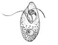
| Ceratiumsp. | |
| Scientific classification | |
| Domain: | Eukaryota |
| Clade: | Diaphoretickes |
| Clade: | SAR |
| Clade: | Alveolata |
| Phylum: | Myzozoa |
| Subphylum: | Dinozoa |
| Superclass: | Dinoflagellata Bütschli1885 [1880–1889]sensuGomez 2012[2][3][4] |
| Classes | |
| Synonyms | |
| |
In terms of number of species, dinoflagellates are one of the largest groups of marine eukaryotes, although substantially smaller thandiatoms.[8]Some species areendosymbiontsof marine animals and play an important part in the biology ofcoral reefs.Other dinoflagellates are unpigmented predators on other protozoa, and a few forms areparasitic(for example,OodiniumandPfiesteria). Some dinoflagellates produce resting stages, called dinoflagellate cysts ordinocysts,as part of their lifecycles; this occurs in 84 of the 350 described freshwater species and a little more than 10% of the known marine species.[9][10]Dinoflagellates arealveolatespossessing twoflagella,the ancestral condition ofbikonts.
About 1,555 species of free-living marine dinoflagellates are currently described.[11]Another estimate suggests about 2,000 living species, of which more than 1,700 are marine (free-living, as well as benthic) and about 220 are from fresh water.[12]The latest estimates suggest a total of 2,294 living dinoflagellate species, which includes marine, freshwater, and parasitic dinoflagellates.[2]
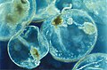
A rapid accumulation of certain dinoflagellates can result in a visible coloration of the water, colloquially known asred tide(aharmful algal bloom), which can causeshellfish poisoningif humans eat contaminated shellfish. Some dinoflagellates also exhibitbioluminescence,primarily emitting blue-green light, which may be visible in oceanic areas under certain conditions.
Etymology
editThe term "dinoflagellate" is a combination of the Greekdinosand the Latinflagellum.Dinosmeans "whirling" and signifies the distinctive way in which dinoflagellates were observed to swim.Flagellummeans "whip" and this refers to theirflagella.[13]
History
editIn 1753, the first modern dinoflagellates were described byHenry Bakeras "Animalcules which cause the Sparkling Light in Sea Water",[14]and named byOtto Friedrich Müllerin 1773.[15]The term derives from the Greek word δῖνος (dînos), meaning whirling, and Latinflagellum,a diminutive term for a whip or scourge.
In the 1830s, the German microscopistChristian Gottfried Ehrenbergexamined many water and plankton samples and proposed several dinoflagellate genera that are still used today includingPeridinium, Prorocentrum,andDinophysis.[16]
These same dinoflagellates were first defined byOtto Bütschliin 1885 as theflagellateorder Dinoflagellida.[17]Botanists treated them as a division of algae, namedPyrrophytaorPyrrhophyta( "fire algae"; Greekpyrr(h)os,fire) after the bioluminescent forms, orDinophyta.At various times, thecryptomonads,ebriids,andellobiopsidshave been included here, but only the last are now considered close relatives. Dinoflagellates have a known ability to transform from noncyst to cyst-forming strategies, which makes recreating their evolutionary history extremely difficult.
Morphology
edit
- Plastidmembranes (3, secondary red)
- Thylakoids,site of thelight-dependent reactionsofphotosynthesis
- Pyrenoid,center ofcarbon fixation
- Trichocyst
- Alveolus, surface cavity or pit
- Thecal plate
- Sac pusule
- Vacuome
- Golgi apparatus;modifiesproteinsand sends them out of the cell
- Endoplasmic reticulum,the transport network for molecules going to specific parts of the cell
- Transverse flagellum
- Striated strand
- Collecting pusule
- Mitochondrion,createsATP(energy) for the cell
- Nucleus
- Nucleolus
- Condensed chromosome
- Starch granule
- Lysosome,holds enzymes
- Phagosome,vesicle formed around a particle
- Mastigoneme,"hairs" that attached to flagellum
- Longitudinalflagellum
Dinoflagellates are unicellular and possess two dissimilar flagella arising from the ventral cell side (dinokont flagellation). They have a ribbon-like transverse flagellum with multiple waves that beats to the cell's left, and a more conventional one, the longitudinal flagellum, that beats posteriorly.[18][19][20]The transverse flagellum is a wavy ribbon in which only the outer edge undulates from base to tip, due to the action of the axoneme which runs along it. The axonemal edge has simple hairs that can be of varying lengths. The flagellar movement produces forward propulsion and also a turning force. The longitudinal flagellum is relatively conventional in appearance, with few or no hairs. It beats with only one or two periods to its wave. The flagella lie in surface grooves: the transverse one in the cingulum and the longitudinal one in the sulcus, although its distal portion projects freely behind the cell. In dinoflagellate species with desmokont flagellation (e.g.,Prorocentrum), the two flagella are differentiated as in dinokonts, but they are not associated with grooves.
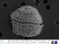
Dinoflagellates have a complex cell covering called anamphiesmaor cortex, composed of a series of membranes, flattenedvesiclescalled alveoli (= amphiesmal vesicles) and related structures.[21][22]In thecate ( "armoured" ) dinoflagellates, these support overlappingcelluloseplates to create a sort of armor called thetheca or lorica,as opposed to athecate ( "nude" ) dinoflagellates. These occur in various shapes and arrangements, depending on the species and sometimes on the stage of the dinoflagellate. Conventionally, the term tabulation has been used to refer to this arrangement ofthecal plates.The plate configuration can be denoted with the plate formula or tabulation formula. Fibrousextrusomesare also found in many forms.[23][24]
A transverse groove, the so-called cingulum (or cigulum) runs around the cell, thus dividing it into an anterior (episoma) and posterior (hyposoma). If and only if a theca is present, the parts are called epitheca and hypotheca, respectively. Posteriorly, starting from the transverse groove, there is a longitudinal furrow called the sulcus. The transverse flagellum strikes in the cingulum, the longitudinal flagellum in the sulcus.[25][24]
Together with various other structural and genetic details, this organization indicates a close relationship between the dinoflagellates, theApicomplexa,andciliates,collectively referred to as thealveolates.[23]
Dinoflagellate tabulations can be grouped into six "tabulation types":gymnodinoid,suessoid,gonyaulacoid–peridinioid,nannoceratopsioid,dinophysioid,andprorocentroid.[26]
Most Dinoflagellates have a plastid derived from secondary endosymbiosis of red algae, however dinoflagellates with plastids derived from green algae and tertiary endosymbiosis of diatoms have also been discovered.[27]Similar to other photosynthetic organisms, dinoflagellates containchlorophyllsaand c2 and the carotenoid beta-carotene. Dinoflagellates also produce thexanthophyllsincludingperidinin,dinoxanthin,anddiadinoxanthin.Thesepigmentsgive many dinoflagellates their typical golden brown color. However, the dinoflagellatesKarenia brevis,Karenia mikimotoi,andKarlodinium micrumhave acquired other pigments through endosymbiosis, includingfucoxanthin.[28] This suggests their chloroplasts were incorporated by severalendosymbiotic eventsinvolving already colored or secondarily colorless forms. The discovery ofplastidsin theApicomplexahas led some to suggest they were inherited from an ancestor common to the two groups, but none of the more basal lines has them. All the same, the dinoflagellate cell consists of the more common organelles such as rough and smoothendoplasmic reticulum,Golgi apparatus,mitochondria,lipidandstarchgrains, and foodvacuoles.Some have even been found with a light-sensitive organelle, theeyespot or stigma,or a larger nucleus containing a prominentnucleolus.The dinoflagellateErythropsidiniumhas the smallest known eye.[29]
Some athecate species have an internal skeleton consisting of two star-likesiliceouselements that has an unknown function, and can be found asmicrofossils.Tappan[30]gave a survey of dinoflagellates withinternal skeletons.This included the first detailed description of thepentastersinActiniscus pentasterias,based on scanningelectron microscopy.They are placed within the orderGymnodiniales,suborderActiniscineae.[5]
Theca structure and formation
editThe formation of thecal plates has been studied in detail through ultrastructural studies.[22]
The dinoflagellate nucleus: dinokaryon
edit'Core dinoflagellates' (dinokaryotes) have a peculiar form ofnucleus,called adinokaryon,in which thechromosomesare attached to thenuclear membrane.These carry reduced number ofhistones.In place of histones, dinoflagellate nuclei contain a novel, dominant family of nuclear proteins that appear to be of viral origin, thus are calledDinoflagellate viral nucleoproteins(DVNPs) which are highly basic, bind DNA with similar affinity to histones, and occur in multiple posttranslationally modified forms.[31]Dinoflagellate nuclei remain condensed throughout interphase rather than just duringmitosis,which is closed and involves a uniquely extranuclearmitotic spindle.[32]This sort of nucleus was once considered to be an intermediate between the nucleoid region ofprokaryotesand the true nuclei ofeukaryotes,so were termed "mesokaryotic",but now are considered derived rather than primitive traits (i. e. ancestors of dinoflagellates had typical eukaryotic nuclei). In addition to dinokaryotes, DVNPs can be found in a group of basal dinoflagellates (known as MarineAlveolates,"MALVs" ) that branch as sister to dinokaryotes (Syndiniales).[33]
Classification
editGenerality
edit
Dinoflagellates are protists and have been classified using both theInternational Code of Botanical Nomenclature(ICBN, now renamed as ICN) and theInternational Code of Zoological Nomenclature(ICZN). About half of living dinoflagellate species are autotrophs possessing chloroplasts and half are nonphotosynthesising heterotrophs.
Theperidinindinoflagellates, named after their peridinin plastids, appear to be ancestral for the dinoflagellate lineage. Almost half of all known species have chloroplasts, which are either the original peridinin plastids or new plastids acquired from other lineages of unicellular algae through endosymbiosis. The remaining species have lost their photosynthetic abilities and have adapted to a heterotrophic, parasitic orkleptoplasticlifestyle.[34][35]
Most (but not all) dinoflagellates have adinokaryon,described below (see:Life cycle,below). Dinoflagellates with a dinokaryon are classified underDinokaryota,while dinoflagellates without a dinokaryon are classified underSyndiniales.
Although classified aseukaryotes,the dinoflagellate nuclei are not characteristically eukaryotic, as some of them lackhistonesandnucleosomes,and maintain continually condensed chromosomes duringmitosis.The dinoflagellate nucleus was termed 'mesokaryotic' by Dodge (1966),[36]due to its possession of intermediate characteristics between the coiled DNA areas of prokaryotic bacteria and the well-defined eukaryotic nucleus. This group, however, does contain typically eukaryoticorganelles,such as Golgi bodies, mitochondria, and chloroplasts.[37]

Jakob Schiller (1931–1937) provided a description of all the species, both marine and freshwater, known at that time.[38]Later, Alain Sournia (1973, 1978, 1982, 1990, 1993) listed the new taxonomic entries published after Schiller (1931–1937).[39][40][41][42][43]Sournia (1986) gave descriptions and illustrations of the marine genera of dinoflagellates, excluding information at the species level.[44]The latest index is written by Gómez.[2]
Identification
editEnglish-language taxonomic monographs covering large numbers of species are published for the Gulf of Mexico,[45]the Indian Ocean,[46]the British Isles,[47]the Mediterranean[48]and the North Sea.[49]
The main source for identification of freshwater dinoflagellates is theSüsswasser Flora.[50]
Calcofluor-whitecan be used to stain thecal plates in armoured dinoflagellates.[51]
Ecology and physiology
editHabitats
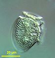
editDinoflagellates are found in all aquatic environments: marine, brackish, and fresh water, including in snow or ice. They are also common in benthic environments and sea ice.
Endosymbionts
editAllZooxanthellaeare dinoflagellates and most of them are members within Symbiodiniaceae (e.g. the genusSymbiodinium).[52]The association betweenSymbiodiniumand reef-buildingcoralsis widely known. However, endosymbionticZooxanthellaeinhabit a great number of other invertebrates and protists, for example manysea anemones,jellyfish,nudibranchs,the giant clamTridacna,and several species ofradiolariansandforaminiferans.[53]Many extant dinoflagellates areparasites(here defined as organisms that eat their prey from the inside, i.e.endoparasites,or that remain attached to their prey for longer periods of time, i.e. ectoparasites). They can parasitize animal or protist hosts.Protoodinium, Crepidoodinium, Piscinoodinium,andBlastodiniumretain their plastids while feeding on their zooplanktonic or fish hosts. In most parasitic dinoflagellates, the infective stage resembles a typical motile dinoflagellate cell.
Nutritional strategies
editThree nutritional strategies are seen in dinoflagellates:phototrophy,mixotrophy,andheterotrophy.Phototrophs can bephotoautotrophsorauxotrophs.Mixotrophic dinoflagellatesare photosynthetically active, but are also heterotrophic. Facultative mixotrophs, in which autotrophy or heterotrophy is sufficient for nutrition, are classified as amphitrophic. If both forms are required, the organisms are mixotrophicsensu stricto.Some free-living dinoflagellates do not have chloroplasts, but host a phototrophic endosymbiont. A few dinoflagellates may use alien chloroplasts (cleptochloroplasts), obtained from food (kleptoplasty). Some dinoflagellates may feed on other organisms as predators or parasites.[54]
Food inclusions contain bacteria, bluegreen algae, diatoms, ciliates, and other dinoflagellates.[55][56][57][58][59][60][61]
Mechanisms of capture and ingestion in dinoflagellates are quite diverse. Several dinoflagellates, both thecate (e.g.Ceratium hirundinella,[60]Peridinium globulus[58]) and nonthecate (e.g.Oxyrrhis marina,[56]Gymnodiniumsp.[62]andKofoidiniumspp.[63]), draw prey to the sulcal region of the cell (either via water currents set up by the flagella or via pseudopodial extensions) and ingest the prey through the sulcus. In severalProtoperidiniumspp., e.g.P. conicum,a large feeding veil—a pseudopod called the pallium—is extruded to capture prey which is subsequently digestedextracellularly(= pallium-feeding).[64][65]Oblea,Zygabikodinium,andDiplopsalisare the only other dinoflagellate genera known to use this particular feeding mechanism.[65][66][67] Gymnodinium fungiforme,commonly found as a contaminant in algal or ciliate cultures, feeds by attaching to its prey and ingesting prey cytoplasm through an extensible peduncle.[68]Two related genera,PolykrikosandNeatodinium,shoot out a harpoon-like organelle to capture prey.[69]
Some mixotrophic dinoflagellates are able to produce neurotoxins that have anti-grazing effects on larger copepods and enhance the ability of the dinoflagellate to prey upon larger copepods. Toxic strains ofKarlodinium veneficumproduce karlotoxin that kills predators who ingest them, thus reducing predatory populations and allowing blooms of both toxic and non-toxic strains ofK. veneficum.Further, the production of karlotoxin enhances the predatory ability ofK. veneficumby immobilizing its larger prey.[70]K. armigerare more inclined to prey upon copepods by releasing a potent neurotoxin that immobilizes its prey upon contact. WhenK. armigerare present in large enough quantities, they are able to cull whole populations of their copepod prey.[71]
The feeding mechanisms of the oceanic dinoflagellates remain unknown, although pseudopodial extensions were observed inPodolampas bipes.[72]
Pigments in dinoflagellates
editDinoflagellates possess a distinctive suite of photosynthetic pigments that allow them to survive and grow in a variety of aquatic environments. Like other phytoplankton, many dinoflagellates contain chlorophyll a and chlorophyll c, which are essential for photosynthesis and light energy capture.[73]However, unlike green algae and higher plants, they lack chlorophyll b. Instead, they utilize chlorophyll c2, which is more efficient for absorbing blue-green light, making them well adapted to low-light or deeper water conditions.[74]These pigments, along with carotenoids, contribute to the characteristic coloration of dinoflagellates, which can range from golden-brown to red.
A unique pigment in dinoflagellates is peridinin, a specialized carotenoid that plays a key role in light harvesting and energy transfer to chlorophyll a.[75]Peridinin is highly efficient in capturing blue light, which penetrates deeper into the water column, giving many dinoflagellates a competitive advantage in stratified or turbid environments.[76]Additionally, dinoflagellates contain other carotenoids such as diadinoxanthin and dinoxanthin, which play important roles in photoprotection by dissipating excess light energy and preventing oxidative stress under high irradiance.[77]These pigments are necessary for photoacclimation, allowing dinoflagellates to survive under fluctuating light conditions.
Not all dinoflagellates rely solely on photosynthetic pigments for energy. Many species are heterotrophic or mixotrophic, meaning they can acquire nutrients through both photosynthesis and predation.[78]Symbiotic dinoflagellates, such as Symbiodinium, play a important ecological role by forming mutualistic relationships with corals, where their pigments drive photosynthesis and energy production that sustain coral reef ecosystems.[79]The unique pigment composition of dinoflagellates also contributes to large-scale phenomena such as harmful algal blooms and red tides, where high concentrations of pigmented cells cause dramatic discoloration of coastal waters and can produce toxic effects.[80]
Blooms
editIntroduction
editDinoflagellate blooms are generally unpredictable, short, with low species diversity, and with little species succession.[81]The low species diversity can be due to multiple factors. One way a lack of diversity may occur in a bloom is through a reduction in predation and a decreased competition. The first may be achieved by having predators reject the dinoflagellate, by, for example, decreasing the amount of food it can eat. This additionally helps prevent a future increase in predation pressure by causing predators that reject it to lack the energy to breed. A species can then inhibit the growth of its competitors, thus achieving dominance.[82]
Harmful algal blooms
editDinoflagellates sometimes bloom in concentrations of more than a million cells per millilitre. Under such circumstances, they can produce toxins (generally calleddinotoxins) in quantities capable of killing fish and accumulating in filter feeders such asshellfish,which in turn may be passed on to people who eat them. This phenomenon is called ared tide,from the color the bloom imparts to the water. Some colorless dinoflagellates may also form toxic blooms, such asPfiesteria.Some dinoflagellate blooms are not dangerous. Bluish flickers visible in ocean water at night often come from blooms ofbioluminescentdinoflagellates, which emit short flashes of light when disturbed.

A red tide occurs because dinoflagellates are able to reproduce rapidly and copiously as a result of the abundant nutrients in the water. Although the resulting red waves are an interesting visual phenomenon, they containtoxinsthat not only affect all marine life in the ocean, but the people who consume them as well.[83]A specific carrier isshellfish.This can introduce both nonfatal and fatal illnesses. One such poison issaxitoxin,a powerfulparalyticneurotoxin.[84][85][86]
Human inputs ofphosphatefurther encourage these red tides, so strong interest exists in learning more about dinoflagellates, from both medical and economic perspectives. Dinoflagellates are known to be particularly capable of scavenging dissolved organic phosphorus for P-nutrient, several HAS species have been found to be highly versatile and mechanistically diversified in utilizing different types of DOPs.[84][85][86]The ecology ofharmful algal bloomsis extensively studied.[87]
Bioluminescence
edit
At night, water can have an appearance of sparkling light due to the bioluminescence of dinoflagellates.[88][89]More than 18 genera of dinoflagellates are bioluminescent,[90]and the majority of them emit a blue-green light.[91]These species containscintillons,individual cytoplasmic bodies (about 0.5 μm in diameter) distributed mainly in the cortical region of the cell, outpockets of the main cell vacuole. They containdinoflagellate luciferase,the main enzyme involved in dinoflagellate bioluminescence, andluciferin,a chlorophyll-derived tetrapyrrole ring that acts as the substrate to the light-producing reaction. The luminescence occurs as a brief (0.1 sec) blue flash (max 476 nm) when stimulated, usually by mechanical disturbance. Therefore, when mechanically stimulated—by boat, swimming, or waves, for example—a blue sparkling light can be seen emanating from the sea surface.[92]
Dinoflagellate bioluminescence is controlled by a circadian clock and only occurs at night.[93]Luminescent and nonluminescent strains can occur in the same species. The number of scintillons is higher during night than during day, and breaks down during the end of the night, at the time of maximal bioluminescence.[94]
The luciferin-luciferase reaction responsible for the bioluminescence is pH sensitive.[92]When the pH drops, luciferase changes its shape, allowing luciferin, more specifically tetrapyrrole, to bind.[92]Dinoflagellates can use bioluminescence as a defense mechanism. They can startle their predators by their flashing light or they can ward off potential predators by an indirect effect such as the "burglar alarm". The bioluminescence attracts attention to the dinoflagellate and its attacker, making the predator more vulnerable to predation from higher trophic levels.[92]
Bioluminescent dinoflagellate ecosystem bays are among the rarest and most fragile,[95]with the most famous ones being the Bioluminescent Bay inLa Parguera, Lajas,Puerto Rico; Mosquito Bay inVieques, Puerto Rico;and Las Cabezas de San Juan Reserva NaturalFajardo, Puerto Rico.Also, a bioluminescent lagoon is near Montego Bay, Jamaica, and bioluminescent harbors surround Castine, Maine.[96]Within the United States, Central Florida is home to theIndian River Lagoonwhich is abundant with dinoflagellates in the summer and bioluminescent ctenophore in the winter.[97]
Lipid and sterol production
editDinoflagellates produce characteristic lipids and sterols.[98]One of these sterols is typical of dinoflagellates and is calleddinosterol.
Transport
editDinoflagellatethecacan sink rapidly to the seafloor inmarine snow.[99]
Life cycle
editIntroduction
editDinoflagellates have ahaplontic life cycle,with the possible exception ofNoctilucaand its relatives.[5] The life cycle usually involves asexual reproduction by means of mitosis, either throughdesmoschisisoreleuteroschisis.More complex life cycles occur, more particularly with parasitic dinoflagellates. Sexual reproduction also occurs,[100]though this mode of reproduction is only known in a small percentage of dinoflagellates.[101]This takes place by fusion of two individuals to form azygote,which may remain mobile in typical dinoflagellate fashion and is then called a planozygote. This zygote may later form a resting stage orhypnozygote,which is called adinoflagellate cystordinocyst.After (or before) germination of the cyst, the hatchling undergoesmeiosisto produce newhaploid cells.Dinoflagellates appear to be capable of carrying out severalDNA repairprocesses that can deal with different types ofDNA damage.[102]


Dinoflagellate cysts
editThe life cycle of many dinoflagellates includes at least one nonflagellated benthic stage as acyst.Different types of dinoflagellate cysts are mainly defined based on morphological (number and type of layers in the cell wall) and functional (long- or short-term endurance) differences. These characteristics were initially thought to clearly distinguishpellicle(thin-walled) cysts fromresting(double-walled) dinoflagellate cysts. The former were considered short-term (temporal) and the latter long-term (resting) cysts. However, during the last two decades further knowledge has highlighted the great intricacy of dinoflagellate life histories.[103]

More than 10% of the approximately 2000 known marine dinoflagellate species produce cysts as part of their life cycle (see diagram on the right). These benthic phases play an important role in the ecology of the species, as part of a planktonic-benthic link in which the cysts remain in the sediment layer during conditions unfavorable for vegetative growth and, from there, reinoculate the water column when favorable conditions are restored.[103]
Indeed, during dinoflagellate evolution the need to adapt to fluctuating environments and/or to seasonality is thought to have driven the development of this life cycle stage. Most protists form dormant cysts in order to withstand starvation and UV damage.[104]However, there are enormous differences in the main phenotypic, physiological and resistance properties of each dinoflagellate species cysts. Unlike in higher plants most of this variability, for example indormancyperiods, has not been proven yet to be attributed to latitude adaptation or to depend on other life cycle traits.[105][106]Thus, despite recent advances in the understanding of the life histories of many dinoflagellate species, including the role of cyst stages, many gaps remain in knowledge about their origin and functionality.[103]
Recognition of the capacity of dinoflagellates to encyst dates back to the early 20th century, inbiostratigraphicstudies of fossil dinoflagellate cysts.Paul Reinschwas the first to identify cysts as the fossilized remains of dinoflagellates.[107]Later, cyst formation from gamete fusion was reported, which led to the conclusion that encystment is associated with sexual reproduction.[100]These observations also gave credence to the idea that microalgal encystment is essentially a process whereby zygotes prepare themselves for a dormant period.[108]Because the resting cysts studied until that time came from sexual processes, dormancy was associated with sexuality, a presumption that was maintained for many years. This attribution was coincident with evolutionary theories about the origin of eukaryotic cell fusion and sexuality, which postulated advantages for species with diploid resting stages, in their ability to withstand nutrient stress and mutational UV radiation through recombinational repair, and for those with haploid vegetative stages, as asexual division doubles the number of cells.[104]Nonetheless, certain environmental conditions may limit the advantages of recombination and sexuality,[109]such that in fungi, for example, complex combinations of haploid and diploid cycles have evolved that include asexual and sexual resting stages.[110][103]
However, in the general life cycle of cyst-producing dinoflagellates as outlined in the 1960s and 1970s, resting cysts were assumed to be the fate of sexuality,[100][111]which itself was regarded as a response to stress or unfavorable conditions. Sexuality involves the fusion of haploid gametes from motile planktonic vegetative stages to produce diploidplanozygotesthat eventually form cysts, orhypnozygotes,whose germination is subject to bothendogenousandexogenouscontrols. Endogenously, a species-specific physiological maturation minimum period (dormancy) is mandatory before germination can occur. Thus, hypnozygotes were also referred to as "resting" or "resistant" cysts, in reference to this physiological trait and their capacity following dormancy to remain viable in the sediments for long periods of time. Exogenously, germination is only possible within a window of favorable environmental conditions.[103]
Yet, with the discovery that planozygotes were also able to divide it became apparent that the complexity of dinoflagellate life cycles was greater than originally thought.[112][113]Following corroboration of this behavior in several species, the capacity of dinoflagellate sexual phases to restore the vegetative phase, bypassing cyst formation, became well accepted.[114][115]Further, in 2006 Kremp and Parrow showed the dormant resting cysts of the Baltic cold water dinoflagellatesScrippsiella hangoeiandGymnodiniumsp. were formed by the direct encystment of haploid vegetative cells, i.e., asexually.[116]In addition, for the zygotic cysts ofPfiesteria piscicidadormancy was not essential.[117][103]
Genomics
editOne of the most striking features of dinoflagellates is the large amount of cellular DNA that they contain. Most eukaryotic algae contain on average about 0.54 pg DNA/cell, whereas estimates of dinoflagellate DNA content range from 3–250 pg/cell,[32]corresponding to roughly 3000–215 000 Mb (in comparison, the haploid human genome is 3180 Mb and hexaploidTriticumwheat is 16 000 Mb).Polyploidyor polyteny may account for this large cellular DNA content,[118]but earlier studies of DNA reassociation kinetics and recent genome analyses do not support this hypothesis.[119]Rather, this has been attributed, hypothetically, to the rampant retroposition found in dinoflagellate genomes.[120][121]
In addition to their disproportionately large genomes, dinoflagellate nuclei are unique in their morphology, regulation, and composition. Their DNA is so tightly packed that exactly how many chromosomes they have is still uncertain.[122]
The dinoflagellates share an unusual mitochondrial genome organisation with their relatives, theApicomplexa.[123]Both groups have very reduced mitochondrial genomes (around 6 kilobases (kb) in the Apicomplexa vs ~16kb for human mitochondria). One species,Amoebophryaceratii,has lost its mitochondrial genome completely, yet still has functional mitochondria.[124]The genes on the dinoflagellate genomes have undergone a number of reorganisations, including massive genome amplification and recombination which have resulted in multiple copies of each gene and gene fragments linked in numerous combinations. Loss of the standard stop codons, trans-splicing of mRNAs for the mRNA of cox3, and extensive RNA editing recoding of most genes has occurred.[125][126]The reasons for this transformation are unknown. In a small group of dinoflagellates, called 'dinotoms' (Durinskia and Kryptoperidinium), the endosymbionts (diatoms) still have mitochondria, making them the only organisms with two evolutionarily distinct mitochondria.[127]
In most of the species, the plastid genome consist of just 14 genes.[128]
The DNA of the plastid in the peridinin-containing dinoflagellates is contained in a series of small circles calledminicircles.[129]Each circle contains one or two polypeptide genes. The genes for these polypeptides are chloroplast-specific because their homologs from other photosynthetic eukaryotes are exclusively encoded in the chloroplast genome. Within each circle is a distinguishable 'core' region. Genes are always in the same orientation with respect to this core region.
In terms ofDNA barcoding,ITS sequences can be used to identify species,[130]where a genetic distance of p≥0.04 can be used to delimit species,[131]which has been successfully applied to resolve long-standing taxonomic confusion as in the case of resolving the Alexandrium tamarense complex into five species.[132]A recent study[133]revealed a substantial proportion of dinoflagellate genes encode for unknown functions, and that these genes could be conserved and lineage-specific.
Evolutionary history
editDinoflagellates are mainly represented as fossils bydinocysts,which have a long geological record with lowest occurrences during the mid-Triassic,[134]whilst geochemical markers suggest a presence to the Early Cambrian.[135]Some evidence indicates dinosteroids in manyPaleozoicandPrecambrianrocks might be the product of ancestral dinoflagellates (protodinoflagellates).[136][137]Dinoflagellates show a classic radiation of morphologies during the Late Triassic through the MiddleJurassic.[138][139][140]More modern-looking forms proliferate during the later Jurassic andCretaceous.[138]This trend continues into theCenozoic,albeit with some loss of diversity.[138][134]
Molecular phylogenetics show that dinoflagellates are grouped withciliatesandapicomplexans(=Sporozoa) in a well-supported clade, thealveolates.The closest relatives to dinokaryotic dinoflagellates appear to beapicomplexans,Perkinsus, Parvilucifera,syndinians, andOxyrrhis.[141]Molecular phylogenies are similar to phylogenies based on morphology.[142][143]
The earliest stages of dinoflagellate evolution appear to be dominated by parasitic lineages, such as perkinsids and syndinians (e.g.AmoebophryaandHematodinium).[144][145][146][147]
All dinoflagellates contain red algal plastids or remnant (nonphotosynthetic) organelles of red algal origin.[148]The parasitic dinoflagellateHematodiniumhowever lacks a plastid entirely.[149]Some groups that have lost the photosynthetic properties of their original red algae plastids has obtained new photosynthetic plastids (chloroplasts) through so-called serial endosymbiosis, both secondary and tertiary:
- Lepidodiniumunusually possesses a green algae-derived plastid (all other serially-acquired plastids can be traced back to red algae).[150]The plastid is most related to free-livingPedinomonas(hence likely secondary). Two previously undescribed dinoflagellates ( "MGD" and "TGD" ) contain a closely-related plastid.[151]
- Karenia,Karlodinium,andTakayamapossess plastids ofhaptophyteorigin, produced in three separate events.[152]
- "Dinotoms" (DurinskiaandKryptoperidinium) have plastids derived fromdiatoms.[153][154]
Some species also performkleptoplasty:
- Dinophysishave plastids from acryptomonad,due to kleptoplasty from a cilate prey.[155]
- The Kareniaceae (which contains the three haptophyte-having genera) contains two separate cases of kleptoplasty.[156][152]
Dinoflagellate evolution has been summarized into five principal organizational types: prorocentroid, dinophysoid, gonyaulacoid, peridinioid, and gymnodinoid.[157] The transitions of marine species into fresh water have been frequent events during the diversification of dinoflagellates and have occurred recently.[158]
Many dinoflagellates also have a symbiotic relationship with cyanobacteria, called cyanobionts, which have a reduced genome and has not been found outside their hosts. The Dinophysoid dinoflagellates have two genera, Amphisolenia and Triposolenia, that contain intracellular cyanobionts, and four genera; Citharistes, Histioneis, Parahistioneis, and Ornithocercus, that contain extracellular cyanobionts.[159]Most of the cyanobionts are used for nitrogen fixation, not for photosynthesis, but some don't have the ability to fix nitrogen. The dinoflagellateOrnithocercus magnificusis host for symbionts which resides in an extracellular chamber. While it is not fully known how the dinoflagellate benefit from it, it has been suggested it is farming the cyanobacteria in specialized chambers and regularly digest some of them.[160]
Recently, theliving fossilDapsilidinium pastielsiiwas found inhabiting theIndo-Pacific Warm Pool,which served as arefugiumfor thermophilic dinoflagellates,[161]and others such asCalciodinellum operosumandPosoniella tricarinelloideswere also described from fossils before later being found alive.[162][163]
Examples
edit-
-
-
 Ceratium macroceros(Dinophyceae)
 -
 Ceratium furcoides(Dinophyceae)
 -
 Unknown dinoflagellate underSEM(Dinophyceae)
 -
 Pfiesteria shumwayae(Dinophyceae)
 -
 Symbiodiniumsp. (Dinophyceae):zooxanthella,a coral endosymbiont
 -


See also
editReferences
edit- ^Parfrey LW, Lahr DJ, Knoll AH, Katz LA (August 2011)."Estimating the timing of early eukaryotic diversification with multigene molecular clocks".Proceedings of the National Academy of Sciences of the United States of America.108(33):13624–13629.Bibcode:2011PNAS..10813624P.doi:10.1073/pnas.1110633108.PMC3158185.PMID21810989.
- ^abcGómez F (2012)."A checklist and classification of living dinoflagellates (Dinoflagellata, Alveolata)".CICIMAR Oceánides.27(1):65–140.doi:10.37543/oceanides.v27i1.111.
- ^Ruggiero MA, Gordon DP, Orrell TM, Bailly N, Bourgoin T, Brusca RC, et al. (2015)."A higher level classification of all living organisms".PLOS ONE.10(4): e0119248.Bibcode:2015PLoSO..1019248R.doi:10.1371/journal.pone.0119248.PMC4418965.PMID25923521.
- ^Silar P (2016). "Protistes Eucaryotes: Origine, Evolution et Biologie des Microbes Eucaryotes".HAL Archives-ouvertes.Creative Commons. pp.1–462.ISBN9782955584101.OCLC1019558675.Archivedfrom the original on 2016-05-13.Retrieved2016-09-04.
- ^abcFensome RA, Taylor RJ, Norris G, Sarjeant WA, Wharton DI, Williams GL (1993).A classification of living and fossil dinoflagellates.Micropaleontology Special Publication. Vol. 7. Hanover, PA: Sheridan Press.OCLC263894965.
- ^Stoecker DK (1999). "Mixotrophy among Dinoflagellates".The Journal of Eukaryotic Microbiology.46(4):397–401.doi:10.1111/j.1550-7408.1999.tb04619.x.S2CID83885629.
- ^Esser K, Lüttge U, Beyschlag W, Murata J (2012-12-06).Progress in Botany: Genetics Physiology Systematics Ecology.Springer.ISBN978-3-6421-8819-0.Archivedfrom the original on 2022-01-28.Retrieved2020-10-22.
- ^Guiry MD (October 2012). "How many species of algae are there?".Journal of Phycology.48(5):1057–1063.Bibcode:2012JPcgy..48.1057G.doi:10.1111/j.1529-8817.2012.01222.x.PMID27011267.S2CID30911529.
- ^Mertens KN, Rengefors K, Moestrup Ø, Ellegaard M (2012). "A review of recent freshwater dinoflagellate cysts: Taxonomy, phylogeny, ecology and palaeocology".Phycologia.51(6):612–619.Bibcode:2012Phyco..51..612M.doi:10.2216/11-89.1.S2CID86845462.
- ^Bravo I, Figueroa RI (January 2014)."Towards an Ecological Understanding of Dinoflagellate Cyst Functions".Microorganisms.2(1):11–32.doi:10.3390/microorganisms2010011.PMC5029505.PMID27694774.
- ^Gómez F (2005)."A list of free-living dinoflagellate species in the world's oceans".Acta Botanica Croatica.64(1):129–212.
- ^Taylor FR, Hoppenrath M, Saldarriaga JF (February 2008). "Dinoflagellate diversity and distribution".Biodivers. Conserv.17(2):407–418.Bibcode:2008BiCon..17..407T.doi:10.1007/s10531-007-9258-3.S2CID9810504.
- ^Carty S, Parrow MW (2015). "Dinoflagellates".Freshwater Algae of North America.Academic Press. pp.773–807.doi:10.1016/B978-0-12-385876-4.00017-7.ISBN978-0-12-385876-4.
- ^Baker M (1753).Employment for the microscope.London: Dodsley.doi:10.5962/bhl.title.45920.OCLC722119426.
- ^Müller, O.F. 1773. Vermium terrestrium et fluviatilium, seu Animalium Infusoriorum, Helmithicorum et Testaceorum, non marinorum, succincta historia, vol. 1. Pars prima. p. 34, 135. Faber, Havniae, et Lipsiae 1773.
- ^Ehrenberg C.G. (1832) Beiträge zur Kenntnis der Organisation der Infusorien und ihrer geographischer Verbreitung, besonders in Sibirien. — Abhandlungen der Königlichen Akademie der Wissenschaften zu Berlin. Aus dem Jahre 1830. Physikalische Abhandlungen 1830: 1–88, Pls 1–8.
- ^Bütschli O. (1885) 3. Unterabtheilung (Ordnung) Dinoflagellata. – In: Dr. H.G. Bronn's Klassen und Ordnungen des Thier-Reichs, wissenschaftlich dargestellt in Wort und Bild. Erster Band Protozoa. – C.F. Winter'sche Verlagshandlung, Leipzig und Heidelberg. Pp. 906–1029; Pl.
- ^Taylor FJR (March 1975). "Non-helical transverse flagella in dinoflagellates".Phycologia.14(1):45–7.Bibcode:1975Phyco..14...45T.doi:10.2216/i0031-8884-14-1-45.1.
- ^Leblond PH, Taylor FJ (April 1976). "The propulsive mechanism of the dinoflagellate transverse flagellum reconsidered".Bio Systems.8(1):33–39.Bibcode:1976BiSys...8...33L.doi:10.1016/0303-2647(76)90005-8.PMID986199.
- ^Gaines G, Taylor FJ (May 1985). "Form and function of the dinoflagellate transverse flagellum".J. Protozool.32(2):290–6.doi:10.1111/j.1550-7408.1985.tb03053.x.
- ^Morrill LC, Loeblich AR (1983).Ultrastructure of the Dinoflagellate Amphiesma.International Review of Cytology. Vol. 82. pp.151–80.doi:10.1016/s0074-7696(08)60825-6.ISBN978-0-1236-4482-4.PMID6684652.
- ^abNetzel H, Dürr G (2012-12-02).Ch. 3: Dinoflagellate cell cortex.Academic Press. pp.43–106.ISBN978-0-3231-3813-0.Archivedfrom the original on 2014-07-07.Retrieved2016-03-05.InSpector 1984
- ^abCavalier-Smith T (1991). "Cell diversification in heterotrophic flagellates". In Patterson DJ, Larsen J (eds.).The biology of free-living heterotrophic flagellates.Systematics Association Publications. Vol. 45. Clarendon Press. pp.113–131.ISBN978-0-1985-7747-8.
- ^abFreudenthal HD (1962). "Symbiodiniumgen. Nov. AndSymbiodinium microadriaticumsp. nov., a Zooxanthella: Taxonomy, Life Cycle, and Morphology ".The Journal of Protozoology.9(1):45–52.doi:10.1111/j.1550-7408.1962.tb02579.x.
- ^Trench RK, Blank RJ (1987). "Symbiodinium microadriaticumFreudenthal,S. goreauiisp. nov.,S. kawagutiisp. nov. AndS. pilosumsp. nov.: Gymnodinioid Dinoflagellate Symbionts of Marine Invertebrates ".Journal of Phycology.23(3):469–81.Bibcode:1987JPcgy..23..469T.doi:10.1111/j.1529-8817.1987.tb02534.x.S2CID83712799.
- ^Medlin LK, Fensome RA (2013). "Dinoflagellate macroevolution".Biological and Geological Perspectives of Dinoflagellates.Geological Society of London. pp.263–274.doi:10.1144/tms5.25.ISBN978-1-86239-368-4.
- ^Keeling PJ (March 2010)."The endosymbiotic origin, diversification and fate of plastids".Philosophical Transactions of the Royal Society of London. Series B, Biological Sciences.365(1541):729–748.doi:10.1098/rstb.2009.0103.PMC2817223.PMID20124341.
- ^Hackett JD, Anderson DM, Erdner DL, Bhattacharya D (October 2004). "Dinoflagellates: a remarkable evolutionary experiment".American Journal of Botany.91(10):1523–1534.doi:10.3732/ajb.91.10.1523.PMID21652307.
- ^Schwab IR (September 2004)."You are what you eat".The British Journal of Ophthalmology.88(9): 1113.doi:10.1136/bjo.2004.049510.PMC1772300.PMID15352316.
- ^Tappan HN (1980).The Paleobiology of Plant Protists.Geology. W.H. Freeman.ISBN978-0-7167-1109-4.
- ^Gornik SG, Ford KL, Mulhern TD, Bacic A, McFadden GI, Waller RF (December 2012)."Loss of nucleosomal DNA condensation coincides with appearance of a novel nuclear protein in dinoflagellates".Current Biology.22(24):2303–2312.Bibcode:2012CBio...22.2303G.doi:10.1016/j.cub.2012.10.036.PMID23159597.
- ^abSpector DL (2012-12-02).Dinoflagellate nuclei.Academic Press. pp.107–147.ISBN978-0-3231-3813-0.Archivedfrom the original on 2014-07-07.Retrieved2016-03-05.InSpector 1984
- ^Strassert JF, Karnkowska A, Hehenberger E, Del Campo J, Kolisko M, Okamoto N, et al. (January 2018)."Single cell genomics of uncultured marine alveolates shows paraphyly of basal dinoflagellates".The ISME Journal(Submitted manuscript).12(1):304–308.Bibcode:2018ISMEJ..12..304S.doi:10.1038/ismej.2017.167.PMC5739020.PMID28994824.Archivedfrom the original on 2018-12-14.Retrieved2018-10-23.
- ^Gabrielsen TM, Minge MA, Espelund M, Tooming-Klunderud A, Patil V, Nederbragt AJ, et al. (April 2011)."Genome evolution of a tertiary dinoflagellate plastid".PLOS ONE.6(4): e19132.Bibcode:2011PLoSO...619132G.doi:10.1371/journal.pone.0019132.PMC3082547.PMID21541332.
- ^Bodył A, Moszczyński K (2006)."Did the peridinin plastid evolve through tertiary endosymbiosis? A hypothesis".European Journal of Phycology.41(4):435–448.Bibcode:2006EJPhy..41..435B.doi:10.1080/09670260600961080.
- ^Dodge (1966). Cited but unreferenced inSteidinger KA, Jangen K (1997)."Dinoflagellates".In Tomas CR (ed.).Identifying Marine Phytoplankton.Academic Press. pp.387–584.ISBN978-0-0805-3442-8.Archivedfrom the original on 2014-07-07.Retrieved2016-03-05.
- ^Steidinger KA, Jangen K (1997)."Dinoflagellates".In Tomas CR (ed.).Identifying Marine Phytoplankton.Academic Press. pp.387–584.ISBN978-0-0805-3442-8.Archivedfrom the original on 2014-07-07.Retrieved2016-03-05.
- ^Schiller, J., 1931–1937: Dinoflagellatae (Peridinineae) in monographischer Behandlung. In: RABENHORST, L. (ed.), Kryptogamen-Flora von Deutschland, Österreichs und der Schweiz. Akad. Verlag., Leipzig. Vol. 10 (3): Teil 1 (1–3) (1931–1933): Teil 2 (1–4)(1935–1937).
- ^Sournia A (1973). "Catalogue des espèces et taxons infraspécifiques de dinoflagellés marins actuels publiés depuis la révision de J. Schiller. I. Dinoflagellés libres".Beih. Nova Hedwigia.48:1–92.
- ^Sournia A (1978)."Catalogue des espèces et taxons infraspécifiques de dinoflagellésmarins actuels publiés depuis la révision de J. Schiller. III (Complément)".Rev. Algol.13:3–40 +erratum 13,186.ISSN0035-0702.
- ^Sournia A (1982). "Catalogue des espèces et taxons infraspécifiques de dinoflagellésmarins actuels publiés depuis la révision de J. Schiller. IV. (Complément)".Arch. Protistenkd.126(2):151–168.doi:10.1016/S0003-9365(82)80046-8.
- ^Sournia A (1990). "Catalogue des espèces et taxons infraspécifiques de dinoflagellésmarins actuels publiés depuis la révision de J. Schiller. V. (Complément)".Acta Protozool.29:321–346.ISSN0065-1583.
- ^Sournia A (1993)."Catalogue des espèces et taxons infraspécifiques de dinoflagellésmarins actuels publiés depuis la révision de J. Schiller. VI. (Complément)".Cryptog. Algol.14:133–144.doi:10.5962/p.309374.ISSN0181-1568.
- ^SOURNIA, A., 1986: Atlas du Phytoplancton Marin. Vol. I: Introduction, Cyanophycées,Dictyochophycées, Dinophycées et Raphidophycées. Editions du CNRS, Paris.
- ^Steidinger KA, Williams J (1970).Dinoflagellates.Memoirs of the Hourglass Cruises. Vol. 2. Florida: Marine Research Laboratory.OCLC6206528.
- ^Taylor FJ, Hart-Jones B (1976).Dinoflagellates from the International Indian Ocean Expedition: A Report on Material Collected by the R.V. "Anton Bruun" 1963–1964.Biblioteca Botanica. Vol. 132. E. Schweizerbart.ISBN978-3-5104-8003-6.OCLC3026853.
- ^Dodge JD (1982).Marine Dinoflagellates of the British Isles.London: Her Majesty's Stationery Office.ISBN9780112411963.OCLC681855348.
- ^Gómez F (April 2003). "Checklist of Mediterranean free-living dinoflagellates".Botanica Marina.46(3):215–242.doi:10.1515/BOT.2003.021.S2CID84744638.
- ^Hoppenrath M, Elbrächter M, Drebes G (2009).Marine phytoplankton: selected microphytoplankton species from the North Sea around Helgoland and Sylt.Stuttgart: E. Schweizerbart'sche Verlagsbuchhandlung (Nägele und Obermiller).ISBN978-3-5106-1392-2.
- ^Popovský J, Pfiester LA (1990).Dinophyceae (Dinoflagellida).Süßwasserflora von Mitteleuropa. Vol. 6. Fischer.ISBN978-3-3340-0247-6.[permanent dead link]
- ^Fritz L, Triemer R (December 1985). "A rapid simple technique utilizing calcofluor white M2R for the visualization of dinoflagellate thecal plates".J. Phycol.21(4):662–664.Bibcode:1985JPcgy..21..662F.doi:10.1111/j.0022-3646.1985.00662.x.S2CID85004940.
- ^Freudenthal et al. 2007
- ^Trench RK (1997)."Diversity of symbiotic dinoflagellates and the evolution of microalgal-invertebrate symbioses".In Lessios HA, MacIntyre IG (eds.).Proceedings of the eighth International Coral Reef Symposium, Panama, June 24–29, 1996.Vol. 2. Balboa, Panama: Smithsonian Tropical Research Institute. pp.1275–86.OCLC833272061.
- ^Schnepf E, Elbrächter M (February 1992). "Nutritional strategies in dinoflagellates: A review with emphasis on cell biological aspects".European Journal of Protistology.28(1):3–24.doi:10.1016/S0932-4739(11)80315-9.PMID23194978.
- ^Kofoid CA,Swezy O(1921)."The free-living unarmoured dinoflagellata".Mere. Univ. Calif.5:1–538.doi:10.5962/bhl.title.24995.Archived(PDF)from the original on 2022-05-15.Retrieved2019-09-24.
- ^abBarker HA (1935). "The culture and physiology of the marine dinoflagellates".Arch. Mikrobiol.6(1–5):157–181.Bibcode:1935ArMic...6..157B.doi:10.1007/BF00407285.S2CID44657010.
- ^Biecheler B (1952). "Recherches sur les Peridiniens".Bull. Biol. Fr. Belg.36(Suppl):1–149.ISSN0994-575X.
- ^abBursa AS (1961). "The annual oceanographic cycle at Igloolik in the Canadian Arctic. II. The phytoplankton".J. Fish. Res. Board Can.18(4):563–615.doi:10.1139/f61-046.
- ^Norris DR (1969)."Possible phagotrophic feeding inCeratium lunulaSchimper ".Limnol. Oceanogr.14(3):448–9.Bibcode:1969LimOc..14..448N.doi:10.4319/lo.1969.14.3.0448.
- ^abDodge JD, Crawford RM (June 1970). "The morphology and fine structure ofCeratium hirundinella(Dinophyceae) ".J. Phycol.6(2):137–149.Bibcode:1970JPcgy...6..137D.doi:10.1111/j.1529-8817.1970.tb02372.x.S2CID84034844.
- ^Elbrachter M (1979). "On the taxonomy of unarmored dinophytes (Dinophyta) from the Northwest African upwelling region".Meteor Forschungsergebnisse.30:1–22.
- ^Frey LC, Stoermer EF (1980). "Dinoflagellate phagotrophy in the upper Great Lakes".Trans. Am. Microsc. Soc.99(4):439–444.doi:10.2307/3225654.JSTOR3225654.
- ^Cachon PJ, Cachon M. "Le systeme stomatopharyngien de Kofoidinium Pavillard. Comparisons avec celui divers Peridiniens fibres et parasites".Protistologica.10:217–222.
- ^Gaines G, Taylor FJ (1984). "Extracellular digestion in marine dinoflagellates".J. Plankton Res.6(6):1057–61.doi:10.1093/plankt/6.6.1057.
- ^abJacobson DM, Anderson DM (September 1986). "Thecate heterotrophic dinoflagellates: feeding behavior and mechanisms".J. Phycol.22(3):249–258.doi:10.1111/j.1529-8817.1986.tb00021.x.S2CID84321400.
- ^Strom SL, Buskey EJ (1993)."Feeding, growth, and behavior of the thecate heterotrophic dinoflagellateOblea rotunda".Limnol. Oceanogr.38(5):965–977.Bibcode:1993LimOc..38..965S.doi:10.4319/lo.1993.38.5.0965.
- ^Naustvoll LJ (January 1998). "Growth and grazing by the thecate heterotrophic dinoflagellatesDiplopsalis lenticula(Diplopsalidaceae, Dinophyceae) ".Phycologia.37(1):1–9.Bibcode:1998Phyco..37....1N.doi:10.2216/i0031-8884-37-1-1.1.
- ^Spero HJ (September 1982). "Phagotrophy inGymnodinium fungiforme(Pyrrophyta): the peduncle as an organelle of ingestion ".J. Phycol.18(3):356–360.Bibcode:1982JPcgy..18..356S.doi:10.1111/j.1529-8817.1982.tb03196.x.S2CID85988790.
- ^"Researchers capture dinoflagellate on video shooting harpoons at prey".Archivedfrom the original on 2019-12-28.Retrieved2019-05-19.
- ^Adolf JE, Krupatkina D, Bachvaroff T, Place AR (2007). "Karlotoxin mediates grazing byOxyrrhis marinaon strains ofKarlodinium veneficum".Harmful Algae.6(3):400–412.Bibcode:2007HAlga...6..400A.doi:10.1016/j.hal.2006.12.003.
- ^Berge T, Poulsen LK, Moldrup M, Daugbjerg N, Juel Hansen P (October 2012)."Marine microalgae attack and feed on metazoans".The ISME Journal.6(10):1926–1936.Bibcode:2012ISMEJ...6.1926B.doi:10.1038/ismej.2012.29.PMC3446796.PMID22513533.
- ^Schütt F (1895). "2. Teil, Studien über die Zellen der Peridineen".Die Peridineen der Plankton-Expedition.Ergebnisse der Plankton-Expedition der Humboldt-Stiftung. Allgemeiner Theil. pp.1–170.OCLC69377189.
- ^Jeffrey SW, Wright S, Zapata M (2011-01-01).Microalgal classes and their signature pigments.University of Tasmania.ISBN978-0-511-73226-3.
- ^Schlüter L, Møhlenberg F, Havskum H, Larsen S (2000)."The use of phytoplankton pigments for identifying and quantifying phytoplankton groups in coastal areas:testing the influence of light and nutrients on pigment/chlorophyll a ratios".Marine Ecology Progress Series.192:49–63.doi:10.3354/meps192049.ISSN0171-8630.
- ^Takaichi S (2011)."Carotenoids in algae: distributions, biosyntheses and functions".Marine Drugs.9(6):1101–1118.doi:10.3390/md9061101.ISSN1660-3397.PMC3131562.PMID21747749.
{{cite journal}}:CS1 maint: unflagged free DOI (link) - ^Jiang J, Zhang H, Kang Y, Bina D, Lo CS, Blankenship RE (July 2012)."Characterization of the peridinin-chlorophyll a-protein complex in the dinoflagellate Symbiodinium".Biochimica Et Biophysica Acta.1817(7):983–989.doi:10.1016/j.bbabio.2012.03.027.ISSN0006-3002.PMC3947849.PMID22497797.
- ^Lavaud J, Rousseau B, van Gorkom HJ, Etienne AL (2002-07-01)."Influence of the Diadinoxanthin Pool Size on Photoprotection in the Marine Planktonic DiatomPhaeodactylum tricornutum".Plant Physiology.129(3):1398–1406.doi:10.1104/pp.002014.ISSN1532-2548.Archived fromthe originalon 2025-02-02.
- ^"(PDF) Acquired phototrophy in aquatic protists".ResearchGate.Archived fromthe originalon 2018-07-01.Retrieved2025-02-16.
- ^Stat M, Carter D, Hoegh-Guldberg O (2006-09-27)."The evolutionary history of Symbiodinium and scleractinian hosts—Symbiosis, diversity, and the effect of climate change".Perspectives in Plant Ecology, Evolution and Systematics.8(1):23–43.doi:10.1016/j.ppees.2006.04.001.ISSN1433-8319.
- ^Anderson DM, Cembella AD, Hallegraeff GM (2012)."Progress in understanding harmful algal blooms: paradigm shifts and new technologies for research, monitoring, and management".Annual Review of Marine Science.4:143–176.doi:10.1146/annurev-marine-120308-081121.ISSN1941-1405.PMC5373096.PMID22457972.
- ^Smayda TJ(2002). "Adaptive ecology, growth strategies and the global bloom expansion of dinoflagellates".Journal of Oceanography.58(2):281–294.Bibcode:2002JOce...58..281S.doi:10.1023/A:1015861725470.ISSN0916-8370.S2CID55024118.
- ^Huntley M, Sykes P, Rohan S, Marin V (1986)."Chemically-mediated rejection of dinoflagellate prey by the copepodsCalanus pacificusandParacalanus parvus:mechanism, occurrence and significance ".Marine Ecology Progress Series.28:105–120.Bibcode:1986MEPS...28..105H.doi:10.3354/meps028105.
- ^Faust M, Gulledge, R.A. (2002).Identifying Harmful Marine Dinoflagellates.Contributions from the United States National Herbarium. Vol. 42. Washington, D.C.: Department of Systematic Biology, Botany, National Museum of Natural History.ISSN0097-1618.Archived fromthe originalon 2007-04-30.Retrieved2007-05-18.
- ^abLin S, Litaker RW, Sunda WG (February 2016). "Phosphorus physiological ecology and molecular mechanisms in marine phytoplankton".Journal of Phycology.52(1):10–36.Bibcode:2016JPcgy..52...10L.doi:10.1111/jpy.12365.PMID26987085.S2CID206147416.
- ^abZhang C, Luo H, Huang L, Lin S (July 2017). "Molecular mechanism of glucose-6-phosphate utilization in the dinoflagellate Karenia mikimotoi".Harmful Algae.67:74–84.Bibcode:2017HAlga..67...74Z.doi:10.1016/j.hal.2017.06.006.PMID28755722.
- ^abLuo H, Lin X, Li L, Lin L, Zhang C, Lin S (November 2017)."Transcriptomic and physiological analyses of the dinoflagellate Karenia mikimotoi reveal non-alkaline phosphatase-based molecular machinery of ATP utilisation".Environmental Microbiology.19(11):4506–4518.Bibcode:2017EnvMi..19.4506L.doi:10.1111/1462-2920.13899.PMID28856827.S2CID3598741.
- ^Granéli E, Turner JT (2007).Ecology of Harmful Algae.Ecological Studies: Analysis and Synthesis. Vol. 189. Springer.ISBN978-3-5407-4009-4.ISSN0070-8356.Archivedfrom the original on 2014-07-07.Retrieved2016-03-05.
- ^Castro P, Huber ME (2010).Marine Biology(8th ed.). McGraw Hill. p. 95.ISBN978-0-0711-1302-1.
- ^Hastings JW (1996). "Chemistries and colors of bioluminescent reactions: a review".Gene.173(1 Spec No):5–11.doi:10.1016/0378-1119(95)00676-1.PMID8707056.
- ^Poupin, J., A.-S. Cussatlegras, and P. Geistdoerfer. 1999. Plancton marin bioluminescent. Rapport scientifique du Laboratoire d'Océanographie de l'École Navale LOEN, Brest, France, 83 pp.
- ^Sweeney B.Bioluminescence and circadian rhythms.In:Taylor 1987,pp. 269–281
- ^abcdHaddock SH, Moline MA, Case JF (1 October 2009). "Bioluminescence in the sea".Annual Review of Marine Science.2:443–493.Bibcode:2010ARMS....2..443H.doi:10.1146/annurev-marine-120308-081028.PMID21141672.S2CID3872860.
- ^Knaust R, Urbig T, Li L, Taylor W, Hastings JW (February 1998). "The circadian rhythm of bioluminescence in Pyrocystis is not due to differences in the amount of luciferase: a comparative study of three bioluminescent marine dinoflagellates".J. Phycol.34(1):167–172.Bibcode:1998JPcgy..34..167K.doi:10.1046/j.1529-8817.1998.340167.x.S2CID84990824.
- ^Fritz L, Morse D, Hastings JW (February 1990). "The circadian bioluminescence rhythm of Gonyaulax is related to daily variations in the number of light-emitting organelles".Journal of Cell Science.95(Pt 2):321–328.doi:10.1242/jcs.95.2.321.PMID2196272.
- ^Dybas CL (May 2012). "Bright Microbes—Scientists uncover new clues to bioluminescence".Scientific American.360(5): 19.Bibcode:2012SciAm.306e..19D.doi:10.1038/scientificamerican0512-19.
- ^Castine Kayak (2015)."Castine Kayak Bioluminescent Bay Night Kayak Excursion".visitmaine.Archived fromthe originalon 2 July 2015.Retrieved1 July2015.
- ^Kennedy Duckett M (2015-02-10)."Florida by Water: Experience Bioluminescence".National Geographic Society.Archived fromthe originalon 2018-07-31.Retrieved31 July2018.
- ^Withers N.Dinoflagellate sterols.In:Taylor 1987,pp. 316–359
- ^Alldredge AL, Passow U, Haddock SH (1998)."The characteristics and transparent exopolymer particle (TEP) content of marine snow formed from thecate dinoflagellates".J. Plankton Res.20(3):393–406.doi:10.1093/plankt/20.3.393.
- ^abcvon Stosch HA (1973)."Observations on vegetative reproduction and sexual life cycles of two freshwater dinoflagellates,Gymnodinium pseudopalustreSchiller andWoloszynskia apiculatasp. nov ".British Phycological Journal.8(2):105–34.doi:10.1080/00071617300650141.Archivedfrom the original on 2019-02-13.Retrieved2014-05-13.
- ^Pfiester, Anderson (1987)."Ch. 14".In Taylor F (ed.).The Biology of dinoflagellates.Botanical monographs. Vol. 21. Blackwell Scientific.ISBN978-0-6320-0915-2.
- ^Li C, Wong JT (July 2019)."DNA Damage Response Pathways in Dinoflagellates".Microorganisms.7(7): 191.doi:10.3390/microorganisms7070191.PMC6680887.PMID31284474.
- ^abcdefghBravo I, Figueroa RI (January 2014)."Towards an Ecological Understanding of Dinoflagellate Cyst Functions".Microorganisms.2(1):11–32.doi:10.3390/microorganisms2010011.PMC5029505.PMID27694774.
 Material was copied from this source, which is available under aCreative Commons Attribution 3.0 International LicenseArchived2011-02-23 at theWayback Machine.
- ^abCavalier-Smith T (February 2002)."Origins of the machinery of recombination and sex".Heredity.88(2):125–141.doi:10.1038/sj.hdy.6800034.PMID11932771.S2CID13213957.
- ^Wagmann K, Hautekèete NC, Piquot Y, Meunier C, Schmitt SE, Van Dijk H (November 2012)."Seed dormancy distribution: explanatory ecological factors".Annals of Botany.110(6):1205–1219.doi:10.1093/aob/mcs194.PMC3478053.PMID22952378.
- ^Debieu M, Tang C, Stich B, Sikosek T, Effgen S, Josephs E, et al. (2013)."Co-variation between seed dormancy, growth rate and flowering time changes with latitude in Arabidopsis thaliana".PLOS ONE.8(5): e61075.Bibcode:2013PLoSO...861075D.doi:10.1371/journal.pone.0061075.PMC3662791.PMID23717385.
- ^Reinsch, P.F. (1905) "Die palinosphärien, ein mikroskopischer vegetabile organismus in der mukronatenkreide"...Cent. Miner. Geol. Palaeontol..., 402–407.
- ^Loeblich A, Loeblich L (2012) [1984]."Dinoflagellate cysts".In Spector D (ed.).Dinoflagellates.Academic Press. pp.444–474.ISBN978-0-323-13813-0.
- ^Lenormand T, Otto SP (September 2000)."The evolution of recombination in a heterogeneous environment".Genetics.156(1):423–438.doi:10.1093/genetics/156.1.423.PMC1461255.PMID10978305.
- ^Lee SC, Ni M, Li W, Shertz C, Heitman J (June 2010)."The evolution of sex: a perspective from the fungal kingdom".Microbiology and Molecular Biology Reviews.74(2):298–340.doi:10.1128/MMBR.00005-10.PMC2884414.PMID20508251.
- ^Anderson DM, Wall D (1978). "Potential Importance of Benthic Cysts of Gonyaulax Tamarensis and G. Excavata in Initiating Toxic Dinoflagellate Blooms1, 2, 3".Journal of Phycology.14(2):224–234.Bibcode:1978JPcgy..14..224A.doi:10.1111/j.1529-8817.1978.tb02452.x.S2CID85372383.
- ^Uchida T, Yoshikawa K, Arai A, Arai S (1991)."Life-Cycle and Its Control of Sargassum muticum (Phaeophyta) in Batch Cultures".Nippon Suisan Gakkaishi.57(12):2249–2253.doi:10.2331/suisan.57.2249.
- ^Uchida T, Matsuyama Y, Yamaguchi M, Honjo T (1996). "The life cycle of Gyrodinium instriatum (Dinophyceae) in culture".Phycological Research.44(3):119–123.doi:10.1111/j.1440-1835.1996.tb00040.x.S2CID84051080.
- ^Figueroa RI, Bravo I (2005). "Sexual Reproduction and Two Different Encystment Strategies of Lingulodinium Polyedrum (Dinophyceae) in Culture1".Journal of Phycology.41(2):370–379.Bibcode:2005JPcgy..41..370F.doi:10.1111/j.1529-8817.2005.04150.x.hdl:10508/11133.S2CID85652450.
- ^Figueroa RI, Bravo I, Garcés E (2006). "Multiple Routes of Sexuality in Alexandrium Taylori (Dinophyceae) in Culture".Journal of Phycology.42(5):1028–1039.Bibcode:2006JPcgy..42.1028F.doi:10.1111/j.1529-8817.2006.00262.x.S2CID85188678.
- ^Kremp A, Parrow MW (2006). "Evidence for Asexual Resting Cysts in the Life Cycle of the Marine Peridinoid Dinoflagellate, Scrippsiella Hangoei1".Journal of Phycology.42(2):400–409.Bibcode:2006JPcgy..42..400K.doi:10.1111/j.1529-8817.2006.00205.x.S2CID84228384.
- ^Parrow MW, Burkholder JM (2004). "The Sexual Life Cycles of Pfiesteria Piscicida and Cryptoperidiniopsoids (Dinophyceae)1".Journal of Phycology.40(4):664–673.Bibcode:2004JPcgy..40..664P.doi:10.1111/j.1529-8817.2004.03202.x.S2CID83695348.
- ^Beam CA, Himes M (2012-12-02).Ch. 8: Dinoflagellate genetics.Academic Press. pp.263–298.ISBN978-0-3231-3813-0.Archivedfrom the original on 2014-07-07.Retrieved2016-03-05.InSpector 1984
- ^Lin S, Cheng S, Song B, Zhong X, Lin X, Li W, et al. (November 2015)."The Symbiodinium kawagutii genome illuminates dinoflagellate gene expression and coral symbiosis".Science.350(6261):691–694.Bibcode:2015Sci...350..691L.doi:10.1126/science.aad0408.PMID26542574.
- ^Song B, Morse D, Song Y, Fu Y, Lin X, Wang W, et al. (August 2017)."Comparative Genomics Reveals Two Major Bouts of Gene Retroposition Coinciding with Crucial Periods of Symbiodinium Evolution".Genome Biology and Evolution.9(8):2037–2047.doi:10.1093/gbe/evx144.PMC5585692.PMID28903461.
- ^Hou Y, Ji N, Zhang H, Shi X, Han H, Lin S (February 2019)."Genome size-dependent pcna gene copy number in dinoflagellates and molecular evidence of retroposition as a major evolutionary mechanism".Journal of Phycology.55(1):37–46.Bibcode:2019JPcgy..55...37H.doi:10.1111/jpy.12815.PMID30468510.
- ^University of Queensland (13 May 2019)."Understanding relationship break-ups to protect the reef".ScienceDaily.Archivedfrom the original on 13 May 2019.Retrieved16 May2019.
- ^Jackson CJ, Gornik SG, Waller RF (2012)."The mitochondrial genome and transcriptome of the basal dinoflagellate Hematodinium sp.: character evolution within the highly derived mitochondrial genomes of dinoflagellates".Genome Biology and Evolution.4(1):59–72.doi:10.1093/gbe/evr122.PMC3268668.PMID22113794.
- ^John U, Lu Y, Wohlrab S, Groth M, Janouškovec J, Kohli GS, et al. (April 2019)."An aerobic eukaryotic parasite with functional mitochondria that likely lacks a mitochondrial genome".Science Advances.5(4): eaav1110.Bibcode:2019SciA....5.1110J.doi:10.1126/sciadv.aav1110.PMC6482013.PMID31032404.
- ^Lin S, Zhang H, Spencer DF, Norman JE, Gray MW (July 2002). "Widespread and extensive editing of mitochondrial mRNAS in dinoflagellates".Journal of Molecular Biology.320(4):727–739.doi:10.1016/S0022-2836(02)00468-0.PMID12095251.
- ^Lin S, Zhang H, Gray MW (2008)."RNA editing in dinoflagellates and its implications for the evolutionary history of the editing machinery".In Smith H (ed.).RNA and DNA editing: Molecular Mechanisms and Their Integration into Biological Systems.Wiley. pp.280–309.ISBN978-0-470-26225-2.
- ^Imanian B, Pombert JF, Dorrell RG, Burki F, Keeling PJ (2012)."Tertiary endosymbiosis in two dinotoms has generated little change in the mitochondrial genomes of their dinoflagellate hosts and diatom endosymbionts".PLOS ONE.7(8): e43763.Bibcode:2012PLoSO...743763I.doi:10.1371/journal.pone.0043763.PMC3423374.PMID22916303.
- ^Das B (2019). "Marine parasite survives without key genes".Nature Middle East.doi:10.1038/nmiddleeast.2019.63.S2CID149458671.
- ^Laatsch T, Zauner S, Stoebe-Maier B, Kowallik KV, Maier UG (July 2004)."Plastid-derived single gene minicircles of the dinoflagellate Ceratium horridum are localized in the nucleus".Molecular Biology and Evolution.21(7):1318–1322.doi:10.1093/molbev/msh127.PMID15034134.
- ^Stern RF, Andersen RA, Jameson I, Küpper FC,Coffroth MA,Vaulot D, et al. (2012)."Evaluating the ribosomal internal transcribed spacer (ITS) as a candidate dinoflagellate barcode marker".PLOS ONE.7(8): e42780.Bibcode:2012PLoSO...742780S.doi:10.1371/journal.pone.0042780.PMC3420951.PMID22916158.
- ^Litaker RW, Vandersea MW, Kibler SR, Reece KS, Stokes NA, Lutzoni FM, et al. (April 2007). "Recognizing dinoflagellate species using ITS rDNA sequences".J. Phycol.43(2):344–355.Bibcode:2007JPcgy..43..344W.doi:10.1111/j.1529-8817.2007.00320.x.S2CID85929661.
- ^Wang L, Zhuang Y, Zhang H, Lin X, Lin S (January 2014). "DNA barcoding species in Alexandrium tamarense complex using ITS and proposing designation of five species".Harmful Algae.31:100–113.Bibcode:2014HAlga..31..100W.doi:10.1016/j.hal.2013.10.013.PMID28040099.
- ^Stephens TG, Ragan MA, Bhattacharya D, Chan CX (November 2018)."Core genes in diverse dinoflagellate lineages include a wealth of conserved dark genes with unknown functions".Scientific Reports.8(1): 17175.Bibcode:2018NatSR...817175S.doi:10.1038/s41598-018-35620-z.PMC6249206.PMID30464192.
- ^abMacRae RA, Fensome RA, Williams GL (1996). "Fossil dinoflagellate diversity, originations, and extinctions and their significance".Can. J. Bot.74(11):1687–94.doi:10.1139/b96-205.ISSN0008-4026.
- ^Moldowan JM, Talyzina NM (August 1998). "Biogeochemical evidence for dinoflagellate ancestors in the early cambrian".Science.281(5380):1168–1170.Bibcode:1998Sci...281.1168M.doi:10.1126/science.281.5380.1168.PMID9712575.
- ^Moldowan JM, Dahl JE, Jacobson SR, Huizinga BJ, Fago FJ, Shetty R, et al. (February 1996). "Chemostratigraphic reconstruction of biofacies: molecular evidence linking cyst-forming dinoflagellates with Pre-Triassic ancestors".Geology.24(2):159–162.Bibcode:1996Geo....24..159M.doi:10.1130/0091-7613(1996)024<0159:CROBME>2.3.CO;2.
- ^Talyzina NM, Moldowan JM, Johannisson A, Fago FJ (January 2000). "Affinities of early Cambrian acritarchs studied by using microscopy, fluorescence flow cytometry and biomarkers".Rev. Palaeobot. Palynol.108(1–2):37–53.Bibcode:2000RPaPa.108...37T.doi:10.1016/S0034-6667(99)00032-9.
- ^abcFensome R (8 June 2022)."Dinoflagellates".AASP-The Palynological Society.[permanent dead link]
- ^Fensome RA, MacRae RA, Moldowan JM, Taylor FJ, Williams GL (1996). "The Early Mesozoic Radiation of Dinoflagellates".Paleobiology.22(3):329–338.Bibcode:1996Pbio...22..329F.doi:10.1017/S0094837300016316.ISSN0094-8373.JSTOR2401092.S2CID133043109.
- ^Chacón J, Gottschling M (July 2020). "Dawn of the dinophytes: A first attempt to date origin and diversification of harmful algae".Harmful Algae.97:101871.Bibcode:2020HAlga..9701871C.doi:10.1016/j.hal.2020.101871.PMID32732051.S2CID220891202.
- ^Saldarriaga J, Taylor MFJR, Cavalier-Smith T,Menden-Deuer S,Keeling PJ (2004). "Molecular data and the evolutionary history of dinoflagellates".Eur J Protistol.40(1):85–111.doi:10.1016/j.ejop.2003.11.003.hdl:2429/16056.
- ^Fensome RA, Saldarriaga JF, Taylor FJ (1999)."Dinoflagellate phylogeny revisited: reconciling morphological and molecular based phylogenies".Grana.38(2–3):66–80.Bibcode:1999Grana..38...66F.doi:10.1080/00173139908559216.
- ^Gottschling M, Chacón J, Žerdoner Čalasan A, Neuhaus S, Kretschmann J, Stibor H, et al. (2020)."Phylogenetic placement of environmental sequences using taxonomically reliable databases helps to rigorously assess dinophyte biodiversity in Bavarian lakes (Germany)".Freshwater Biology.65(2):193–208.Bibcode:2020FrBio..65..193G.doi:10.1111/fwb.13413.
- ^Gunderson JH, Goss SH, Coats DW (1999). "The phylogenetic position of Amoebophrya sp. infecting Gymnodinium sanguineum".The Journal of Eukaryotic Microbiology.46(2):194–197.doi:10.1111/j.1550-7408.1999.tb04603.x.PMID10361739.S2CID7836479.
Gunderson JH, John SA, Boman WC, Coats DW (2002). "Multiple strains of the parasitic dinoflagellate Amoebophrya exist in Chesapeake Bay".The Journal of Eukaryotic Microbiology.49(6):469–474.doi:10.1111/j.1550-7408.2002.tb00230.x.PMID12503682.S2CID30051291. - ^López-García P, Rodríguez-Valera F, Pedrós-Alió C, Moreira D (February 2001). "Unexpected diversity of small eukaryotes in deep-sea Antarctic plankton".Nature.409(6820):603–607.Bibcode:2001Natur.409..603L.doi:10.1038/35054537.PMID11214316.S2CID11550698.
- ^Moon-van der Staay SY, De Wachter R, Vaulot D (February 2001). "Oceanic 18S rDNA sequences from picoplankton reveal unsuspected eukaryotic diversity".Nature.409(6820):607–610.Bibcode:2001Natur.409..607M.doi:10.1038/35054541.PMID11214317.S2CID4362835.
- ^Saldarriaga JF, Taylor FJ, Keeling PJ, Cavalier-Smith T (September 2001). "Dinoflagellate nuclear SSU rRNA phylogeny suggests multiple plastid losses and replacements".Journal of Molecular Evolution.53(3):204–213.Bibcode:2001JMolE..53..204S.doi:10.1007/s002390010210.PMID11523007.S2CID28522930.
- ^Janouskovec J, Horák A, Oborník M, Lukes J, Keeling PJ (June 2010)."A common red algal origin of the apicomplexan, dinoflagellate, and heterokont plastids".Proceedings of the National Academy of Sciences of the United States of America.107(24):10949–10954.Bibcode:2010PNAS..10710949J.doi:10.1073/pnas.1003335107.PMC2890776.PMID20534454.
- ^Gornik SG, Cassin AM, MacRae JI, Ramaprasad A, Rchiad Z, McConville MJ, et al. (May 2015)."Endosymbiosis undone by stepwise elimination of the plastid in a parasitic dinoflagellate".Proceedings of the National Academy of Sciences of the United States of America.112(18):5767–5772.Bibcode:2015PNAS..112.5767G.doi:10.1073/pnas.1423400112.PMC4426444.PMID25902514.
- ^Dorrell RG, Howe CJ (August 2015)."Integration of plastids with their hosts: Lessons learned from dinoflagellates".Proceedings of the National Academy of Sciences of the United States of America.112(33):10247–10254.Bibcode:2015PNAS..11210247D.doi:10.1073/pnas.1421380112.PMC4547248.PMID25995366.
- ^Sarai C, Tanifuji G, Nakayama T, Kamikawa R, Takahashi K, Yazaki E, et al. (10 March 2020)."Dinoflagellates with relic endosymbiont nuclei as models for elucidating organellogenesis".Proceedings of the National Academy of Sciences of the United States of America.117(10):5364–5375.doi:10.1073/pnas.1911884117.PMC7071878.PMID32094181.
- ^abNovák Vanclová AM, Nef C, Füssy Z, Vancl A, Liu F, Bowler C, et al. (18 March 2024)."New plastids, old proteins: repeated endosymbiotic acquisitions in kareniacean dinoflagellates".EMBO Reports.25(4):1859–1885.doi:10.1038/s44319-024-00103-y.PMC11014865.
- ^Kretschmann J, Žerdoner Čalasan A, Gottschling M (January 2018). "Molecular phylogenetics of dinophytes harboring diatoms as endosymbionts (Kryptoperidiniaceae, Peridiniales), with evolutionary interpretations and a focus on the identity of Durinskia oculata from Prague".Molecular Phylogenetics and Evolution.118:392–402.Bibcode:2018MolPE.118..392K.doi:10.1016/j.ympev.2017.10.011.PMID29066288.
- ^Imanian B, Keeling PJ (September 2007)."The dinoflagellates Durinskia baltica and Kryptoperidinium foliaceum retain functionally overlapping mitochondria from two evolutionarily distinct lineages".BMC Evolutionary Biology.7(1): 172.Bibcode:2007BMCEE...7..172I.doi:10.1186/1471-2148-7-172.PMC2096628.PMID17892581..
- ^Kim, M., Nam, S. W., Shin, W., Coats, D. W. and Park, M. G. 2012:Dinophysis caudata(Dinophyceae) sequesters and retains plastids from the mixotrophic ciliate preyMesodinium Rubrum.Journal of Phycology, 48: 569-579. doi:10.1111/j.1529-8817.2012.01150.x
- ^Gast RJ, Moran DM, Dennett MR, Caron DA (January 2007). "Kleptoplasty in an Antarctic dinoflagellate: caught in evolutionary transition?".Environmental Microbiology.9(1):39–45.Bibcode:2007EnvMi...9...39G.doi:10.1111/j.1462-2920.2006.01109.x.PMID17227410.
- ^Taylor FJ (1980). "On dinoflagellate evolution".Bio Systems.13(1–2):65–108.Bibcode:1980BiSys..13...65T.doi:10.1016/0303-2647(80)90006-4.PMID7002229.
- ^Žerdoner Čalasan A, Kretschmann J, Gottschling M (November 2019)."They are young, and they are many: dating freshwater lineages in unicellular dinophytes".Environmental Microbiology.21(11):4125–4135.Bibcode:2019EnvMi..21.4125Z.doi:10.1111/1462-2920.14766.PMID31369197.
- ^Kim M, Choi DH, Park MG (May 2021)."Cyanobiont genetic diversity and host specificity of cyanobiont-bearing dinoflagellate Ornithocercus in temperate coastal waters".Scientific Reports.11(1): 9458.Bibcode:2021NatSR..11.9458K.doi:10.1038/s41598-021-89072-z.PMC8097063.PMID33947914.
- ^Nakayama T, Nomura M, Takano Y, Tanifuji G, Shiba K, Inaba K, et al. (August 2019)."Single-cell genomics unveiled a cryptic cyanobacterial lineage with a worldwide distribution hidden by a dinoflagellate host".Proceedings of the National Academy of Sciences of the United States of America.116(32):15973–15978.Bibcode:2019PNAS..11615973N.doi:10.1073/pnas.1902538116.PMC6689939.PMID31235587.
- ^Mertens KN, Takano Y, Head MJ, Matsuoka K (2014). "Living fossils in the Indo-Pacific warm pool: A refuge for thermophilic dinoflagellates during glaciations".Geology.42(6):531–534.Bibcode:2014Geo....42..531M.doi:10.1130/G35456.1.
- ^Montresor M, Janofske D, Willems H (1997). "The cyst-theca relationship inCalciodinellum operosumemend. (Peridiniales, Dinophyceae) and a new approach for the study of calcareous cysts ".Journal of Phycology.33(1):122–131.Bibcode:1997JPcgy..33..122M.doi:10.1111/j.0022-3646.1997.00122.x.S2CID84169394.
- ^Gu H, Kirsch M, Zinssmeister C, Soehner S, Meier KJ, Liu T, et al. (September 2013). "Waking the dead: morphological and molecular characterization of extant †Posoniella tricarinelloides (Thoracosphaeraceae, Dinophyceae)".Protist.164(5):583–597.doi:10.1016/j.protis.2013.06.001.PMID23850812.
Bibliography
edit- Spector D (1984).Dinoflagellates.Academic Press.ISBN978-0-3231-3813-0.Archivedfrom the original on 2014-07-07.Retrieved2016-03-05.
- Taylor F (1987).The Biology of Dinoflagellates.Botanical monographs. Vol. 21. Blackwell Scientific.ISBN978-0-6320-0915-2.
External links
edit
- International Society for the Study of Harmful Algae
- Classic dinoflagellate monographs
- Japanese dinoflagellate siteArchived2013-05-12 at theWayback Machine
- Noctiluca scintillans—Guide to the Marine Zooplankton of south eastern Australia,Tasmanian Aquaculture & Fisheries Institute
- Tree of Life DinoflagellatesArchived2012-10-13 at theWayback Machine
- Centre of Excellence for Dinophyte Taxonomy CEDiT
- DinoflagellatesArchived2012-10-13 at theWayback Machine
- Judson O(5 January 2010)."A Tale of Two Flagella".New York Times.
- ^Tomas CK (1997),"Introduction",Identifying Marine Phytoplankton,Elsevier, pp.585–589,ISBN978-0-12-693018-4,retrieved2025-02-16