Theforearmis the region of theupper limbbetween theelbowand thewrist.[1]The term forearm is used inanatomyto distinguish it from thearm,a word which is used to describe the entire appendage of the upper limb, but which in anatomy, technically, means only the region of the upper arm, whereas the lower "arm" is called the forearm. It ishomologousto the region of thelegthat lies between thekneeand theanklejoints, thecrus.
| Forearm | |
|---|---|
 The forearm is highlighted in magenta | |
| Details | |
| Identifiers | |
| Latin | antebrachium |
| MeSH | D005542 |
| TA98 | A01.1.00.024 |
| TA2 | 146 |
| FMA | 9663 |
| Anatomical terminology | |
The forearm contains two longbones,theradiusand theulna,[2]forming the two radioulnar joints. Theinterosseous membraneconnects these bones. Ultimately, the forearm is covered by skin, the anterior surface usually being less hairy than the posterior surface.
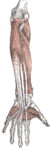
The forearm contains many muscles, including the flexors and extensors of thewrist,flexors and extensors of the digits, a flexor of the elbow (brachioradialis), andpronatorsandsupinatorsthat turn the hand to face down or upwards, respectively. In cross-section, the forearm can be divided into twofascial compartments.The posterior compartment contains the extensors of the hands, which are supplied by theradial nerve.The anterior compartment contains the flexors and is mainly supplied by themedian nerve.The flexor muscles are more massive than the extensors because they work against gravity and act as anti-gravity muscles. Theulnar nervealso runs the length of the forearm.[3]
Theradialandulnar arteriesand their branches supply the blood to the forearm. These usually run on the anterior face of the radius and ulna down the whole forearm. The main superficial veins of the forearm are thecephalic,median antebrachialand thebasilic vein.These veins can be used for cannularisation orvenipuncture,although thecubital fossais a preferred site for getting blood.
Structure
editBones and joints
editThe bones of the forearm are theradius(located on the lateral side) and theulna(located on the medial side)

Radius
editProximally, the head of the radius articulates with thecapitulum of the humerusand the radial notch of the ulna at theelbow.Thearticulationbetween the radius and the ulna at the elbow is known as theproximal radioulnar joint.
Distally, it articulates with the ulna again at thedistal radioulnar joint.It forms part of thewrist jointby articulating with thescaphoidat its lateral aspect and with thelunateat its medial aspect.
Ulna
editProximally, thetrochlear notchof the ulna articulates with thetrochlea of the humerusand theradial notcharticulates with the head of the radius at theelbow.[4]
Distally it forms part of thedistal radioulnar jointand also articulates with thewrist.[5]
Muscles
edit| Compartment | Level | Muscle | E/I | Nerve |
|---|---|---|---|---|
| Anterior | superficial | flexor carpi radialis | E | median |
| Anterior | superficial | palmaris longus | E | median |
| Anterior | superficial | flexor carpi ulnaris | E | ulnar |
| Anterior | superficial | pronator teres | I | median |
| Anterior | superficial (or intermediate) | flexor digitorum superficialis (sublimis) | E | median |
| Anterior | deep | flexor digitorum profundus | E | ulnar + median |
| Anterior | deep | flexor pollicis longus | E | median |
| Anterior | deep | pronator quadratus | I | median |
| Posterior | (see below) | brachioradialis | I | radial |
| Posterior | superficial | extensor carpi radialis longus | E | radial |
| Posterior | superficial | extensor carpi radialis brevis | E | radial |
| Posterior | intermediate | extensor digitorum (communis) | E | radial |
| Posterior | intermediate | extensor digiti minimi (proprius) | E | radial |
| Posterior | superficial | extensor carpi ulnaris | E | radial |
| Posterior | deep | abductor pollicis longus | E | radial |
| Posterior | deep | extensor pollicis brevis | E | radial |
| Posterior | deep | extensor pollicis longus | E | radial |
| Posterior | deep | extensor indicis (proprius) | E | radial |
| Posterior | deep | supinator | I | radial |
| Posterior | deep | anconeus | I | radial |
- "E/I" refers to "extrinsic" or "intrinsic". The intrinsic muscles of the forearm act on the forearm, meaning, across the elbow joint and theproximalanddistalradioulnar joints (resulting inpronationorsupination), whereas the extrinsic muscles act upon the hand and wrist. In most cases, the extrinsic anterior muscles areflexors,while the extrinsic posterior muscles areextensors.
- The brachioradialis, flexor of the forearm, is unusual in that it is located in theposterior compartment,but it is actually in the anterior portion of the forearm.
- Theanconeusis considered by some as a part of theposterior compartment of the arm.[6]
Nerves
edit- See separate nerve articles for details on divisions proximal to the elbow and distal to the wrist; seeBrachial plexusfor the origins of the median, radial and ulnar nerves.
- Median nerve– interior nerve of the anterior compartment (PT,FCR,PL,FDS).
- anterior interosseous nerve(suppliesFPL,lat. 1/2 ofFDP,PQ).
- Radial nerve– supplies muscles of the posterior compartment (ECRL,ECRB).
- Superficial branch of radial nerve
- Deep branch of radial nerve,becomesPosterior interosseus nerveand supplies muscles of the posterior compartment (ED,EDM,ECU,APL,EPB,EPL,EI).
- Ulnar nerve– supplies some medial muscles (FCU,med. 1/2 ofFDP).
Vessels
edit
Other structures
editFunction
editThe forearm can be brought closer to the upper arm (flexed) and brought away from the upper arm (extended) due to movement at theelbow.The forearm can also be rotated so that the palm of thehandrotates inwards (pronated) and rotated back so that the palm rotates outwards (supinated) due to movement at the elbow and thedistal radioulnar joint.[5]
Clinical significance
edit
Afractureof the forearm can be classified as to whether it involves only the ulna (ulnar fracture), only the radius (radius fracture), or both radioulnar fracture.
For treatment of children withtorus fracturesof the forearm splinting appears to work better than casting.[7] Genetically determined disorders likehereditary multiple exostosescan lead to hand and forearm deformities. Hereditary multiple exostoses is due growth disturbance of the epiphyses of the radius and ulna, the two bones of the forearm.[8]
Additional images
edit-
 Superficial muscles of the forearm
 -
 Deep muscles of the anterior forearm
 -
 Deep muscles of the posterior forearm
 -
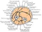 Cross-section through the middle of the forearm.
 -
 Bones of the forearm - ulna (left) and radius (right)


See also
editReferences
edit- ^WebMD (2009)."forearm".Webster's New World Medical Dictionary(3rd ed.). Houghton Mifflin Harcourt. p. 166.ISBN978-0-544-18897-6.
- ^"Forearm".The Lecturio Medical Concept Library.Retrieved2021-06-22.
- ^Mitchell, Brittney; Whited, Lacey (2020-08-15)."Anatomy, Shoulder and Upper Limb, Forearm Muscles".National Center for Biotechnology Information, U.S. National Library of Medicine.StatPearls Publishing LLC.Retrieved22 June2021.
- ^"Structure of The Forearm".The Lecturio Medical Concept Library.Retrieved2021-06-22.
- ^abStandring, Susan (2016).Gray's anatomy: the anatomical basis of clinical practice(Forty-first ed.). [Philadelphia].ISBN9780702052309.OCLC920806541.
{{cite book}}:CS1 maint: location missing publisher (link) - ^"Dissector Answers — Axilla & Arm".The University of Michigan. Archived fromthe originalon 3 January 2008.Retrieved2008-01-17.
- ^Jiang N, Cao ZH, Ma YF, Lin Z, Yu B (November 2016). "Management of Pediatric Forearm Torus Fractures: A Systematic Review and Meta-Analysis".Pediatric Emergency Care.32(11):773–778.doi:10.1097/pec.0000000000000579.PMID26555307.S2CID25796224.
- ^El-Sobky TA, Samir S, Atiyya AN, Mahmoud S, Aly AS, Soliman R (2018)."Current paediatric orthopaedic practice in hereditary multiple osteochondromas of the forearm: a systematic review".SICOT-J.4:10.doi:10.1051/sicotj/2018002.PMC5863686.PMID29565244.