
Beta sheet

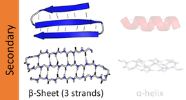
Thebeta sheet(β-sheet,alsoβ-pleated sheet) is a commonmotifof the regularprotein secondary structure.Beta sheets consist ofbeta strands(β-strands) connected laterally by at least two or threebackbonehydrogen bonds,forming a generally twisted, pleated sheet. A β-strand is a stretch ofpolypeptidechain typically 3 to 10amino acidslong with backbone in an extendedconformation.The supramolecular association of β-sheets has been implicated in the formation of thefibrilsandprotein aggregatesobserved inamyloidosis,Alzheimer's diseaseand otherproteinopathies.
History[edit]

The first β-sheet structure was proposed byWilliam Astburyin the 1930s. He proposed the idea of hydrogen bonding between thepeptide bondsof parallel or antiparallel extended β-strands. However, Astbury did not have the necessary data on the bond geometry of the amino acids in order to build accurate models, especially since he did not then know that thepeptide bondwas planar. A refined version was proposed byLinus PaulingandRobert Coreyin 1951. Their model incorporated the planarity of the peptide bond which they previously explained as resulting from keto-enoltautomerization.
Structure and orientation[edit]
Geometry[edit]
The majority of β-strands are arranged adjacent to other strands and form an extensivehydrogen bondnetwork with their neighbors in which theN−Hgroups in the backbone of one strand establishhydrogen bondswith theC=Ogroups in the backbone of the adjacent strands. In the fully extended β-strand, successive side chains point straight up and straight down in an alternating pattern. Adjacent β-strands in a β-sheet are aligned so that their Cαatoms are adjacent and their side chains point in the same direction. The "pleated" appearance of β-strands arises from tetrahedral chemical bonding at the Cαatom; for example, if a side chain points straight up, then the bonds to the C′ must point slightly downwards, since its bond angle is approximately 109.5°. The pleating causes the distance between Cα
iand Cα
i+ 2to be approximately 6Å(0.60nm), rather than the 7.6 Å (0.76 nm) expected from two fully extendedtranspeptides.The "sideways" distance between adjacent Cαatoms inhydrogen-bondedβ-strands is roughly 5 Å (0.50 nm).

However, β-strands are rarely perfectly extended; rather, they exhibit a twist. The energetically preferreddihedral anglesnear (φ,ψ) = (–135°, 135°) (broadly, the upper left region of theRamachandran plot) diverge significantly from the fully extended conformation (φ,ψ) = (–180°, 180°).[1]The twist is often associated with alternating fluctuations in thedihedral anglesto prevent the individual β-strands in a larger sheet from splaying apart. A good example of a strongly twisted β-hairpin can be seen in the proteinBPTI.
The side chains point outwards from the folds of the pleats, roughly perpendicularly to the plane of the sheet; successive amino acid residues point outwards on alternating faces of the sheet.
Hydrogen bonding patterns[edit]


Because peptide chains have a directionality conferred by theirN-terminusandC-terminus,β-strands too can be said to be directional. They are usually represented in protein topology diagrams by an arrow pointing toward the C-terminus. Adjacent β-strands can formhydrogen bondsin antiparallel, parallel, or mixed arrangements.
In an antiparallel arrangement, the successive β-strands alternate directions so that the N-terminus of one strand is adjacent to the C-terminus of the next. This is the arrangement that produces the strongest inter-strand stability because it allows the inter-strand hydrogen bonds between carbonyls and amines to be planar, which is their preferred orientation. The peptide backbone dihedral angles (φ,ψ) are about (–140°, 135°) in antiparallel sheets. In this case, if two atoms Cα
iand Cα
jare adjacent in twohydrogen-bondedβ-strands, then they form two mutual backbone hydrogen bonds to each other's flankingpeptide groups;this is known as aclose pairof hydrogen bonds.
In a parallel arrangement, all of the N-termini of successive strands are oriented in the same direction; this orientation may be slightly less stable because it introduces nonplanarity in the inter-strand hydrogen bonding pattern. The dihedral angles (φ,ψ) are about (–120°, 115°) in parallel sheets. It is rare to find less than five interacting parallel strands in a motif, suggesting that a smaller number of strands may be unstable, however it is also fundamentally more difficult for parallel β-sheets to form because strands with N and C termini aligned necessarily must be very distant in sequence[citation needed].There is also evidence that parallel β-sheet may be more stable since small amyloidogenic sequences appear to generally aggregate into β-sheet fibrils composed of primarily parallel β-sheet strands, where one would expect anti-parallel fibrils if anti-parallel were more stable.
In parallel β-sheet structure, if two atoms Cα
iand Cα
jare adjacent in twohydrogen-bondedβ-strands, then they donothydrogen bond to each other; rather, one residue forms hydrogen bonds to the residues that flank the other (but not vice versa). For example, residueimay form hydrogen bonds to residuesj− 1 andj+ 1; this is known as awide pairof hydrogen bonds. By contrast, residuejmay hydrogen-bond to different residues altogether, or to none at all.
The hydrogen bond arrangement in parallel beta sheet resembles that in anamide ringmotif with 11 atoms.
Finally, an individual strand may exhibit a mixed bonding pattern, with a parallel strand on one side and an antiparallel strand on the other. Such arrangements are less common than a random distribution of orientations would suggest, suggesting that this pattern is less stable than the anti-parallel arrangement, however bioinformatic analysis always struggles with extracting structural thermodynamics since there are always numerous other structural features present in whole proteins. Also proteins are inherently constrained by folding kinetics as well as folding thermodynamics, so one must always be careful in concluding stability from bioinformatic analysis.
Thehydrogen bondingof β-strands need not be perfect, but can exhibit localized disruptions known asβ-bulges.
The hydrogen bonds lie roughly in the plane of the sheet, with thepeptidecarbonylgroups pointing in alternating directions with successive residues; for comparison, successive carbonyls point in thesamedirection in thealpha helix.
Amino acid propensities[edit]
Large aromatic residues (tyrosine,phenylalanine,tryptophan) and β-branched amino acids (threonine,valine,isoleucine) are favored to be found in β-strands in themiddleof β-sheets. Different types of residues (such asproline) are likely to be found in theedgestrands in β-sheets, presumably to avoid the "edge-to-edge" association between proteins that might lead to aggregation andamyloidformation.[2]
Common structural motifs[edit]


β-hairpin motif[edit]
A very simplestructural motifinvolving β-sheets is theβ-hairpin,in which two antiparallel strands are linked by a short loop of two to five residues, of which one is frequently aglycineor aproline,both of which can assume the dihedral-angle conformations required for a tightturnor aβ-bulge loop.Individual strands can also be linked in more elaborate ways with longer loops that may containα-helices.
Greek key motif[edit]
The Greek key motif consists of four adjacent antiparallel strands and their linking loops. It consists of three antiparallel strands connected by hairpins, while the fourth is adjacent to the first and linked to the third by a longer loop. This type of structure forms easily during theprotein foldingprocess.[3][4]It was named after a pattern common to Greek ornamental artwork (seemeander).
β-α-β motif[edit]
Due to the chirality of their component amino acids, all strands exhibit right-handed twist evident in most higher-order β-sheet structures. In particular, the linking loop between two parallel strands almost always has a right-handed crossover chirality, which is strongly favored by the inherent twist of the sheet.[5]This linking loop frequently contains a helical region, in which case it is called aβ-α-βmotif. A closely related motif called a β-α-β-α motif forms the basic component of the most commonly observed proteintertiary structure,theTIM barrel.


β-meander motif[edit]
A simplesupersecondaryprotein topology composed of two or more consecutive antiparallel β-strands linked together byhairpinloops.[7][8]This motif is common in β-sheets and can be found in several structural architectures includingβ-barrelsandβ-propellers.
The vast majority of β-meander regions in proteins are found packed against other motifs or sections of the polypeptide chain, forming portions of the hydrophobic core that canonically drives formation of the folded structure.[9]However, several notable exceptions include the Outer Surface Protein A (OspA) variants[6]and the Single Layer β-sheet Proteins (SLBPs)[10]which contain single-layer β-sheets in the absence of a traditional hydrophobic core. These β-rich proteins feature an extended single-layer β-meander β-sheets that are primarily stabilized via inter-β-strand interactions and hydrophobic interactions present in the turn regions connecting individual strands.
Psi-loop motif[edit]
The psi-loop (Ψ-loop) motif consists of two antiparallel strands with one strand in between that is connected to both by hydrogen bonds.[11]There are four possible strand topologies for single Ψ-loops.[12]This motif is rare as the process resulting in its formation seems unlikely to occur during protein folding. The Ψ-loop was first identified in theaspartic proteasefamily.[12]
Structural architectures of proteins with β-sheets[edit]
β-sheets are present inall-β,α+βandα/βdomains, and in manypeptidesor small proteins with poorly defined overall architecture.[13][14]All-β domainsmay formβ-barrels,β-sandwiches,β-prisms,β-propellers,andβ-helices.
Structural topology[edit]
Thetopologyof a β-sheet describes the order ofhydrogen-bondedβ-strands along the backbone. For example, theflavodoxin foldhas a five-stranded, parallel β-sheet with topology 21345; thus, the edge strands are β-strand 2 and β-strand 5 along the backbone. Spelled out explicitly, β-strand 2 is H-bonded to β-strand 1, which is H-bonded to β-strand 3, which is H-bonded to β-strand 4, which is H-bonded to β-strand 5, the other edge strand. In the same system, the Greek key motif described above has a 4123 topology. Thesecondary structureof a β-sheet can be described roughly by giving the number of strands, their topology, and whether theirhydrogen bondsare parallel or antiparallel.
β-sheets can beopen,meaning that they have two edge strands (as in theflavodoxin foldor theimmunoglobulin fold) or they can beclosedβ-barrels(such as theTIM barrel).β-Barrelsare often described by theirstaggerorshear.Some open β-sheets are very curved and fold over on themselves (as in theSH3 domain) or form horseshoe shapes (as in theribonuclease inhibitor). Open β-sheets can assemble face-to-face (such as theβ-propeller domainorimmunoglobulin fold) or edge-to-edge, forming one big β-sheet.
Dynamic features[edit]
β-pleated sheet structures are made from extended β-strand polypeptide chains, with strands linked to their neighbours byhydrogen bonds.Due to this extended backbone conformation, β-sheets resiststretching.β-sheets in proteins may carry outlow-frequencyaccordion-like motion as observed by theRaman spectroscopy[15]and analyzed with the quasi-continuum model.[16]
Parallel β-helices[edit]

Aβ-helixis formed from repeating structural units consisting of two or three short β-strands linked by short loops. These units "stack" atop one another in a helical fashion so that successive repetitions of the same strand hydrogen-bond with each other in a parallel orientation. See theβ-helixarticle for further information.
In lefthanded β-helices, the strands themselves are quite straight and untwisted; the resulting helical surfaces are nearly flat, forming a regulartriangular prismshape, as shown for the 1QRE archaeal carbonic anhydrase at right. Other examples are the lipid A synthesis enzymeLpxAand insect antifreeze proteins with a regular array of Thr sidechains on one face that mimic the structure of ice.[17]

Righthanded β-helices, typified by thepectate lyaseenzyme shown at left orP22 phage tailspike protein,have a less regular cross-section, longer and indented on one of the sides; of the three linker loops, one is consistently just two residues long and the others are variable, often elaborated to form a binding or active site.[18]
A two-sided β-helix (right-handed) is found in some bacterialmetalloproteases;its two loops are each six residues long and bind stabilizing calcium ions to maintain the integrity of the structure, using the backbone and the Asp side chain oxygens of a GGXGXD sequence motif.[19]This fold is called a β-roll in the SCOP classification.
In pathology[edit]
Some proteins that are disordered or helical as monomers, such as amyloid β (seeamyloid plaque) can form β-sheet-rich oligomeric structures associated with pathological states. The amyloid β protein's oligomeric form is implicated as a cause ofAlzheimer's.Its structure has yet to be determined in full, but recent data suggest that it may resemble an unusual two-strand β-helix.[20]
The side chains from the amino acid residues found in a β-sheet structure may also be arranged such that many of the adjacent sidechains on one side of the sheet are hydrophobic, while many of those adjacent to each other on the alternate side of the sheet are polar or charged (hydrophilic),[21]which can be useful if the sheet is to form a boundary between polar/watery and nonpolar/greasy environments.
See also[edit]
References[edit]
- ^Voet D, Voet JG (2004).Biochemistry(3rd ed.). Hoboken, NJ: Wiley. pp.227–231.ISBN0-471-19350-X.
- ^Richardson JS, Richardson DC (March 2002)."Natural beta-sheet proteins use negative design to avoid edge-to-edge aggregation".Proceedings of the National Academy of Sciences of the United States of America.99(5): 2754–9.Bibcode:2002PNAS...99.2754R.doi:10.1073/pnas.052706099.PMC122420.PMID11880627.
- ^Tertiary Protein Structure and Folds: section 4.3.2.1.FromPrinciples of Protein Structure, Comparative Protein Modelling, and Visualisation
- ^Hutchinson EG, Thornton JM (April 1993). "The Greek key motif: extraction, classification and analysis".Protein Engineering.6(3): 233–45.doi:10.1093/protein/6.3.233.PMID8506258.
- ^See sections II B and III C, D inRichardson JS (1981). "The Anatomy and Taxonomy of Protein Structure".Anatomy and Taxonomy of Protein Structures.Vol. 34. pp. 167–339.doi:10.1016/s0065-3233(08)60520-3.ISBN0-12-034234-0.PMID7020376.
{{cite book}}:|journal=ignored (help) - ^abMakabe K, McElheny D, Tereshko V, Hilyard A, Gawlak G, Yan S, et al. (November 2006)."Atomic structures of peptide self-assembly mimics".Proceedings of the National Academy of Sciences of the United States of America.103(47): 17753–8.Bibcode:2006PNAS..10317753M.doi:10.1073/pnas.0606690103.PMC1693819.PMID17093048.
- ^"SCOP: Fold: WW domain-like".Archived fromthe originalon 2012-02-04.Retrieved2007-06-01.
- ^PPS '96 – Super Secondary Structure
- ^Biancalana M, Makabe K, Koide S (February 2010)."Minimalist design of water-soluble cross-beta architecture".Proceedings of the National Academy of Sciences of the United States of America.107(8): 3469–74.Bibcode:2010PNAS..107.3469B.doi:10.1073/pnas.0912654107.PMC2840449.PMID20133689.
- ^Xu, Qingping; Biancalana, Matthew; Grant, Joanna C.; Chiu, Hsiu-Ju; Jaroszewski, Lukasz; Knuth, Mark W.; Lesley, Scott A.; Godzik, Adam; Elsliger, Marc-André; Deacon, Ashley M.; Wilson, Ian A. (September 2019)."Structures of single-layer β-sheet proteins evolved from β-hairpin repeats".Protein Science.28(9): 1676–1689.doi:10.1002/pro.3683.ISSN1469-896X.PMC6699103.PMID31306512.
- ^Hutchinson EG, Thornton JM (February 1996)."PROMOTIF--a program to identify and analyze structural motifs in proteins".Protein Science.5(2): 212–20.doi:10.1002/pro.5560050204.PMC2143354.PMID8745398.
- ^abHutchinson EG, Thornton JM (1990). "HERA--a program to draw schematic diagrams of protein secondary structures".Proteins.8(3): 203–12.doi:10.1002/prot.340080303.PMID2281084.S2CID28921557.
- ^Hubbard TJ, Murzin AG, Brenner SE, Chothia C (January 1997)."SCOP: a structural classification of proteins database".Nucleic Acids Research.25(1): 236–9.doi:10.1093/nar/25.1.236.PMC146380.PMID9016544.
- ^Fox NK, Brenner SE, Chandonia JM (January 2014)."SCOPe: Structural Classification of Proteins--extended, integrating SCOP and ASTRAL data and classification of new structures".Nucleic Acids Research.42(Database issue): D304-9.doi:10.1093/nar/gkt1240.PMC3965108.PMID24304899.
- ^Painter PC, Mosher LE, Rhoads C (July 1982). "Low-frequency modes in the Raman spectra of proteins".Biopolymers.21(7): 1469–72.doi:10.1002/bip.360210715.PMID7115900.
- ^Chou KC (August 1985)."Low-frequency motions in protein molecules. Beta-sheet and beta-barrel".Biophysical Journal.48(2): 289–97.Bibcode:1985BpJ....48..289C.doi:10.1016/S0006-3495(85)83782-6.PMC1329320.PMID4052563.
- ^Liou YC, Tocilj A, Davies PL, Jia Z (July 2000). "Mimicry of ice structure by surface hydroxyls and water of a beta-helix antifreeze protein".Nature.406(6793): 322–4.Bibcode:2000Natur.406..322L.doi:10.1038/35018604.PMID10917536.S2CID4385352.
- ^Branden C, Tooze J (1999).Introduction to Protein Structure.New York: Garland. pp. 20–32.ISBN0-8153-2305-0.
- ^Baumann U, Wu S, Flaherty KM, McKay DB (September 1993)."Three-dimensional structure of the alkaline protease of Pseudomonas aeruginosa: a two-domain protein with a calcium binding parallel beta roll motif".The EMBO Journal.12(9): 3357–64.doi:10.1002/j.1460-2075.1993.tb06009.x.PMC413609.PMID8253063.
- ^Nelson R, Sawaya MR, Balbirnie M, Madsen AØ, Riekel C, Grothe R, Eisenberg D (June 2005)."Structure of the cross-beta pine of amyloid-like fibrils".Nature.435(7043): 773–8.Bibcode:2005Natur.435..773N.doi:10.1038/nature03680.PMC1479801.PMID15944695.
- ^Zhang S, Holmes T, Lockshin C, Rich A (April 1993)."Spontaneous assembly of a self-complementary oligopeptide to form a stable macroscopic membrane".Proceedings of the National Academy of Sciences of the United States of America.90(8): 3334–8.Bibcode:1993PNAS...90.3334Z.doi:10.1073/pnas.90.8.3334.PMC46294.PMID7682699.
Further reading[edit]
- Cooper J (31 May 1996)."Super Secondary Structure - Part II".Principles of Protein Structure Using the Internet.Retrieved25 May2007.
- "Open-sided Beta-meander".Structural Classification of Proteins (SCOP).20 October 2006. Archived fromthe originalon 4 February 2012.Retrieved31 May2007.