CD83

CD83(Cluster of Differentiation 83) is a humanproteinencoded by theCD83gene.[5]
Structure
[edit]The membrane-bound form of CD83 consists of an extracellularV-type immunoglobulin-like domain,atransmembrane domainand a cytoplasmicsignalingtail. A free soluble form consists of the immunoglobulin-like domain alone. Membrane-bound CD83 is expected to form trimers. Soluble CD83 is able to assemble into dodecameric complexes.[6]
Gene
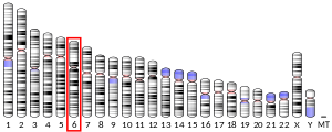
[edit]The CD83 gene is located on humanchromosome6p23 and mouse chromosome 13. In humans, apromoter261 bp upstream consists of fiveNF-κBand threeinterferon regulatory factorbinding sites, reflecting the involvement of CD83 in inflammation,[7]as well as binding sites for thearyl hydrocarbon receptor.The latter also occur in anenhancersequence located 185 bp downstream, inside the secondintron,[8]and may suggest negative regulation of transcription by microbial metabolites produced in the gut.
Function
[edit]The transmembrane domain of membrane-bound CD83 stabilizesMHC II,costimulatory moleculesandCD28in the membrane by antagonizing MARCH-familyE3 ubiquitin ligases.[9][10]
Ligands
[edit]It is not clear what ligands interact with CD83, but membrane-bound CD83 may homotypically interact with the soluble form, suggestingautocrineimmune regulation.[11]However, it contrasts with differences between the single expression of soluble CD83 onmonocytesand membrane-bound CD83 on activateddendritic cellsseems also as their good marker.[clarification needed][12]Soluble CD83 also binds toCD154,leading toT helpertype 2 lymphocyteapoptosisby suppression ofBcl-2 inhibitors.[13]
Positive selection
[edit]The development ofthymocytesduring the positive-selection stage may be guided by CD83 expression oncortical thymic epithelial cells(cTECs).CD4+CD8+double-positive thymocytes surrounded by specially differentiated cTECs called thymic nurse cells are tested for function of their αβT cell receptor(TCR); a nonreactive TCR leads to thymocyte death by neglect. Successful rearrangement of a reactive TCR supports survival and restriction of expression to CD4 or CD8 alone on single-positive thymocytes, depending on the ability to recognize MHC II orMHC I,respectively. Upregulation of MHC II turnover on thymic nurse cells by CD83 may enlarge the population of CD4+single-positive thymocytes.[14][10]
Regulatory T cells
[edit]
T regulatory cells(Tregcells) are present in two major populations: thymically induced and peripherally induced Tregcells. All Tregcells express theFoxp3transcription factor, establishing their suppressive phenotype. Foxp3 expression is not affected by loss of CD83 in a CD83 knockout mouse. In contrast, CD83 seems important for peripheral Tregcell induction, as suggested by reduction of this population in a conditional knockout mouse lacking CD83 specifically in Tregcells, which results in a proinflammatory phenotype.[15]
CD83 deficiency also results in an imbalances in effector function of Tregcells, as decreased expression of the T helper type 2 cell transcription factorGATA3is also important forST2production.[16]
Activated Tregcells produce large amounts of soluble CD83, leading to downregulation ofIRAK-1at inflamed sites, downregulation oftoll-like receptorsignaling, and switching of inflammatory signals to tolerance establishment.[16]
Dendritic cells
[edit]CD83 expression is a marker for mature dendritic cells.[12]CD83 stabilizes MHC II on membrane by antagonizing MARCH E3 ubiquitin ligases. A MARCH1 knockout mouse shows accumulation of MHC II, which leads to reducedCD4+T lymphocyte activation and reducedIL-12production.[17]Conversely, a CD83 knockout mouse shows a reduction of MHC II andCD86,better response to bacterial infection, and higher production of IL-12 than in the wild type. CD83 seems to be an important regulator of dendritic cell phenotype and MHC II turnover, mediated by CD83-dependentendosomeprocessing.[11]
B cells
[edit]CD83 expression correlates with rate of activation ofB lymphocytesand it is under control of theB cell receptor,CD40,or Toll-like receptor activation, as in other lymphocytes, where CD83 is expressed upon stimulation. A CD83 knockout mouse shows upregulated proliferation of B lymphocytes, suggesting that CD83 acts as a brake on proliferation.[18]CD83 does not affect affinity maturation of antibodies, but its deficiency enhances immunoglobulin E class switching, suggesting that CD83 may be involved in allergy development and could be a therapeutic target for allergy treatment.[19]
See also
[edit]References
[edit]- ^abcGRCh38: Ensembl release 89: ENSG00000112149–Ensembl,May 2017
- ^abcGRCm38: Ensembl release 89: ENSMUSG00000015396–Ensembl,May 2017
- ^"Human PubMed Reference:".National Center for Biotechnology Information, U.S. National Library of Medicine.
- ^"Mouse PubMed Reference:".National Center for Biotechnology Information, U.S. National Library of Medicine.
- ^"Entrez Gene: CD83 CD83 molecule".
- ^Berchtold S, Jones T, Mühl-Zürbes P, Sheer D, Schuler G, Steinkasserer A (March 1999). "The human dendritic cell marker CD83 maps to chromosome 6p23".Annals of Human Genetics.63(Pt 2): 181–3.doi:10.1046/j.1469-1809.1999.6320181.x.PMID10738529.S2CID25338621.
- ^Stein MF, Lang S, Winkler TH, Deinzer A, Erber S, Nettelbeck DM, et al. (April 2013)."Multiple interferon regulatory factor and NF-κB sites cooperate in mediating cell-type- and maturation-specific activation of the human CD83 promoter in dendritic cells".Molecular and Cellular Biology.33(7): 1331–44.doi:10.1128/MCB.01051-12.PMC3624272.PMID23339870.
- ^Michalski J, Deinzer A, Stich L, Zinser E, Steinkasserer A, Knippertz I (July 2020)."Quercetin induces an immunoregulatory phenotype in maturing human dendritic cells".Immunobiology.225(4): 151929.doi:10.1016/j.imbio.2020.151929.PMID32115260.
- ^Grosche, Linda; Knippertz, Ilka; König, Christina; Royzman, Dmytro; Wild, Andreas B.; Zinser, Elisabeth; Sticht, Heinrich; Muller, Yves A.; Steinkasserer, Alexander; Lechmann, Matthias (17 April 2020)."The CD83 Molecule – An Important Immune Checkpoint".Frontiers in Immunology.11:721.doi:10.3389/fimmu.2020.00721.PMC7181454.PMID32362900.
- ^abvon Rohrscheidt, Julia; Petrozziello, Elisabetta; Nedjic, Jelena; Federle, Christine; Krzyzak, Lena; Ploegh, Hidde L.; Ishido, Satoshi; Steinkasserer, Alexander; Klein, Ludger (22 August 2016)."Thymic CD4 T cell selection requires attenuation of March8-mediated MHCII turnover in cortical epithelial cells through CD83".Journal of Experimental Medicine.213(9): 1685–1694.doi:10.1084/jem.20160316.PMC4995086.PMID27503071.
- ^abBates, J M; Flanagan, K; Mo, L; Ota, N; Ding, J; Ho, S; Liu, S; Roose-Girma, M; Warming, S; Diehl, L (March 2015)."Dendritic cell CD83 homotypic interactions regulate inflammation and promote mucosal homeostasis".Mucosal Immunology.8(2): 414–428.doi:10.1038/mi.2014.79.PMC4326976.PMID25204675.
- ^abChen, Liwen; Zhu, Yibei; Zhang, Guangbo; Gao, Chao; Zhong, Weixue; Zhang, Xueguang (15 November 2011)."CD83-stimulated monocytes suppress T-cell immune responses through production of prostaglandin E2".Proceedings of the National Academy of Sciences of the United States of America.108(46): 18778–18783.doi:10.1073/pnas.1018994108.ISSN1091-6490.PMC3219128.PMID22065790.
- ^Wu, Yong-Jin; Song, Yan-Nan; Geng, Xiao-Rui; Ma, Fei; Mo, Li-Hua; Zhang, Xiao-Wen; Liu, Da-Bo; Liu, Zhi-Gang; Yang, Ping-Chang (2020)."Soluble CD83 alleviates experimental allergic rhinitis through modulating antigen-specific Th2 cell property".International Journal of Biological Sciences.16(2): 216–227.doi:10.7150/ijbs.38722.PMC6949156.PMID31929750.
- ^Kadouri, Noam; Nevo, Shir; Goldfarb, Yael; Abramson, Jakub (April 2020). "Thymic epithelial cell heterogeneity: TEC by TEC".Nature Reviews Immunology.20(4): 239–253.doi:10.1038/s41577-019-0238-0.PMID31804611.S2CID208622435.
- ^Doebbeler, Marina; Koenig, Christina; Krzyzak, Lena; Seitz, Christine; Wild, Andreas; Ulas, Thomas; Baßler, Kevin; Kopelyanskiy, Dmitry; Butterhof, Alina; Kuhnt, Christine; Kreiser, Simon; Stich, Lena; Zinser, Elisabeth; Knippertz, Ilka; Wirtz, Stefan; Riegel, Christin; Hoffmann, Petra; Edinger, Matthias; Nitschke, Lars; Winkler, Thomas; Schultze, Joachim L.; Steinkasserer, Alexander; Lechmann, Matthias (7 June 2018)."CD83 expression is essential for Treg cell differentiation and stability".JCI Insight.3(11): e99712.doi:10.1172/jci.insight.99712.PMC6124443.PMID29875316.
- ^abMaitra, Urmila; Davis, Sarah; Reilly, Christopher M.; Li, Liwu (1 May 2009)."Differential Regulation of Foxp3 and IL-17 Expression in CD4 T Helper Cells by IRAK-1".The Journal of Immunology.182(9): 5763–5769.doi:10.4049/jimmunol.0900124.PMC4773027.PMID19380824.
- ^Ishido, Satoshi; Matsuki, Yohei; Goto, Eiji; Kajikawa, Mizuho; Ohmura-Hoshino, Mari (March 2010)."MARCH-I: A new regulator of dendritic cell function".Molecules and Cells.29(3): 229–232.doi:10.1007/s10059-010-0051-x.PMID20213309.S2CID10403102.
- ^Kretschmer, Birte; Kühl, Svenja; Fleischer, Bernhard; Breloer, Minka (May 2011). "Activated T cells induce rapid CD83 expression on B cells by engagement of CD40".Immunology Letters.136(2): 221–227.doi:10.1016/j.imlet.2011.01.013.PMID21277328.
- ^Krzyzak, Lena; Seitz, Christine; Urbat, Anne; Hutzler, Stefan; Ostalecki, Christian; Gläsner, Joachim; Hiergeist, Andreas; Gessner, André; Winkler, Thomas H.; Steinkasserer, Alexander; Nitschke, Lars (1 May 2016)."CD83 Modulates B Cell Activation and Germinal Center Responses".The Journal of Immunology.196(9): 3581–3594.doi:10.4049/jimmunol.1502163.PMID26983787.
Further reading
[edit]- Lechmann M, Berchtold S, Hauber J, Steinkasserer A (June 2002). "CD83 on dendritic cells: more than just a marker for maturation".Trends in Immunology.23(6): 273–5.doi:10.1016/S1471-4906(02)02214-7.PMID12072358.
- Zhou LJ, Schwarting R, Smith HM, Tedder TF (July 1992)."A novel cell-surface molecule expressed by human interdigitating reticulum cells, Langerhans cells, and activated lymphocytes is a new member of the Ig superfamily".Journal of Immunology.149(2): 735–42.doi:10.4049/jimmunol.149.2.735.PMID1378080.S2CID24475187.
- Zhou LJ, Tedder TF (April 1995). "Human blood dendritic cells selectively express CD83, a member of the immunoglobulin superfamily".Journal of Immunology.154(8): 3821–35.doi:10.4049/jimmunol.154.8.3821.PMID7706722.S2CID24672615.
- Maruyama K, Sugano S (January 1994). "Oligo-capping: a simple method to replace the cap structure of eukaryotic mRNAs with oligoribonucleotides".Gene.138(1–2): 171–4.doi:10.1016/0378-1119(94)90802-8.PMID8125298.
- Kozlow EJ, Wilson GL, Fox CH, Kehrl JH (January 1993)."Subtractive cDNA cloning of a novel member of the Ig gene superfamily expressed at high levels in activated B lymphocytes".Blood.81(2): 454–61.doi:10.1182/blood.V81.2.454.454.PMID8422464.
- de la Fuente MA, Pizcueta P, Nadal M, Bosch J, Engel P (September 1997)."CD84 leukocyte antigen is a new member of the Ig superfamily".Blood.90(6): 2398–405.doi:10.1182/blood.V90.6.2398.PMID9310491.
- Suzuki Y, Yoshitomo-Nakagawa K, Maruyama K, Suyama A, Sugano S (October 1997). "Construction and characterization of a full length-enriched and a 5'-end-enriched cDNA library".Gene.200(1–2): 149–56.doi:10.1016/S0378-1119(97)00411-3.PMID9373149.
- Olavesen MG, Bentley E, Mason RV, Stephens RJ, Ragoussis J (December 1997). "Fine mapping of 39 ESTs on human chromosome 6p23-p25".Genomics.46(2): 303–6.doi:10.1006/geno.1997.5032.PMID9417921.
- Twist CJ, Beier DR, Disteche CM, Edelhoff S, Tedder TF (1998). "The mouse Cd83 gene: structure, domain organization, and chromosome localization".Immunogenetics.48(6): 383–93.doi:10.1007/s002510050449.PMID9799334.S2CID19869850.
- Berchtold S, Mühl-Zürbes P, Heufler C, Winklehner P, Schuler G, Steinkasserer A (November 1999). "Cloning, recombinant expression and biochemical characterization of the murine CD83 molecule which is specifically upregulated during dendritic cell maturation".FEBS Letters.461(3): 211–6.doi:10.1016/S0014-5793(99)01465-9.PMID10567699.S2CID28053654.
- Scholler N, Hayden-Ledbetter M, Hellström KE, Hellström I, Ledbetter JA (March 2001)."CD83 is an I-type lectin adhesion receptor that binds monocytes and a subset of activated CD8+ T cells [corrected]".Journal of Immunology.166(6): 3865–72.doi:10.4049/jimmunol.166.6.3865.PMID11238630.
- Fanales-Belasio E, Moretti S, Nappi F, Barillari G, Micheletti F, Cafaro A, Ensoli B (January 2002)."Native HIV-1 Tat protein targets monocyte-derived dendritic cells and enhances their maturation, function, and antigen-specific T cell responses".Journal of Immunology.168(1): 197–206.doi:10.4049/jimmunol.168.1.197.hdl:2108/188460.PMID11751963.S2CID25473321.
- Berchtold S, Mühl-Zürbes P, Maczek E, Golka A, Schuler G, Steinkasserer A (July 2002). "Cloning and characterization of the promoter region of the human CD83 gene".Immunobiology.205(3): 231–46.doi:10.1078/0171-2985-00128.PMID12182451.
- te Velde AA, van Kooyk Y, Braat H, Hommes DW, Dellemijn TA, Slors JF, et al. (January 2003)."Increased expression of DC-SIGN+IL-12+IL-18+ and CD83+IL-12-IL-18- dendritic cell populations in the colonic mucosa of patients with Crohn's disease".European Journal of Immunology.33(1): 143–51.doi:10.1002/immu.200390017.PMID12594843.S2CID44959575.
- Dudziak D, Kieser A, Dirmeier U, Nimmerjahn F, Berchtold S, Steinkasserer A, et al. (August 2003)."Latent membrane protein 1 of Epstein-Barr virus induces CD83 by the NF-kappaB signaling pathway".Journal of Virology.77(15): 8290–8.doi:10.1128/JVI.77.15.8290-8298.2003.PMC165234.PMID12857898.
- Hock BD, Haring LF, Steinkasserer A, Taylor KG, Patton WN, McKenzie JL (March 2004). "The soluble form of CD83 is present at elevated levels in a number of hematological malignancies".Leukemia Research.28(3): 237–41.doi:10.1016/S0145-2126(03)00255-8.PMID14687618.
- Sénéchal B, Boruchov AM, Reagan JL, Hart DN, Young JW (June 2004)."Infection of mature monocyte-derived dendritic cells with human cytomegalovirus inhibits stimulation of T-cell proliferation via the release of soluble CD83".Blood.103(11): 4207–15.doi:10.1182/blood-2003-12-4350.PMID14962896.
- Cao W, Lee SH, Lu J (January 2005)."CD83 is preformed inside monocytes, macrophages and dendritic cells, but it is only stably expressed on activated dendritic cells".The Biochemical Journal.385(Pt 1): 85–93.doi:10.1042/BJ20040741.PMC1134676.PMID15320871.
External links
[edit]- CD83+protein,+humanat the U.S. National Library of MedicineMedical Subject Headings(MeSH)
- HumanCD83genome location andCD83gene details page in theUCSC Genome Browser.


