
Lingual papillae
| Lingual papillae | |
|---|---|
 Anatomic landmarks of the tongue. Filiform papillae cover most of the dorsal surface of the anterior 2/3 of the tongue, with fungiform interspaced. Just in front of the sulcus terminalis lies a V-shaped line of circumvallate papillae, and on the posterior aspects of the lateral margins of the tongue lie the foliate papillae. | |
 Semidiagrammatic view of a portion of the mucous membrane of the tongue. Two fungiform papillae are shown. On some of the filiform papillae the epithelial prolongations stand erect, in one they are spread out, and in three they are folded in. | |
| Details | |
| Part of | Tongue |
| Identifiers | |
| Latin | papillae linguales |
| NeuroLexID | birnlex_4102 |
| TA98 | A05.1.04.013 |
| TA2 | 2837 |
| TH | H3.04.01.0.03006 |
| FMA | 54819 |
| Anatomical terminology | |
Lingual papillae(sg.:papilla) are small structures on the upper surface of thetonguethat give it its characteristic rough texture. The four types of papillae on the human tongue have different structures and are accordingly classified as circumvallate (or vallate), fungiform, filiform, and foliate. All except the filiform papillae are associated withtaste buds.[1]
Structure
[edit]In living subjects, lingual papillae are more readily seen when the tongue is dry.[2]There are four types of papillae present on the tongue in humans:
Filiform papillae
[edit]
Filiform papillaeare the most numerous of the lingual papillae.[1]They are fine, small, cone-shaped papillae found on the anterior surface of the tongue.[3]They are responsible for giving the tongue its texture and are responsible for the sensation of touch. Unlike the other kinds of papillae, filiform papillae do not contain taste buds.[1]They cover most of the front two-thirds of the tongue's surface.[2]
They appear as very small, conical or cylindrical surface projections,[2]and are arranged in rows which lie parallel to thesulcus terminalis.At the tip of the tongue, these rows become more transverse.[2]
Histologically,they are made up of irregularconnective tissuecores with a keratin–containingepitheliumwhich has fine secondary threads.[2]Heavy keratinization of filiform papillae, occurring for instance in cats, gives the tongue a roughness that is characteristic of these animals.
These papillae have a whitish tint, owing to the thickness and density of their epithelium. This epithelium has undergone a peculiar modification as the cells have become cone–like and elongated into dense, overlapping, brush-like threads. They also contain a number of elastic fibers, which render them firmer and more elastic than the other types of papillae. The larger and longer papillae of this group are sometimes termed papillae conicae.
Fungiform papillae
[edit]
Thefungiform papillaeare club shaped projections on thetongue,generally red in color. They are found on the tip of the tongue, scattered amongst thefiliform papillaebut are mostly present on the tip and sides of the tongue. They havetaste budson their upper surface which can distinguish the five tastes:sweet,sour,bitter,salty,andumami.They have a core ofconnective tissue.The fungiform papillae are innervated by theseventh cranial nerve,more specifically via thesubmandibular ganglion,chorda tympani,andgeniculate ganglionascending to thesolitary nucleusin thebrainstem.
Foliate papillae
[edit]
Foliate papillaeare short vertical folds and are present on each side of the tongue.[2]They are located on the sides at the back of the tongue, just in front of thepalatoglossal archof thefauces,[4][2]There are four or five vertical folds,[2]and their size and shape is variable.[4]The foliate papillae appear as a series of red colored, leaf–like ridges ofmucosa.[2]They are covered withepithelium,lackkeratinand so are softer, and bear many taste buds.[2]They are usually bilaterally symmetrical. Sometimes they appear small and inconspicuous, and at other times they are prominent. Because their location is a high risk site fororal cancer,and their tendency to occasionally swell, they may be mistaken astumorsorinflammatory disease.Taste buds, thereceptorsof thegustatory sense,are scattered over the mucous membrane of their surface. Serous glands drain into the folds and clean the taste buds.Lingual tonsilsare found immediately behind the foliate papillae and, whenhyperplastic,cause a prominence of the papillae.
Circumvallate papillae
[edit]
Thecircumvallate papillae(orvallate papillae) are dome-shaped structures on the human tongue that vary in number from 8 to 12. They are situated on the surface of the tongue immediately in front of the foramen cecum and sulcus terminalis, forming a row on either side; the two rows run backward and medially, and meet in the midline. Each papillae consists of a projection of mucous membrane from 1 to 2 mm. wide, attached to the bottom of a circular depression of the mucous membrane; the margin of the depression is elevated to form a wall (vallum), and between this and the papilla is a circular sulcus termed the fossa. The papilla is shaped like a truncated cone, the smaller end being directed downward and attached to the tongue, the broader part or base projecting a little above the surface of the tongue and being studded with numerous small secondary papillae and covered by stratifiedsquamous epithelium. Ducts of lingualsalivary glands,known asVon Ebner's glandsempty aseroussecretion into the base of the circular depression, which acts like amoat.The function of the secretion is presumed to flush materials from the base of circular depression to ensure that taste buds can respond to changing stimuli rapidly.[5]The circumvallate papillae get special afferent taste innervation from cranial nerve IX, the glossopharyngeal nerve, even though they are anterior to the sulcus terminalis. The rest of the anterior two-thirds of the tongue gets taste innervation from the chorda tympani of cranial nerve VII, distributed with the lingual nerve of cranial nerve V.
Function
[edit]Lingual papillae, particularly filiform papillae, are thought to increase the surface area of the tongue and to increase the area of contact and friction between the tongue and food.[2]This may increase the tongue's ability to manipulate a bolus of food, and also to position food between the teeth duringmastication(chewing) andswallowing.
Clinical significance
[edit]Depapillation
[edit]In some diseases, there can be depapillation of the tongue, where the lingual papillae are lost, leaving a smooth, red and possibly sore area. Examples of depapillating oral conditions includegeographic tongue,median rhomboid glossitisand other types ofglossitis.The term glossitis, particularlyatrophic glossitisis often used synonymously with depapillation. Where the entire dorsal surface of the tongue has lost its papillae, this is sometimes termed "bald tongue".[4]Nutritional deficienciesof iron,folic acid,andB vitaminsmay cause depapillation of the tongue.[4]
Papillitis/hypertrophy
[edit]Papillitis refers to inflammation of the papillae, and sometimes the termhypertrophyis used interchangeably.[citation needed]
Infoliate papillitisthe foliate papillae appear swollen. This may occur due to mechanical irritation, or as a reaction to anupper respiratory tract infection.[4]Other sources state that foliate papilitis refers to inflammation of thelingual tonsil,which islymphoid tissue.[6]
History
[edit]Etymology
[edit]The termlingualis derived from the Latin wordlinguameaning "tongue" or "speech".Papillais from Latin, meaning "nipple".
Vallate(pronounced/ˈvæleɪt/VAL-ayt) is from the Latin wordvallum(rampart, wall), and means "having a raised edge surrounding a depression". This refers to the circular mucosal elevation which surrounds the circumvallate papillae.
Fungiform(pronounced/ˈfʌndʒɪfɔːrm/FUN-jif-orm) is from the Latin wordsfungus(mushroom) andforma,and means "shaped like a mushroom or fungus".
Foliate(pronounced/ˈfoʊliət/FOH-lee-ət) is from the Latin wordfoliatus(leafy), and means "shaped like a leaf".
Filiform(pronounced/ˈfɪlɪfɔːrm/FIL-if-orm) is from the Latin wordfilum(thread), and means "shaped like a filament or thread".
Other animals
[edit]Seven types of papillae are described in domestic mammals, with their presence and distribution being species-specific:[7] -Mechanical papillae: filiform, conical, lentiform, marginal; -Taste papillae: fungiform, circumvallate, foliate
Foliate papillae are fairly rudimentary structures in humans,[1]representing evolutionary vestiges of similar structures in many other mammals.[2]
Additional images
[edit]-
 The mouth. The cheeks have been slit transversely and the tongue pulled forward.
 -
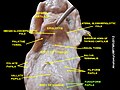 Papillae and other tongue landmarks
 -
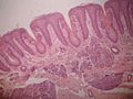 Foliate papillae
 -
 Floor of mouth. Deep dissection. Anterior view.
 -
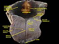 Floor of mouth. Deep dissection. Anterior view.
 -
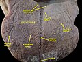
 A picture showing filiform papillae taken using a USB microscope.


References
[edit]- ^abcdNorton N (2007).Netter's head and neck anatomy for dentistry.illustrations by Netter FH. Philadelphia, Pa.: Saunders Elsevier. p. 402.ISBN978-1929007882.
- ^abcdefghijklSusan Standring (editor in chief)] (2008). "Chapter 33: NECK AND UPPER AERODIGESTIVE TRACT".Gray's anatomy: the anatomical basis of clinical practice(40th ed.). [Edinburgh]: Churchill Livingstone/Elsevier.ISBN978-0443066849.
{{cite book}}:|last=has generic name (help) - ^"Tongue | Gastrointestinal Tract".histologyguide.com.Retrieved2023-02-19.
- ^abcdeScully C (2013).Oral and maxillofacial medicine: the basis of diagnosis and treatment(3rd ed.). Edinburgh: Churchill Livingstone. pp. 401, 402.ISBN9780702049484.
- ^Ross, H R; Pawlina, W (2011).Histology: A text and atlas.Baltimore, MD.: Lippincott, Williams, and Wilkins.ISBN978-0-7817-7200-6.
- ^Rajendran A; Sundaram S (10 February 2014).Shafer's Textbook of Oral Pathology(7th ed.). Elsevier Health Sciences APAC. p. 34.ISBN978-81-312-3800-4.
- ^König, Liebich (2020).Veterinary anatomy of domestic animals: textbook and colour atlas(7th, updated and extended ed.). Stuttgart; New York: Georg Thieme Verlag.ISBN978-3-13-242933-8.