Melanopsin
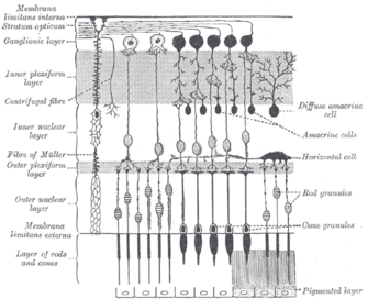
Melanopsinis a type ofphotopigmentbelonging to a larger family of light-sensitiveretinal proteinscalledopsinsand encoded by the geneOpn4.[5]In the mammalianretina,there are two additional categories of opsins, both involved in the formation of visual images:rhodopsinandphotopsin(types I, II, and III) in therodandconephotoreceptor cells, respectively.
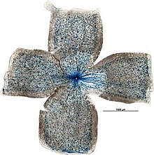
In humans, melanopsin is found inintrinsically photosensitive retinal ganglion cells(ipRGCs).[6]It is also found in the iris of mice and primates.[7]Melanopsin is also found in rats,amphioxus,and other chordates.[8]ipRGCs are photoreceptor cells which are particularly sensitive to the absorption of short-wavelength (blue) visible light and communicate information directly to the area of the brain called thesuprachiasmatic nucleus(SCN), also known as the central "body clock", in mammals.[9]Melanopsin plays an important non-image-forming role in thesettingof circadian rhythms as well as other functions. Mutations in theOpn4gene can lead to clinical disorders, such asSeasonal Affective Disorder(SAD).[10]According to one study, melanopsin has been found in eighteen sites in the human brain (outside the retinohypothalamic tract), intracellularly, in a granular pattern, in the cerebral cortex, the cerebellar cortex and several phylogenetically old regions, primarily in neuronal soma, not in nuclei.[11]Melanopsin is also expressed in human cones. However, only 0.11% to 0.55% of human cones express melanopsin and are exclusively found in the peripheral regions of the retina.[12]The human peripheral retina senses light at high intensities that is best explained by four different photopigment classes.[13]
Discovery[edit]

Melanopsin was discovered byIgnacio Provencioas a newopsinin themelanophores,or light-sensitive skin cells, of theAfrican clawed frogin 1998.[14]A year later, researchers found that mice without anyrodsorcones,the cells involved in image-forming vision, stillentrainedto a light-dark cycle.[15]This observation led to the conclusion that neither rods nor cones, located in the outerretina,are necessary for circadian entrainment and that a third class of photoreceptor exists in the mammalian eye.[5]Provencio and colleagues then found in 2000 that melanopsin is also present in mouse retina, specifically inganglion cells,and that it mediates non-visual photoreceptive tasks.[16]Melanopsin is encoded by theOpn4gene withorthologsin a variety of organisms.[5]
These retinal ganglion cells were found to be innately photosensitive, since they responded to light even while isolated, and were thus namedintrinsically photosensitive Retinal Ganglion Cells (ipRGCs).[17]They constitute a third class ofphotoreceptor cellsin the mammalian retina, besides the already known rods and cones, and were shown to be the principal conduit for light input to circadianphotoentrainment.[16]In fact, it was later demonstrated by Satchidananda Panda and colleagues that melanopsin pigment may be involved in entrainment of acircadian oscillatorto light cycles in mammals since melanopsin was necessary for blind mice to respond to light.[18]
Species distribution[edit]
Mammals haveorthologousmelanopsin genes namedOpn4m,which are derived from one branch of theOpn4family, and are approximately 50-55% conserved.[19]However, non-mammalian vertebrates, including chickens and zebrafish, have another version of the melanopsin gene,Opn4x,which appears to have a distinct lineage that diverged fromOpn4mabout 360 million years ago.[20]Mammals lost the geneOpn4xrelatively early in their evolution, leading to a general reduction in photosensory capability. It is thought that this event can be explained by the fact that this occurred during the time in which nocturnal mammals were evolving.[19]
Structure[edit]
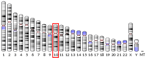
The human melanopsin gene,opn4,is expressed inipRGCs,which comprises only 1-2% ofRGCsin the inner mammalian retina, as studied bySamer Hattarand colleagues.[9]The gene spans approximately 11.8 kb and is mapped to the long arm ofchromosome 10.The gene includes nineintronsand tenexonscompared to the four to seven exons typically found in other human opsins.[16]In non-mammalian vertebrates, melanopsin is found in a wider subset of retinal cells, as well as in photosensitive structures outside the retina, such as theirismuscle of the eye, deep brain regions, thepineal gland,and the skin.[19]ParalogsofOpn4includeOPN1LW,OPN1MW,rhodopsinandencephalopsin.[21]
Melanopsin, like all other animalopsins(e.g.rhodopsin), is aG-protein-coupled receptor (GPCR).The melanopsin protein has an extarcellularN-terminal domain,an intracellularC-terminal domain,and sevenalpha helicesspanning through the plasma membrane.[14]The seventh helix has alysinethat corresponds to Lys2967.43incattlerhodopsin[14]and that is conserved in almost all opsins.[22]This lysine binds covalentlyretinalvia aSchiff-base,[23][24]which makes melanopsin light sensitive. In fact this is abolished if the lysine is replaced by analanine.[25]
Melanopsin is more closely related toinvertebratevisual opsins, which are rhabdomeric opsin, than tovertebratevisual opsins, which are cliary opsins.[14][26][27]This is also reflected by the downstreamsignaling cascade,melanopsin couples in ipRGCs to theG-proteinsG(q),G(11),andG(14),which are all of the G(q)-type.[28]In fact, they can functionally replace each other, as aknocking outonly two of them has nophenotypicaleffect.[29]The G-proteins activate thephospholipase CPLCB4,[7]which causes theTRP-channelsTRPC6andTRPC7mediate to open so that the celldepolarizes.[17][7]This is like in the photoreceptor cells of theDrosophilaeye, and in contrast to the vertebraterodandconecells, wherephototransductioneventually makes the cellshyperpolarize.[30]Like other rhabdomeric opsins, Melanopsin has intrinsicphotoisomeraseactivity.[31]
Function[edit]

Melanopsin-containing ganglion cells,[32]like rods and cones, exhibit both light and darkadaptation;they adjust their sensitivity according to the recent history of light exposure.[33]However, while rods and cones are responsible for the reception of images, patterns, motion, and color, melanopsin-containingipRGCscontribute to various reflexive responses of the brain and body to the presence of light.[17]
Evidence for melanopsin's physiological light detection has been tested in mice. A mouse cell line that is not normally photosensitive,Neuro-2a,is rendered light-sensitive by the addition of human melanopsin. The photoresponse is selectively sensitive to short-wavelength light (peak absorption ~479 nm),[34][35]and has an intrinsicphotoisomeraseregeneration function that is chromatically shifted to longer wavelengths.[36]
Melanopsin photoreceptors are sensitive to a range of wavelengths and reach peak light absorption at blue light wavelengths around 480 nanometers.[37]Other wavelengths of light activate the melanopsin signaling system with decreasing efficiency as they move away from the optimum 480 nm. For example, shorter wavelengths around 445 nm (closer to violet in thevisible spectrum) are half as effective for melanopsin photoreceptor stimulation as light at 480 nm.[37]
Melanopsin in the iris of some, primarily nocturnal, mammals closes the iris when it is exposed to light. This local pupil light reflex (PLR) is absent from primates, even though their irises express melanopsin.[7]
Mechanism[edit]
When light with an appropriate frequency enters the eye, it activates the melanopsin contained inintrinsically photosensitive retinal ganglion cells(ipRGCs), triggering anaction potential.These neuronal electrical signals travel through neuronalaxonsto specific brain targets, such as the center of pupillary control called theolivary pretectal nucleus(OPN) of the midbrain. Consequently, stimulation of melanopsin in ipRGCs mediates behavioral and physiological responses to light, such as pupil constriction and inhibition ofmelatoninrelease from thepineal gland.[38][39]The ipRGCs in the mammalian retina are one terminus of theretinohypothalamic tractthat projects to thesuprachiasmatic nucleus(SCN) of thehypothalamus.The suprachiasmatic nucleus is sometimes described as the brain's "master clock",[40]as it maintains thecircadian rhythm,and nerve signals from ipRGCs to the SCN entrain the internal circadian rhythm to the rising and setting of the sun.[9]The SCN also receives input from rods and cones through the retinohypothalamic tract, so information from all three photosensitive cell types (rods, cones, and ipRGCs) in the mammalian retina are transmitted to the (SCN) SCN.[41]
Melanopsin-containing ganglion cells are thought to influence these targets by releasing theneurotransmittersglutamateandpituitary adenylate cyclase activating polypeptide(PACAP) from their axon terminals.[42]Melanopsin-containing ganglion cells also receive input from rods and cones that can add to the input to these pathways.
Effects on circadian rhythm[edit]
Melanopsin serves an important role in thephotoentrainmentof circadian rhythms in mammals. An organism that isphotoentrainedhas aligned its activity to an approximately 24-hour cycle, thesolar cycleon Earth.[43]In mammals, melanopsin expressing axons target thesuprachiasmatic nucleus (SCN)through theretinohypothalamic tract(RHT).[9]
In mammals, the eye is the main photosensitive organ for the transmission of light signals to the brain. However, blind humans are still able to entrain to the environmental light-dark cycle, despite having no conscious perception of the light. One study exposed subjects to bright light for a prolonged duration of time and measured theirmelatoninconcentrations. Melatonin was not only suppressed in visually unimpaired humans, but also in blind participants, suggesting that the photic pathway used by the circadian system is functionally intact despite blindness.[44]Therefore, physicians no longer practiceenucleationof blind patients, or removal of the eyes at birth, since the eyes play a critical role in the photoentrainment of the circadian pacemaker.
In mutant breeds of mice that lacked only rods, only cones, or both rods and cones, all breeds of mice still entrained to changing light stimuli in the environment, but with a limited response, suggesting thatrodsandconesare not necessary for circadian photoentrainment and that the mammalian eye must have another photopigment required for the regulation of thecircadianclock.[15]
Melanopsin-knockout micedisplay reduced photoentrainment. In comparison to wild-type mice that expressed melanopsin normally, deficits in light-induced phase shifts in locomotion activity were noted in melanopsin-null mice (Opn4 -/-).[18]These melanopsin-deficient mice did not completely lose their circadian rhythms, as they were still able to entrain to changing environmental stimuli, albeit more slowly than normal.[45]This indicated that, although melanopsin is sufficient for entrainment, it must work in conjunction with other photopigments for normal photoentrainment activity. Triple-mutant mice that were rod-less, cone-less, and melanopsin-less display a complete loss in the circadian rhythms, so all three photopigments in these photoreceptors,rhodopsin,photopsinand melanopsin, are necessary for photoentrainment.[46]Therefore, there is a functional redundancy between the three photopigments in the photoentrainment pathway of mammals. Deletion of only one photopigment does not eliminate the organism's ability to entrain to environmental light-dark cycles, but it does reduce the intensity of the response.
Regulation[edit]
Melanopsin undergoesphosphorylationon its intracellularcarboxy tailas a way to deactivate its function. Compared to other opsins, melanopsin has an unusually long carboxy tail that contains 37serineandthreonineamino acid sites that could undergo phosphorylation.[47]However, a cluster of seven amino acids are sufficient to deactivate zebrafish melanopsin. These sites are dephosphorylated when melanopsin is exposed to light and are unique from those that regulate rhodopsin.[48]They are important for proper response to calcium ions in ipRGCs; lack of functional phosphorylation sites, particularly at serine-381 and serine-398, reduce the cell's response to light-induced calcium ion influx when voltage-gated calcium ion channels open.[49]
In terms of the gene Opn4,Dopamine(DA) is a factor in the regulation of melanopsinmRNAin ipRGCs.[50]
Clinical significance[edit]
The discovery of the role of melanopsin in non-image forming vision has led to a growth inoptogenetics.This field has shown promise in clinical applications, including the treatment of human eye diseases such asretinitis pigmentosaanddiabetes.[51]Amissense mutationin Opn4, P10L, has been implicated in 5% of patients withSeasonal Affective Disorder(SAD).[10]This is a condition in which people experience depressive thoughts in the winter due to decreased available light. Additionally, a melanopsin based receptor has been linked tomigrainepain.[52]
Restoration of vision[edit]
There has been recent research on the role of melanopsin inoptogenetictherapy for patients with the degenerative eye diseaseretinitis pigmentosa(RP).[53]Reintroducing functional melanopsin into the eyes of mice with retinal degeneration restores thepupillary light reflex (PLR).These same mice could also distinguish light stimuli from dark stimuli and showed increased sensitivity to room light. The higher sensitivity demonstrated by these mice shows promise for vision restoration that may be applicable to humans and human eye diseases.[51][54]
Control of sleep/wake patterns[edit]
Melanopsin may aid in controlling sleep cycles and wakefulness. Tsunematsu and colleagues createdtransgenicmice that expressed melanopsin inhypothalamicorexinneurons. With a short 4-second pulse of blue light (guided byoptical fibers), the transgenic mice could successfully transition fromslow-wave sleep(SWS), which is commonly known as "deep sleep," to long-lasting wakefulness. After switching off the blue light, the hypothalamicorexinneurons showed activity for several tens of seconds.[51][55]It has been shown that rods and cones play no role in the onset of sleep by light, distinguishing them from ipRGCs and melanopsin. This provides strong evidence that there is a link between ipRGCs in humans and alertness, particularly with high frequency light (e.g. blue light). Therefore, melanopsin can be used as a therapeutic target for controlling the sleep-wake cycle.[56]
Regulation of blood glucose levels[edit]
In a paper published by Ye and colleagues in 2011, melanopsin was utilized to create an optogenetic synthetic transcription device that was tested in a therapeutic setting to produceFc-glucagon-like peptide 1(Fc-GLP-1), a fusion protein that helps control blood glucose levels in mammals withType II Diabetes.The researchers subcutaneously implanted mice with microencapsulated transgenicHEK 293 cellsthat were cotransfected with two vectors including the melanopsin gene and the gene of interest under an NFAT (nuclear factor of activated T cells) promoter, respectively. It is through this engineered pathway that they successfully controlled the expression of Fc-GLP-1 in doubly recessive diabetic mice and reducedhyperglycemia,or high blood glucose levels, in these mice. This shows promise for the use of melanopsin as an optogenetic tool for the treatment of Type II diabetes.[51][57]
See also[edit]
- Light effects on circadian rhythm
- Opsins
- Intrinsically Photosensitive Retinal Ganglion Cells (ipRGCs)
- Suprachiasmatic nucleus (SCN)
- Retinohypothalamic tract
References[edit]
- ^abcGRCh38: Ensembl release 89: ENSG00000122375–Ensembl,May 2017
- ^abcGRCm38: Ensembl release 89: ENSMUSG00000021799–Ensembl,May 2017
- ^"Human PubMed Reference:".National Center for Biotechnology Information, U.S. National Library of Medicine.
- ^"Mouse PubMed Reference:".National Center for Biotechnology Information, U.S. National Library of Medicine.
- ^abcHankins MW, Peirson SN, Foster RG (January 2008). "Melanopsin: an exciting photopigment".Trends in Neurosciences.31(1): 27–36.doi:10.1016/j.tins.2007.11.002.PMID18054803.S2CID1645433.
- ^Provencio I, Warthen DM (2012). "Melanopsin, the photopigment of intrinsically photosensitive retinal ganglion cells".Wiley Interdisciplinary Reviews: Membrane Transport and Signaling.1(2): 228–237.doi:10.1002/wmts.29.
- ^abcdXue T, Do MT, Riccio A, Jiang Z, Hsieh J, Wang HC, et al. (November 2011)."Melanopsin signalling in mammalian iris and retina".Nature.479(7371): 67–73.Bibcode:2011Natur.479...67X.doi:10.1038/nature10567.PMC3270891.PMID22051675.
- ^Angueyra JM, Pulido C, Malagón G, Nasi E, Gomez M (2012)."Melanopsin-expressing amphioxus photoreceptors transduce light via a phospholipase C signaling cascade".PLOS ONE.7(1): e29813.Bibcode:2012PLoSO...729813A.doi:10.1371/journal.pone.0029813.PMC3250494.PMID22235344.
- ^abcdHattar S, Liao HW, Takao M, Berson DM, Yau KW (February 2002)."Melanopsin-containing retinal ganglion cells: architecture, projections, and intrinsic photosensitivity".Science.295(5557): 1065–1070.Bibcode:2002Sci...295.1065H.doi:10.1126/science.1069609.PMC2885915.PMID11834834.
- ^abRoecklein KA, Rohan KJ, Duncan WC, Rollag MD, Rosenthal NE, Lipsky RH, et al. (April 2009)."A missense variant (P10L) of the melanopsin (OPN4) gene in seasonal affective disorder".Journal of Affective Disorders.114(1–3): 279–285.doi:10.1016/j.jad.2008.08.005.PMC2647333.PMID18804284.
- ^Nissilä J, Mänttäri S, Tuominen H, Särkioja T, Takala T, Saarela S, et al. (2012). "P-780 – The abundance and distribution of melanopsin (OPN4) protein in human brain".European Psychiatry.27:1–8.doi:10.1016/S0924-9338(12)74947-7.S2CID82045589.
- ^Dkhissi-Benyahya O, Rieux C, Hut RA, Cooper HM (April 2006)."Immunohistochemical evidence of a melanopsin cone in human retina".Investigative Ophthalmology & Visual Science.47(4): 1636–1641.doi:10.1167/iovs.05-1459.PMID16565403.
- ^Horiguchi H, Winawer J, Dougherty RF, Wandell BA (January 2013)."Human trichromacy revisited".Proceedings of the National Academy of Sciences of the United States of America.110(3): E260–E269.Bibcode:2013PNAS..110E.260H.doi:10.1073/pnas.1214240110.PMC3549098.PMID23256158.
- ^abcdProvencio I, Jiang G, De Grip WJ, Hayes WP, Rollag MD (January 1998)."Melanopsin: An opsin in melanophores, brain, and eye".Proceedings of the National Academy of Sciences of the United States of America.95(1): 340–345.Bibcode:1998PNAS...95..340P.doi:10.1073/pnas.95.1.340.PMC18217.PMID9419377.
- ^abFreedman MS, Lucas RJ, Soni B, von Schantz M, Muñoz M, David-Gray Z, et al. (April 1999). "Regulation of mammalian circadian behavior by non-rod, non-cone, ocular photoreceptors".Science.284(5413): 502–504.Bibcode:1999Sci...284..502F.doi:10.1126/science.284.5413.502.PMID10205061.
- ^abcProvencio I, Rodriguez IR, Jiang G, Hayes WP, Moreira EF, Rollag MD (January 2000)."A novel human opsin in the inner retina".The Journal of Neuroscience.20(2): 600–605.doi:10.1523/JNEUROSCI.20-02-00600.2000.PMC6772411.PMID10632589.
- ^abcBerson DM, Dunn FA, Takao M (February 2002). "Phototransduction by retinal ganglion cells that set the circadian clock".Science.295(5557): 1070–1073.Bibcode:2002Sci...295.1070B.doi:10.1126/science.1067262.PMID11834835.S2CID30745140.
- ^abPanda S, Sato TK, Castrucci AM, Rollag MD, DeGrip WJ, Hogenesch JB, et al. (December 2002). "Melanopsin (Opn4) requirement for normal light-induced circadian phase shifting".Science.298(5601): 2213–2216.Bibcode:2002Sci...298.2213P.doi:10.1126/science.1076848.PMID12481141.S2CID20602808.
- ^abcBellingham J, Chaurasia SS, Melyan Z, Liu C, Cameron MA, Tarttelin EE, et al. (July 2006)."Evolution of melanopsin photoreceptors: discovery and characterization of a new melanopsin in nonmammalian vertebrates".PLOS Biology.4(8): e254.doi:10.1371/journal.pbio.0040254.PMC1514791.PMID16856781.

- ^Benton MJ (May 1990). "Phylogeny of the major tetrapod groups: morphological data and divergence dates".Journal of Molecular Evolution.30(5): 409–424.Bibcode:1990JMolE..30..409B.doi:10.1007/BF02101113.PMID2111854.S2CID35082873.
- ^Terakita A (1 March 2005)."The opsins".Genome Biology.6(3): 213.doi:10.1186/gb-2005-6-3-213.PMC1088937.PMID15774036.
- ^Gühmann M, Porter ML, Bok MJ (August 2022)."The Gluopsins: Opsins without the Retinal Binding Lysine".Cells.11(15): 2441.doi:10.3390/cells11152441.PMC9368030.PMID35954284.
- ^Collins FD (March 1953). "Rhodopsin and indicator yellow".Nature.171(4350): 469–471.Bibcode:1953Natur.171..469C.doi:10.1038/171469a0.PMID13046517.S2CID4152360.
- ^Pitt GA, Collins FD, Morton RA, Stok P (January 1955)."Studies on rhodopsin. VIII. Retinylidenemethylamine, an indicator yellow analogue".The Biochemical Journal.59(1): 122–128.doi:10.1042/bj0590122.PMC1216098.PMID14351151.
- ^Kumbalasiri T, Rollag MD, Isoldi MC, Castrucci AM, Provencio I (March 2007). "Melanopsin triggers the release of internal calcium stores in response to light".Photochemistry and Photobiology.83(2): 273–279.doi:10.1562/2006-07-11-RA-964.PMID16961436.S2CID23060331.
- ^Porter ML, Blasic JR, Bok MJ, Cameron EG, Pringle T, Cronin TW, et al. (January 2012)."Shedding new light on opsin evolution".Proceedings. Biological Sciences.279(1726): 3–14.doi:10.1098/rspb.2011.1819.PMC3223661.PMID22012981.
- ^Ramirez MD, Pairett AN, Pankey MS, Serb JM, Speiser DI, Swafford AJ, et al. (26 October 2016)."The last common ancestor of most bilaterian animals possessed at least 9 opsins".Genome Biology and Evolution:evw248.doi:10.1093/gbe/evw248.PMC5521729.PMID27797948.
- ^Hughes S, Jagannath A, Hickey D, Gatti S, Wood M, Peirson SN, et al. (January 2015)."Using siRNA to define functional interactions between melanopsin and multiple G Protein partners".Cellular and Molecular Life Sciences.72(1): 165–179.doi:10.1007/s00018-014-1664-6.PMC4282707.PMID24958088.
- ^Chew KS, Schmidt TM, Rupp AC, Kofuji P, Trimarchi JM (28 May 2014)."Loss of gq/11 genes does not abolish melanopsin phototransduction".PLOS ONE.9(5): e98356.Bibcode:2014PLoSO...998356C.doi:10.1371/journal.pone.0098356.PMC4037210.PMID24870805.
- ^Sexton T, Buhr E, Van Gelder RN (January 2012)."Melanopsin and mechanisms of non-visual ocular photoreception".The Journal of Biological Chemistry.287(3): 1649–1656.doi:10.1074/jbc.r111.301226.PMC3265846.PMID22074930.
- ^Panda S, Nayak SK, Campo B, Walker JR, Hogenesch JB, Jegla T (January 2005). "Illumination of the melanopsin signaling pathway".Science.307(5709): 600–604.Bibcode:2005Sci...307..600P.doi:10.1126/science.1105121.PMID15681390.S2CID22713904.
- ^Feigl B, Zele AJ (August 2014)."Melanopsin-expressing intrinsically photosensitive retinal ganglion cells in retinal disease"(PDF).Optometry and Vision Science.91(8): 894–903.doi:10.1097/OPX.0000000000000284.PMID24879087.S2CID34057255.
- ^Wong KY, Dunn FA, Berson DM (December 2005)."Photoreceptor adaptation in intrinsically photosensitive retinal ganglion cells".Neuron.48(6): 1001–1010.doi:10.1016/j.neuron.2005.11.016.PMID16364903.
- ^Bailes HJ, Lucas RJ (May 2013)."Human melanopsin forms a pigment maximally sensitive to blue light (λmax ≈ 479 nm) supporting activation of G(q/11) and G(i/o) signalling cascades".Proceedings. Biological Sciences.280(1759): 20122987.doi:10.1098/rspb.2012.2987.PMC3619500.PMID23554393.
- ^Berson DM (August 2007)."Phototransduction in ganglion-cell photoreceptors".Pflügers Archiv.454(5): 849–855.doi:10.1007/s00424-007-0242-2.PMID17351786.
- ^Melyan Z, Tarttelin EE, Bellingham J, Lucas RJ, Hankins MW (February 2005). "Addition of human melanopsin renders mammalian cells photoresponsive".Nature.433(7027): 741–745.Bibcode:2005Natur.433..741M.doi:10.1038/nature03344.PMID15674244.S2CID4426682.
- ^abEnezi J, Revell V, Brown T, Wynne J, Schlangen L, Lucas R (August 2011)."A" melanopic "spectral efficiency function predicts the sensitivity of melanopsin photoreceptors to polychromatic lights".Journal of Biological Rhythms.26(4): 314–323.doi:10.1177/0748730411409719.PMID21775290.S2CID22369861.
- ^Markwell EL, Feigl B, Zele AJ (May 2010)."Intrinsically photosensitive melanopsin retinal ganglion cell contributions to the pupillary light reflex and circadian rhythm".Clinical & Experimental Optometry.93(3): 137–149.doi:10.1111/j.1444-0938.2010.00479.x.PMID20557555.S2CID21778407.
- ^Zaidi FH, Hull JT, Peirson SN, Wulff K, Aeschbach D, Gooley JJ, et al. (December 2007)."Short-wavelength light sensitivity of circadian, pupillary, and visual awareness in humans lacking an outer retina".Current Biology.17(24): 2122–2128.Bibcode:2007CBio...17.2122Z.doi:10.1016/j.cub.2007.11.034.PMC2151130.PMID18082405.
- ^Evans JA (July 2016)."Collective timekeeping among cells of the master circadian clock".The Journal of Endocrinology.230(1): R27–R49.doi:10.1530/JOE-16-0054.PMC4938744.PMID27154335.
- ^Reppert SM, Weaver DR (August 2002). "Coordination of circadian timing in mammals".Nature.418(6901): 935–941.Bibcode:2002Natur.418..935R.doi:10.1038/nature00965.PMID12198538.S2CID4430366.
- ^Hannibal J, Fahrenkrug J (April 2004). "Target areas innervated by PACAP-immunoreactive retinal ganglion cells".Cell and Tissue Research.316(1): 99–113.doi:10.1007/s00441-004-0858-x.PMID14991397.S2CID24148323.
- ^Allada R, Emery P, Takahashi JS, Rosbash M (2001). "Stopping time: the genetics of fly and mouse circadian clocks".Annual Review of Neuroscience.24(1): 1091–1119.doi:10.1146/annurev.neuro.24.1.1091.PMID11520929.
- ^Czeisler CA, Shanahan TL, Klerman EB, Martens H, Brotman DJ, Emens JS, et al. (January 1995)."Suppression of melatonin secretion in some blind patients by exposure to bright light".The New England Journal of Medicine.332(1): 6–11.doi:10.1056/NEJM199501053320102.PMID7990870.
- ^Rollag MD, Berson DM, Provencio I (June 2003)."Melanopsin, ganglion-cell photoreceptors, and mammalian photoentrainment".Journal of Biological Rhythms.18(3): 227–234.doi:10.1177/0748730403018003005.PMID12828280.S2CID9034442.
- ^Panda S, Provencio I, Tu DC, Pires SS, Rollag MD, Castrucci AM, et al. (July 2003). "Melanopsin is required for non-image-forming photic responses in blind mice".Science.301(5632): 525–527.Bibcode:2003Sci...301..525P.doi:10.1126/science.1086179.PMID12829787.S2CID37600812.
- ^Blasic JR, Lane Brown R, Robinson PR (May 2012)."Light-dependent phosphorylation of the carboxy tail of mouse melanopsin".Cellular and Molecular Life Sciences.69(9): 1551–1562.doi:10.1007/s00018-011-0891-3.PMC4045631.PMID22159583.
- ^Blasic JR, Matos-Cruz V, Ujla D, Cameron EG, Hattar S, Halpern ME, et al. (April 2014)."Identification of critical phosphorylation sites on the carboxy tail of melanopsin".Biochemistry.53(16): 2644–2649.doi:10.1021/bi401724r.PMC4010260.PMID24678795.
- ^Fahrenkrug J, Falktoft B, Georg B, Hannibal J, Kristiansen SB, Klausen TK (December 2014)."Phosphorylation of rat melanopsin at Ser-381 and Ser-398 by light/dark and its importance for intrinsically photosensitive ganglion cells (ipRGCs) cellular Ca2+ signaling".The Journal of Biological Chemistry.289(51): 35482–35493.doi:10.1074/jbc.M114.586529.PMC4271233.PMID25378407.
- ^Sakamoto K, Liu C, Kasamatsu M, Pozdeyev NV, Iuvone PM, Tosini G (December 2005). "Dopamine regulates melanopsin mRNA expression in intrinsically photosensitive retinal ganglion cells".The European Journal of Neuroscience.22(12): 3129–3136.doi:10.1111/j.1460-9568.2005.04512.x.PMID16367779.S2CID21517576.
- ^abcdKoizumi A, Tanaka KF, Yamanaka A (January 2013). "The manipulation of neural and cellular activities by ectopic expression of melanopsin".Neuroscience Research.75(1): 3–5.doi:10.1016/j.neures.2012.07.010.PMID22982474.S2CID21771987.
- ^Jennifer Couzin-Frankel (2010)."Why Light Makes Migraines Worse – ScienceNOW".Archivedfrom the original on 31 July 2016.Retrieved3 April2011.
- ^Busskamp V, Picaud S, Sahel JA, Roska B (February 2012)."Optogenetic therapy for retinitis pigmentosa".Gene Therapy.19(2): 169–175.doi:10.1038/gt.2011.155.PMID21993174.
- ^Lin B, Koizumi A, Tanaka N, Panda S, Masland RH (October 2008)."Restoration of visual function in retinal degeneration mice by ectopic expression of melanopsin".Proceedings of the National Academy of Sciences of the United States of America.105(41): 16009–16014.Bibcode:2008PNAS..10516009L.doi:10.1073/pnas.0806114105.PMC2572922.PMID18836071.
- ^Tsunematsu T, Tanaka KF, Yamanaka A, Koizumi A (January 2013). "Ectopic expression of melanopsin in orexin/hypocretin neurons enables control of wakefulness of mice in vivo by blue light".Neuroscience Research.75(1): 23–28.doi:10.1016/j.neures.2012.07.005.PMID22868039.S2CID207152803.
- ^Lupi D, Oster H, Thompson S, Foster RG (September 2008). "The acute light-induction of sleep is mediated by OPN4-based photoreception".Nature Neuroscience.11(9): 1068–1073.doi:10.1038/nn.2179.hdl:11858/00-001M-0000-0012-DD96-A.PMID19160505.S2CID15941500.
- ^Ye H, Daoud-El Baba M, Peng RW, Fussenegger M (June 2011). "A synthetic optogenetic transcription device enhances blood-glucose homeostasis in mice".Science.332(6037): 1565–1568.Bibcode:2011Sci...332.1565Y.doi:10.1126/science.1203535.PMID21700876.S2CID6166189.
Further reading[edit]
- Rovere G, Nadal-Nicolás FM, Wang J, Bernal-Garro JM, García-Carrillo N, Villegas-Pérez MP, et al. (December 2016)."Melanopsin-Containing or Non-Melanopsin-Containing Retinal Ganglion Cells Response to Acute Ocular Hypertension With or Without Brain-Derived Neurotrophic Factor Neuroprotection".Investigative Ophthalmology & Visual Science.57(15): 6652–6661.doi:10.1167/iovs.16-20146.PMID27930778.


