
Microscope slide


Amicroscope slideis a thin flat piece ofglass,typically 75 by 26 mm (3 by 1 inches) and about 1 mm thick, used to hold objects for examination under amicroscope.Typically the object ismounted(secured) on the slide, and then both are inserted together in the microscope for viewing. This arrangement allows several slide-mounted objects to be quickly inserted and removed from the microscope, labeled, transported, and stored in appropriate slide cases or folders etc.
Microscope slides are often used together with a cover slip or cover glass, a smaller and thinner sheet of glass that is placed over the specimen. Slides are held in place on the microscope's stage by slide clips, slide clamps or a cross-table which is used to achieve precise, remote movement of the slide upon the microscope's stage (such as in an automated/computer operated system, or where touching the slide with fingers is inappropriate either due to the risk ofcontaminationor lack of precision).
History
[edit]
The origin of the concept was pieces ofivoryorbone,containing specimens held between disks of transparentmica,that wouldslideinto the gap between the stage and the objective.[1]These "sliders" were popular inVictorianEnglanduntil theRoyal Microscopical Societyintroduced the standardized glass microscope slide.[2]
Dimensions and types
[edit]
A standard microscope slide measures about 75 mm by 25 mm (3″ by 1″) and is about 1 mm thick. A range of other sizes are available for various special purposes, such as 75 x 50 mm forgeologicaluse, 46 x 27 mm forpetrographicstudies, and 48 x 28 mm forthin sections.Slides are usually made of common glass and their edges are oftenfinely groundor polished.
Microscope slides are usually made of optical qualityglass,such assoda lime glassorborosilicate glass,but specialty plastics are also used.Fused quartzslides are often used whenultraviolettransparency is important, e.g. influorescence microscopy.[3][4]
While plain slides are the most common, there are several specialized types. Aconcavity slideorcavity slidehas one or more shallow depressions ( "wells" ), designed to hold slightly thicker objects, and certain samples such as liquids andtissue cultures.[5]Slides may have rounded corners for increased safety or robustness, or a cut-off corner for use with a slide clamp or cross-table, where the slide is secured by a spring-loaded curved arm contacting one corner, forcing the opposing corner of the slide against a right angled arm which does not move. If this system were used with a slide which did not incorporate these cut-off corners, the corners would chip and the slide could shatter.[5]
Agraticule slideis marked with agridof lines (for example, a 1 mm grid) that allows the size of objects seen under magnification to be easily estimated and provides reference areas for counting minute objects. Sometimes one square of the grid will itself be subdivided into a finer grid. Slides for specialized applications, such ashemocytometersfor cell counting, may have various reservoirs, channels and barriersetchedorgroundon their upper surface.[6]Various permanent markings or masks may beprinted,sand-blasted,or deposited on the surface by the manufacturer, usually with inert materials such asPTFE.[7]
-
 ANeubauer slidefor cell counting.
 -
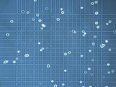 Microscope image of a Neubauer slide's graticule being used to countcells.
 -
 A Neubauer slide held in place on a microscope stand by a slide clamp on a cross-table.
 -
 Standard (75 x 25 mm or 3x1″) and large (75 x 51 mm or 3x2″) microscope slide.


Some slides have afrostedorenamel-coatedarea at one end, for labeling with a pencil or pen.[5]Slides may have special coatings applied by the manufacturer, e.g. for chemical inertness or enhancedcell adhesion.The coating may have a permanentelectric chargeto hold thin or powdery samples. Common coatings includepoly-L-lysine,silanes,epoxy resins,[5][7]or evengold.[8]
Mounting
[edit]

The mounting of specimens on microscope slides is often critical for successful viewing. The problem has been given much attention in the last two centuries and is a well-developed area with many specialized and sometimes quite sophisticated techniques. Specimens are often held into place using the smaller glasscover slips.
The main function of the cover slip is to keep solid specimens pressed flat, and liquid samples shaped into a flat layer of even thickness. This is necessary becausehigh-resolutionmicroscopes have a very narrowregionwithin which they focus.
The cover glass often has several other functions. It holds the specimen in place (either by the weight of the cover slip or, in the case of a wet mount, bysurface tension) and protects the specimen from dust and accidental contact. It protects the microscope'sobjective lensfrom contacting the specimen and vice versa; inoil immersion microscopyorwater immersion microscopythe cover slip prevents contact between the immersion liquid and the specimen. The cover slip can be glued to the slide so as to seal off the specimen, retardingdehydrationandoxidationof the specimen and also preventing contamination. A number of sealants are in use, including commercial sealants, laboratory preparations, or even regular clearnail polish,depending on the sample. A solvent-free sealant that can be used for live cell samples is "valap", a mixture ofvaseline,lanolinandparaffinin equal parts.[9] Microbialandcell culturescan be grown directly on the cover slip before it is placed on the slide, and specimens may be permanently mounted on the slip instead of on the slide.[9]
Cover slips are available in a range of sizes and thicknesses.[10]Using the wrong thickness can result inspherical aberrationand a reduction in resolution and image intensity. Specialty objectives may be used to image specimens without coverslips, or may have correction collars that permit a user to accommodate for alternative coverslip thickness.[11][12]
Dry mount
[edit]In adry mount,the simplest kind of mounting, the object is merely placed on the slide. A cover slip may be placed on top to protect the specimen and the microscope's objective and to keep the specimen still and pressed flat. This mounting can be successfully used for viewing specimens like pollen, feathers, hairs, etc. It is also used to examine particles caught in transparentmembrane filters(e.g., in analysis of airbornedust).
Wet mount or temporary mount
[edit]In awet mount,the specimen is placed in a drop of iodine or other liquid held between the slide and the cover slip by surface tension. This method is commonly used, for example, to view microscopic organisms that grow in pond water or other liquid media, especially lakes.
Prepared mount or permanent mount
[edit]Forpathologicalandbiologicalresearch, the specimen usually undergoes a complexhistologicalpreparation that involvesfixingit to prevent decay,removing any watercontained in it, replacing the water withparaffin,cutting it into very thin sections using amicrotome,placing the sections on a microscope slide,stainingthe tissue using various stains to reveal specific tissue components, clearing the tissue to render it transparent and covering it with a coverslip and mounting medium.
Strewn mount
[edit]Strewn mountingdescribes the production ofpalynologicalmicroscope slides by suspending a concentrated sample indistilled water,placing the samples on a slide, and allowing the water toevaporate.[13]
Mounting media
[edit]Themounting mediumis the solution in which the specimen is embedded, generally under a cover glass. Simple liquids like water orglycerolcan be considered mounting media, though the term generally refers to compounds that harden into a permanent mount. Popular mounting media includePermount,[14]andHoyer's mounting mediumand an alternativeglycerine jelly[15]Properties of a good mounting medium include having arefractive indexclose to that of glass (1.518), non-reactivity with the specimen, stability over time without crystallizing, darkening, or changing refractive index, solubility in the medium the specimen was prepared in (eitheraqueousornon-polar,such asxyleneortoluene), and not causing the specimen stain to fade or leach.[16]
Examples of mounting media
[edit]Aqueous
[edit]Popularly used in immunofluorescent cytochemistry where the fluorescence cannot be archived. The temporary storage must be done in a dark moist chamber. Common examples are:
- Glycerol-PBS (9:1) with antiquench, e.g. any of the following[17]
- p-phenylenediamine
- propyl gallate
- 1,4-Diazabicyclo (2,2,2)-octane (DABCO) (very popular)
- Ascorbic acid
- Mowiol or Gelvatol
- Gelatin
- Mount
- Vectashield
- Prolong Gold
- CyGEL / CyGEL Sustain (to immobilize living, unfixed cells and organisms)
Non-aqueous
[edit]
Used when a permanent mount is required
- Permount (toluene and a polymer of a-pinene, b-pinene, dipentene, b-phellandrene)
- Canada balsam
- DPX (Distrene 80 – a commercialpolystyrene,aplasticizere.g.dibutyl phthalateandxylene)
- DPX new (withxylenebut free of carcinogenicdibutyl phthalate)
- Entellan (with toluene)
- Entellan new
- Hempstead Halide Hoyer's Medium (a proprietary formulation of the traditional Hoyer's medium containing 60%Chloral,but free of known carcinogens)
- Neo-Mount (compatible with aliphatic neo-clear but not compatible with aromatic solvents like xylene)
Contrasting with other types/meanings of "mounting"
[edit]In contrast to mounting necessary for glass coverslips, somewhat similar mounting can be done for bulkierspecimen preservationin glass containers in museums. However an entirely different type of mounting is done forsample preparation,which can be for biological or nonbiological materials and is further subdivided into "hot" (compressive) and "cold" (castable) type mounting processes.[18][19]Though named "mounting", it is more akin to embedding in histology and should not be confused with the mounting described above. The term mounting in other fields has numerous other meanings.
See also
[edit]References
[edit]- ^Adam, George. Essays on the microscope. 1798
- ^Connett, Jess (4 October 2017)."The art of the invisible".Bristol24-7.Retrieved29 March2018.
- ^ Quartz Microscospe Slides and Cover Slipsfrom a commercial website (Ted Pella). Accessed on 2010-01-23.
- ^Quartz Microscope Slides and Cover SlipsArchived2015-08-10 at theWayback Machinecatalog page from a commercial website (SPI Supplies). Accessed on 2010-01-23.
- ^abcd Histology and Light Microscopycatalog page from a commercial website (EMS). Accessed on 2010-01-23.
- ^ "Lentz Microscopy Collection".Retrieved30 May2024.
- ^ab Microscope Slidescatalog page from a commercial website (TEKDON). Accessed on 2010-01-23.
- ^ Gold Coated Microscope Slides and DNA Imaging Kitcatalog page from a commercial website (Asylum Research). Accessed on 2011-08-31.
- ^ab Microscopy – ProtocolsArchived2013-04-03 atarchive.todayteaching webpage by the Moores Cancer Center, University of California at San Diego. Accessed on 2013-02-07.
- ^GE technical specsfrom a commercial website (Ted Pella). Accessed on 2010-01-23.
- ^"Coverslip Correction Collars".Microscopy Resource Center.Olympus.Retrieved4 June2017.
- ^Michael W. Davidson (2010),Optical Aberrations,chapter inMolecular Expressionswebsite at Florida State University. Last edit 2006-06-15 at 02:39 PM, accessed in 2010-01-12.
- ^Zippi, Pierre A. (1991). "SEM and Light Microscope Mounting and Specimen Location Technique for Same-Specimen Study of Palynological Strew Mounts".Micropaleontology.37(4): 407–13.doi:10.2307/1485913.JSTOR1485913.
- ^"Material Safety Data Sheet, Permount Mounting Media"(PDF).Retrieved29 March2018.
- ^"Glycerine Jelly as a Substitute for Hoyer's Solution Mountant".www.mobot.org.Retrieved29 March2018.
- ^Medical Laboratory Science: Theory And PracticeBy Ochei & Kolkatker, p. 446.
- ^Fluorescence specimen mounting, mounting medium, and antiquench agents – UCLA Brain Research Institute
- ^Cold and Hot Mounting Media, Molds, and Pressure Chamber – Electron Microscopy Sciences
- ^Allied High Tech – Allied High Tech Products | Metallographic Mounting Equipment