
Plantaris muscle
| Plantaris muscle | |
|---|---|
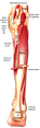
 Plantaris muscle. Seen from behind. | |
Dissection video (1 min 23 s) | |
| Details | |
| Origin | Lateral supracondylar ridgeof femur above lateral head of gastrocnemius <link issue> |
| Insertion | Tendo calcaneus(medial side, deep togastrocnemiustendon) |
| Artery | Sural arteries |
| Nerve | Tibial nervefrom anterior rami of S1-S2 |
| Actions | Plantar flexesfoot and flexesknee |
| Antagonist | Tibialis anterior muscle |
| Identifiers | |
| Latin | musculus plantaris |
| TA98 | A04.7.02.049 |
| TA2 | 2663 |
| FMA | 22543 |
| Anatomical terms of muscle | |
Theplantarisis one of the superficialmusclesof thesuperficial posterior compartment of the leg,one of thefascial compartments of the leg.
It is composed of a thin muscle belly and a long thintendon.While not as thick as theachilles tendon,the plantaris tendon (which tends to be between 30–45 centimetres (12–18 in) in length) is the longest tendon in thehuman body.Not including the tendon, the plantaris muscle is approximately 5–10 centimetres (2.0–3.9 in) long and is absent in 8-12% of the population. It is one of the plantarflexorsin the posterior compartment of the leg, along with thegastrocnemiusandsoleusmuscles. The plantaris is considered to have become an unimportant muscle when human ancestors switched from climbing trees to bipedalism and inanatomically modern humansit mainly acts with the gastrocnemius.[1]
Structure
[edit]The plantaris muscle arises from the inferior part of the lateral supracondylar ridge of thefemurat a position slightly superior to the origin of the lateral head ofgastrocnemius.It passes posterior to theknee jointin an inferomedial direction and becomes tendinous distally to insert into theAchilles tendon.It occasionally separately inserts into the medial side of thecalcaneus.
Innervation
[edit]The plantaris muscle is innervated by thetibial nerve,a branch of thesciatic nervein thesacral plexus.Signaling for contraction begins in thefrontal lobeof thebrainwith thepre-central gyrus(primary motor cortex).Upper motor neuronsare stimulated and send a signal through theinternal capsuleand down thecorticospinal tract.Decussation of thelateral corticospinal tractoccurs in themedullary pyramids,then the fibers continue down the contralateral side of thespinal cord.Upper motor neurons synapse withlower motor neuronsat theanterior horn of the spinal cordin the sacral plexus (formed from theanterior rami of spinal nervesL4, L5, S1–4). The lower motor neuron fibers continue down the sciatic nerve and then diverge into the tibial andcommon fibular nerves.The tibial nerve runs medially at theknee joint.When the tibial nerve receives an action potential, the plantaris muscle contracts, providing weak plantar flexion of the foot and weak flexion of the knee.[2]
Variation
[edit]The muscle may arise from theoblique popliteal ligament.Interdigitations with the lateral head of thegastrocnemiusand a fibrous extension of the muscle to thepatellaare not unusual.[3]
Function
[edit]The plantaris acts to weaklyplantar flextheankle jointand flex theknee joint.
The plantaris muscle may also provideproprioceptivefeedback information to thecentral nervous systemregarding the position of the foot. The unusually high density of proprioceptive receptor end organs supports this notion.[4]
Its motor function is so minimal that its long tendon can readily be harvested for reconstruction elsewhere with little functional deficit. Often mistaken for a nerve by new medical students (and thus called the "freshman's nerve" ), the muscle was useful to otherprimatesfor grasping with their feet.[5]
Clinical significance
[edit]A common injury that is normally attributed to the plantaris muscle is a condition calledtennis leg.Although pain in the calf can be attributed to a rupture of the plantaris muscle, recent ultrasound research has shown that tennis leg more commonly arises from tears in themusculotendinous junctionof the medial gastrocnemius. In one clinical study, 94 out of 141 patients (66.7%) diagnosed with tennis leg were found with a partial rupture of the gastrocnemius muscle, while rupture of the plantaris tendon was only seen in 2 patients (1.4%).[6]
Injury may occur from running, jumping, or pushing off one leg in sports such as tennis, basketball and soccer, which require quick foot movement in a certain direction. Isolated plantaris muscle strains are rare, and ruptures normally occur in conjunction with injury to other muscles in the posterior compartment of the lower leg.[7]Symptoms of a plantaris muscle rupture may include an audible popping sound in the area during physical activity, swelling, pain in the back of the lower leg, and persistent soreness. Ankle flexion may also be painful.[8]
See also
[edit]Additional images
[edit]-
 Animation
 -
 The plantaris is visible under the gastrocnemius.
 -
Dissection video (59 s)
-
 Thesynovial sheathsof the tendons around the ankle. Medial aspect. (Tendon of Plantaris labeled at bottom right.)
 -
 Muscles of the back of the leg. Superficial layer.
 -
 Cross-section through middle of leg. Tendon of plantaris is located betweensoleusandgastrocnemius.
 -
 Plantaris muscle
 -
 Plantaris tendon runs betweensoleusandgastrocnemius.Plantaris tendon is indicated by white arrow-heads.


References
[edit]- ^Sichting, Freddy; Holowka, Nicholas B.; Ebrecht, Florian; Lieberman, Daniel E. (2020-02-26)."Evolutionary anatomy of the plantar aponeurosis in primates, including humans".Journal of Anatomy.237(1). Wiley: 85–104.doi:10.1111/joa.13173.ISSN0021-8782.PMC7309290.PMID32103502.
- ^Saladin, Kenneth S. Anatomy and Physiology The Unity of Form and Function. 6th ed. New York: McGraw-Hill Science Engineering, 2009. Print.[page needed]
- ^Freeman, A. J.; Jacobson, N. A.; Fogg, Q. A. (2008). "Anatomical variations of the plantaris muscle and a potential role in patellofemoral pain syndrome".Clinical Anatomy.21(2): 178–81.doi:10.1002/ca.20594.PMID18266282.S2CID21873763.
- ^Moore, Keith L; & Dalley Arthur R (2008).Clinically Oriented Anatomy(6th ed.). Lippincott Williams and Wilkins.ISBN978-1-60547-652-0[page needed]
- ^Andor, W.J.M., Glaudemans, Rudi A.J.O. Dierckx, Jan L.M.A. Gielen, Johannes (Hans) Zwerver (2015).Nuclear Medicine and Radiologic Imaging in Sports Injuries.Springer. p. 762
- ^Delgado, Gonzalo J.; Chung, Christine B.; Lektrakul, Nitaya; Azocar, Patricio; Botte, Michael J.; Coria, Daniel; Bosch, Enrique; Resnick, Donald (2002). "Tennis Leg: Clinical US Study of 141 Patients and Anatomic Investigation of Four Cadavers with MR Imaging and US1".Radiology.224(1): 112–9.doi:10.1148/radiol.2241011067.PMID12091669.
- ^Spina, A. A. (2007)."The plantaris muscle: Anatomy, injury, imaging, and treatment".The Journal of the Canadian Chiropractic Association.51(3): 158–65.PMC1978447.PMID17885678.
- ^"Running Injuries to the Plantaris & Soleus Muscles".SportsRec.Retrieved20 October2020.
External links
[edit]- Anatomy photo:15:st-0412at the SUNY Downstate Medical Center
- PTCentral