Cerebellar vermis
| Cerebellar vermis | |
|---|---|
 Upper surface of cerebellum. The vermis is highlighted in red. | |
 Vermis (highlighted in red) on the cerebellum. | |
| Details | |
| Part of | Cerebellum |
| Identifiers | |
| Latin | vermis cerebelli |
| MeSH | D065814 |
| NeuroNames | 2463 |
| NeuroLexID | birnlex_1106 |
| TA98 | A14.1.07.006 |
| TA2 | 5819 |
| FMA | 76928 |
| Anatomical terms of neuroanatomy | |
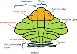
Thecerebellar vermis(fromLatinvermis,"worm" ) is located in the medial, cortico-nuclear zone of thecerebellum,which is in theposterior fossaof thecranium.Theprimary fissurein the vermis curvesventrolaterallyto thesuperiorsurface of thecerebellum,dividing it intoanteriorandposteriorlobes.Functionally, the vermis is associated with bodilypostureandlocomotion.The vermis is included within thespinocerebellumand receives somatic sensory input from theheadand proximal body parts viaascending spinal pathways.[1]
Thecerebellumdevelops in a rostro-caudal manner, withrostralregions in the midline giving rise to the vermis, andcaudalregions developing into thecerebellar hemispheres.[2]By 4 months ofprenatal development,the vermis becomes fullyfoliated,while development of the hemispheres lags by 30–60 days.[3]Postnatally,proliferationand organization of the cellular components of thecerebellumcontinues, with completion of the foliation pattern by 7 months of life[4]and finalmigration,proliferation, andarborizationof cerebellar neurons by20 months.[5]
Inspection of theposterior fossais a common feature ofprenatal ultrasoundand is used primarily to determine whether excess fluid or malformations of the cerebellum exist.[6]Anomalies of the cerebellar vermis are diagnosed in this manner and includephenotypesconsistent withDandy–Walker malformation,rhombencephalosynapsis,displaying no vermis with fusion of thecerebellar hemispheres,pontocerebellar hypoplasia,orstunted growth of the cerebellum,andneoplasms.Inneonates,hypoxic injuryto the cerebellum is fairly common, resulting in neuronal loss andgliosis.Symptoms of these disorders range from mild loss of finemotor controlto severeintellectual disabilityanddeath.Karyotypinghas shown that mostpathologiesassociated with the vermis areinheritedthrough anautosomal recessivepattern, with most knownmutationsoccurring on theX chromosome.[1][7]
The vermis is intimately associated with all regions of thecerebellar cortex,which can be divided into three functional parts, each having distinct connections with thebrainandspinal cord.These regions are thevestibulocerebellum,which is responsible primarily for the control ofeye movements;thespinocerebellum,involved in fine tune body and limb movement; and thecerebrocerebellum,which is associated with planning, initiation and timing ofmovements.[8]
Structure
[edit]
The vermis is the unpaired, median portion of the cerebellum that connects thetwo hemispheres.[9]Both the vermis and the hemispheres are composed oflobulesformed by groups offolia.There are nine lobules of the vermis: lingula, central lobule, culmen, clivus,folium of the vermis,tuber, pyramid, uvula and nodule.[9]These lobules are often difficult to observe during human anatomy classes and may vary in size, shape and number of folia. It has been shown that folia of the cerebellum exhibit frequent variations in form, number and arrangement between individuals.[9]
Lobe anatomy
[edit]
The lingula is the first lobule of the upper portion of the vermis on thesuperoinferioraxis and pertains to thepaleocerebellumtogether with the central lobule, culmen, pyramid and uvula. It is separated from the central lobule by the pre-centralfissure.The central lobule is the second lobule of the upper portion of the vermis on the superoinferior axis. The culmen is the third and largest lobule of the upper portion of the vermis on the superoinferior axis. It is separated from the declive by the primary fissure and is related with theanteriorquadrangular lobule of the hemisphere. The pyramid is the seventh lobule of the vermis on the superoinferior axis. It is separated from the tuber and uvula by the pre-pyramidal and secondary fissures, respectively.[9]This lobule is related with the biventral lobule of the hemisphere. The uvula is the second largest lobule, following the culmen. It pertains to the paleocerebellum and is separated from the nodule by the posterolateral fissure.[9]
Spinocerebellum
[edit]The spinocerebellum receivesproprioceptioninput from the dorsal columns of thespinal cord(including thespinocerebellar tract) and from thetrigeminal nerve,as well as from visual andauditorysystems. It sends fibers to deepcerebellar nucleithat, in turn, project to both thecerebral cortexand thebrain stem,thus providing modulation of descending motor systems.[8]This region comprises the vermis and intermediate parts of the cerebellar hemispheres. Sensory information from the periphery and from theprimary motorandsomatosensory cortexterminate in this region.Purkinje cellsof the vermis project to thefastigial nucleus,controlling theaxialandproximalmusculature involved in the execution of limb movements.[10]Purkinjecells in the intermediate zone of the spinocerebellum project to the interposed nuclei, which control the distal musculature components of thedescending motor pathwaysneeded for limb movement. Both of these nuclei include projections to themotor cortexin thecerebrum.[10]
Nuclei
[edit]Theinterposed nucleusis smaller than thedentate nucleusbut larger than thefastigial nucleusand functions to modulate muscle stretch reflexes of distal musculature.[9]It is locateddorsalto the fourthventricleand lateral to thefastigial nucleus;it receivesafferentneuronal supply from theanterior lobeof the cerebellum and sends output via thesuperior cerebellar peduncleand thered nucleus.[8]
The fastigial nucleus is the most medialefferentcerebellar nucleus, targeting thepontineand medullaryreticular formationas well as thevestibular nuclei.[10]This region deals with antigravity muscle groups and other synergies involved with standing and walking.[11]It is thought that fastigial nucleiaxonsareexcitatoryand project beyond thecerebellum,likely usingglutamateandaspartateasneurotransmitters.[10]
Pathology
[edit]Malformationsof theposterior fossahave been recognized more frequently during the past fewdecadesas the result of recent advances in technology. Malformations of the cerebellar vermis were first identified usingpneumoencephalography,where air is injected into thecerebrospinal fluidspaces of thecerebellum;displaced, occluded ordysplasticstructures could be identified. Upon the advent ofcomputerized tomography(CT) andmagnetic resonance imaging(MRI), the resolution of cranial structures including the mid-hindbrain regions improved dramatically.[12]
Joubert syndrome
[edit]Joubert syndrome(JS) is one of the most commonly diagnosed syndromes associated with the molar tooth sign (MTS),[13]orhypoplasia/dysplasia of the cerebellar vermis accompanied by brainstem abnormalities.JSis defined clinically by features ofhypotoniaininfancywith later development ofataxia,developmental delays,mental retardation,abnormal breathing patterns, abnormal eye movements specific to oculomotorapraxia,or the presence of theMTSon the cranialMRI.[14][15]JSis anautosomal recessivecondition with an estimatedprevalenceof 1: 100,000.[16]
Dandy Walker malformation
[edit]Dandy Walker malformationis a relatively commoncongenitalbrain malformation with a prevalence of 1:30,000 live births.[17]Dandy Walker malformation is characterized by enlargedposterior fossaand in which the cerebellar vermis is completely absent, or present in a rudimentary form, sometimes rotated accompanied by an elevation of the fourthventricle.It is also commonly associated withdysplasiasofbrainstemnuclei.[18]DWM has been reported to be in association with a wide array ofchromosomalanomalies, includingtrisomy 18,trisomy 9,andtrisomy 13.Surveys suggest thatprenatalexposure toteratogenssuch asrubellaoralcoholare correlated with development of Dandy Walker malformation.[19][20]
Rhombencephalosynapsis
[edit]Rhombencephalosynapsisis an anomaly characterized by the absence or severe dysgenesis of the cerebellar vermis with fusion of thecerebellar hemispheres,peduncles,anddentate nuclei.Diagnostic features include fusion of the midbraincolliculi,hydrocephalus,absence of thecorpus callosumother midline structural brain malformations.[21][22][23]
Autism spectrum disorders
[edit]Hypoplasiaand other structural alterations of the vermis have been identified in many patients withautism spectrum disorder(ASD). While the exact nature and extent of the impacts ASD has on the vermis remain in question, it has also been shown that other injuries and malformations of the vermis sometimes produce symptoms closely analogous to ASD. Furthermore, several genetic syndromes known to cause autism (such asfragile X syndrome) have also been shown to cause damage to the vermis.[24]
Damage
[edit]Lesions to the vermis commonly give rise toclinical depression,inappropriate emotional displays (e.g. unwarranted giggling) in addition to movement disorders.[citation needed]
Comparative anatomy
[edit]Earlyneurophysiologistssuggest thatretinalandinertialsignals were selected for about 450 million years ago by primitivebrainstem-cerebellar circuitry because of their relationship with the environment.[25]Microscopically, it is evident thatPurkinje cellprecursors arose fromgranule cells,first forming in irregular patterns, then progressively becoming organized in a layered fashion. Evolutionarily, thePurkinje cellsthen developed extensivedendritic treesthat increasingly became confined to a single plane, through which theaxonsof granule cells threaded, eventually forming a neuronal grid of right angles.[25]The origin of thecerebellumis in close association with that of thenucleiof thevestibular cranial nerveand lateral linenerves,perhaps suggesting that this part of the cerebellum originated as a means of carrying out transformations of thecoordinate systemfrom input data of thevestibular organand thelateral line organs.[26]This suggests that the function of the cerebellumevolvedas a mode of computing and representing an image relating to the position of the body in space. The cerebellar vermis evolved in conjunction with the hemispheres; this is seen inlampreysand highervertebrates.[27]
In fish
[edit]Invertebrates,the cerebellar vermis develops between two bilaterally symmetrical formations locateddorsalto the upper end of themedulla oblongata,orrhombencephalon.This is the region of termination for thefibersof thevestibular nerveand lateral line nerves; thus, these are the oldestafferentpaths to the cerebellum and cerebellar vermis.[27]In bony fish, orteleosts,it has been proposed that the cerebellar auricles, which receive a large amount of input from the vestibulolateral line system, constitute thevestibulocerebellumand arehomologuesof theflocculonodular lobeof higher vertebrates along with thecorpus cerebelli,which receives spinocerebellar and tectocerebellar fibers. Thelabyrinthand the lateral line organs oflampreyshave structural and functional similarity. An important difference between the two structures is that the arrangement of the lateral line organs are such that they are sensitive to relative motion of the fluid surrounding the animal, whereas the labyrinths, having very similar sensing mechanisms, are sensitive toendolymph,providing information concerning the animal's ownequilibriumof the body and orientation in space.[27]
See also
[edit]- cerebellar vermishypoplasia,a geneticciliopathy
- Anatomy of the cerebellum
Additional images
[edit]-
Dissection video. Explains the position of vermis.
-
Animation. Vermis is highlighted in red.
-
Brainstem. Posterior view.
-
Midsagittal view
-
Lobules of the vermis.
References
[edit]- ^abCoffman, K.A.; Dum, R.P.; Strick, P.L. (2011)."Cerebellar vermis is a target of projections from the motor areas in the cerebral cortex".Proceedings of the National Academy of Sciences of the United States of America.108(38): 16068–16073.Bibcode:2011PNAS..10816068C.doi:10.1073/pnas.1107904108.PMC3179064.PMID21911381.
- ^Cho, K. H.; Rodriguez-Vazquez, J. F.; Kim, J. H.; Abe, H.; Murakami, G.; Cho, B. H. (2011). "Early fetal development of the human cerebellum".Surgical and Radiologic Anatomy.33(6): 523–530.doi:10.1007/s00276-011-0796-8.PMID21380713.S2CID25451924.
- ^Parisia, M.; Dobynsb, W. (2003). "Human malformations of the midbrain and hindbrain: review and proposed classification scheme".Molecular Genetics and Metabolism.80(1–2): 36–53.doi:10.1016/j.ymgme.2003.08.010.PMID14567956.
- ^J.D. Loeser; R.J. Lemire; J. Alvord (1973). "The development of the folia in the human cerebellar vermis".Anat. Rec.173(1): 109–114.doi:10.1002/ar.1091730109.PMID5028060.S2CID45169021.
- ^D. Goldowitz; K. Hamre (1998). "The cells and molecules that make a cerebellum".Trends Neurosci.21(9): 375–382.doi:10.1016/S0166-2236(98)01313-7.PMID9735945.S2CID41916018.
- ^Robinson AJ, Blaser S, Toi A, et al. (2007). "The fetal cerebellar vermis: assessment for abnormal development by ultrasonography and magnetic resonance imaging".Ultrasound Quarterly.23(3): 211–223.doi:10.1097/ruq.0b013e31814b162c.PMID17805192.S2CID23068656.
- ^Zanni G, Bertini ES (2011)."X-linked disorders with cerebellar dysgenesis".Orphanet Journal of Rare Diseases.6:24.doi:10.1186/1750-1172-6-24.PMC3115841.PMID21569638.
- ^abcGhez C, Fahn S (1985). "The cerebellum". In Kandel ER, Schwartz JH (eds.).Principles of Neural Science(2nd ed.). New York: Elsevier. pp. 502–522.
- ^abcdefMonte-Bispo, R.F.; et al. (2010)."Cerebellar Vermis: Topography and Variations".Int. J. Morphol.28(2): 439–443.doi:10.4067/s0717-95022010000200018.
- ^abcdRamon-Cajal, S. (1995).Histology of the Nervous System.Oxford University Press.
- ^James D. Geyer; Janice M. Keating; Daniel C. Potts (1998).Neurology for the Boards.Philadelphia: Lippincott-Raven. p. 9.
- ^Patel, Sandeep; Barkovich, A. James (2002)."Analysis and classification of cerebellar malformations".American Journal of Neuroradiology.23(7): 1074–1087.PMC8185716.PMID12169461.
- ^Brancati F, Dallapiccola B, Valente EM (2010)."Joubert Syndrome and related disorders".Orphanet Journal of Rare Diseases.5:20.doi:10.1186/1750-1172-5-20.PMC2913941.PMID20615230.
- ^J.M. Saraiva; M. Baraitser (1992). "Joubert syndrome: a review".American Journal of Medical Genetics.43(4): 726–731.doi:10.1002/ajmg.1320430415.PMID1341417.
- ^B.L. Maria; E. Boltshauser; S.C. Palmer; T.X. Tran (1999). "Clinical features and revised diagnostic criteria in Joubert syndrome".Child Neurology.14(9): 583–590.doi:10.1177/088307389901400906.PMID10488903.S2CID7410607.
- ^D.B. Flannery; J.G. Hudson (1994).A survey of Joubert syndrome.David W. Smith Workshop.
- ^Osenbach, R.K.; Menezes, A.H. (1992). "Diagnosis and management of the Dandy-Walker malformation: 30 years of experience".Pediatric Neurosurgery.18(4): 179–89.doi:10.1159/000120660.PMID1472430.
- ^Kapur, R.; Mahony, B.; Finch, L.; Siebert J. (2009). "Normal and Abnormal Anatomy of the Cerebellar Vermis in Midgestational Human Fetuses".Birth Defects Research.85(8): 700–709.doi:10.1002/bdra.20589.PMID19441098.
- ^J.C. Murray; J.A. Johnson; T.D. Bird (1985). "Dandy–Walker malformation: etiologic heterogeneity and empiric recurrence risks".Clin. Genet.28(4): 272–283.doi:10.1111/j.1399-0004.1985.tb00401.x.PMID4064366.S2CID22883753.
- ^S.K. Clarren; J. Alvord; S.M. Sumi (1978). "Brain malformations related to prenatal exposure to ethanol".Journal of Pediatrics.92(1): 64–67.doi:10.1016/S0022-3476(78)80072-9.PMID619080.
- ^S.P. Toelle; C. Yalcinkaya; N. Kocer; T. Deonna; W.C.G. Overweg-Plandsoen; T. Bast; R. Kalmanchey; P. Barsi; J.F.L. Schneider; A. Capone Mori; E. Boltshauser (2002). "Rhombencephalosynapsis: clinical findings and neuroimaging in 9 children".Neuropediatrics.33(4): 209–214.doi:10.1055/s-2002-34498.PMID12368992.S2CID32510022.
- ^H. Utsunomiya; K. Takano; T. Ogasawara; T. Hashimoto; T. Fukushima; M. Okazaki (1998)."Rhombencephalosynapsis: cerebellar embryogenesis".American Journal of Neuroradiology.19(3): 547–549.PMC8338264.PMID9541316.
- ^C.L. Truwit; A.J. Barkovich; R. Shanahan; T.V. Maroldo (1991)."MR imaging of rhomboencephalosynapsis: report of three cases and review of the literature".American Journal of Neuroradiology.12(5): 957–965.PMC8333516.PMID1950929.
- ^Hampson, David R.; Blatt, Gene J. (2015)."Autism spectrum disorders and neuropathology of the cerebellum".Frontiers in Neuroscience.9:420.doi:10.3389/fnins.2015.00420.ISSN1662-4548.PMC4635214.PMID26594141.
- ^abNieuwenhuys, R.; Voogd, J.; van Huijzen, C. (1988).The Human Central Nervous System: A Synopsis and Atlas(3rd ed.). Heidelberg: Springer-Verlag.
- ^Butler, A.B.; Hodos, W. (1996). "12: The Cerebellum".Comparative Vertebrate Neuroanatomy: Evolution and Adaptation.New York: Wiley-Liss. pp. 180–197.
- ^abcAriens, K.; C.U.; Huber, G.C.; Crosby, E.C. (1960).The Comparative Anatomy of the Nervous System of Vertebrates, Including Man.Vol. 3. New York: Hafner.
External links
[edit]Rhombencephalosynapsis Website Support
- Photo - rollover to see highlightedatUniversity of Texas Southwestern Medical Center at Dallas
- Diagramat medfriendly.com
- Stained brain slice images which include the "Vermis"at theBrainMaps project
- "Anatomy diagram: 13048.000-3".Roche Lexicon - illustrated navigator.Elsevier. Archived fromthe originalon 2012-07-22.
- Atlas image: n2a7p4at the University of Michigan Health System


