
Wrist
This articlemay be too technical for most readers to understand.(June 2015) |
| Wrist | |
|---|---|
 A human showing the wrist in the centre | |
 Thecarpal bones,sometimes included in the definition of the wrist | |
| Details | |
| Identifiers | |
| Latin | articulatio radiocarpalis |
| MeSH | D014953 |
| TA98 | A01.1.00.026 |
| TA2 | 147 |
| FMA | 24922 |
| Anatomical terminology | |
Inhuman anatomy,thewristis variously defined as (1) thecarpusor carpal bones, the complex of eight bones forming the proximal skeletal segment of thehand;[1][2](2) thewrist jointorradiocarpal joint,the joint between theradiusand thecarpus[2]and; (3) the anatomical region surrounding the carpus including the distal parts of the bones of the forearm and the proximal parts of themetacarpusor five metacarpal bones and the series of joints between these bones, thus referred to aswrist joints.[3][4]This region also includes thecarpal tunnel,theanatomical snuff box,bracelet lines, theflexor retinaculum,and theextensor retinaculum.
As a consequence of these various definitions, fractures to the carpal bones are referred to as carpal fractures, while fractures such asdistal radius fractureare often considered fractures to the wrist.
Structure
[edit]


Thedistal radioulnar joint(DRUJ) is apivot jointlocated between thedistalends of theradiusandulna,which make up theforearm.Formed by thehead of the ulnaand theulnar notch of the radius,the DRUJ is separated from the radiocarpal (wrist) joint by anarticular disklying between the radius and thestyloid process of the ulna.Thecapsuleof the joint is lax and extends from theinferior sacciform recessto the ulnar shaft. The DRUJ works with theproximal radioulnar joint(at the elbow) forpronationandsupination.[5]
The radiocarpal (wrist) joint is anellipsoid jointformed by the radius and thearticular discproximally and the proximal row of carpal bones distally. The carpal bones on the ulnar side only make intermittent contact with the proximal side — the triquetrum only makes contact during ulnar abduction. The capsule, lax and un-branched, is thin on the dorsal side and can contain synovial folds. The capsule is continuous with the midcarpal joint and strengthened by numerousligaments,including thepalmaranddorsal radiocarpal ligaments,and theulnarandradial collateral ligaments. [6]
The parts forming the radiocarpal joint are the lower end of theradiusand under surface of thearticular diskabove; and thescaphoid,lunate,andtriquetralbones below. The articular surface of the radius and the undersurface of thearticular diskform together with a transversely elliptical concave surface, the receiving cavity. The superior articular surfaces of the scaphoid, lunate, and triquetrum form a smooth convex surface, thecondyle,which is received into the concavity.[7]
Carpal bonesof thehand:
- Proximal:A=Scaphoid,B=Lunate,C=Triquetrum,D=Pisiform
- Distal:E=Trapezium,F=Trapezoid,G=Capitate,H=Hamate
In the hand proper a total of 13 bones form part of the wrist: eightcarpal bones—scaphoid,lunate,triquetral,pisiform,trapezium,trapezoid,capitate,andhamate— and fivemetacarpal bones—thefirst,second,third,fourth,andfifth metacarpal bones.[8]
Themidcarpal jointis the S-shaped joint space separating the proximal and distal rows of carpal bones. Theintercarpal joints,between the bones of each row, are strengthened by theradiate carpalandpisohamate ligamentsand thepalmar,interosseous,anddorsal intercarpal ligaments.Some degree of mobility is possible between the bones of the proximal row while the bones of the distal row are connected to each other and to the metacarpal bones —at thecarpometacarpal joints— by strong ligaments —thepisometacarpalandpalmaranddorsal carpometacarpal ligament— that makes a functional entity of these bones. Additionally, the joints between the bases of the metacarpal bones —theintermetacarpal articulations— are strengthened bydorsal,interosseous,andpalmar intermetacarpal ligaments.[6]
The earliest carpal bones to ossify arecapitate boneandhamate bonein the first six months of an infant life.[9]
Articulations
[edit]The radiocarpal, intercarpal, midcarpal, carpometacarpal, and intermetacarpal joints often intercommunicate through a common synovial cavity. [10]
Articular surfaces
[edit]It has two articular surfaces named, proximal and distal articular surfaces respectively. The proximal articular surface is made up of the lower end of the radius and a triangular articular disc of the inferior radio-ulnar joint. On the other hand, the distal articular surface is made up of proximal surfaces of the scaphoid, triquetral and lunate bones.[11]

Function
[edit]Movement
[edit]The extrinsic hand muscles are located in the forearm where their bellies form the proximal fleshy roundness. When contracted, most of the tendons of these muscles are prevented from standing up like taut bowstrings around the wrist by passing under theflexor retinaculumon the palmar side and theextensor retinaculumon the dorsal side. On the palmar side the carpal bones form thecarpal tunnel,[12]through which some of the flexor tendons pass intendon sheathsthat enable them to slide back and forth through the narrow passageway (seecarpal tunnel syndrome).[13]
Starting from the mid-position of the hand, themovementspermitted in the wrist proper are (muscles in order of importance):[14][15]
- Marginal movements: radial deviation (abduction, movement towards the thumb) and ulnar deviation (adduction, movement towards the little finger). These movements take place about a dorsopalmar axis (back to front) at the radiocarpal and midcarpal joints passing through the capitate bone.
- Radial abduction (up to 20°):[16]extensor carpi radialis longus,abductor pollicis longus,extensor pollicis longus,flexor carpi radialis,flexor pollicis longus
- Ulnar adduction (up to 30°):[16]extensor carpi ulnaris,flexor carpi ulnaris,extensor digitorum,extensor digiti minimi
- Movements in the plane of the hand: flexion (palmar flexion, tilting towards the palm) and extension (dorsiflexion, tilting towards the back of the hand). These movements take place through a transverse axis passing through the capitate bone. Palmar flexion is the most powerful of these movements because the flexors, especially the finger flexors, are considerably stronger than the extensors.
- Extension (up to 60°):[16]extensor digitorum,extensor carpi radialis longus,extensor carpi radialis brevis,extensor indicis,extensor pollicis longus,extensor digiti minimi,extensor carpi ulnaris
- Palmar flexion (up to 70°):[16]flexor digitorum superficialis,flexor digitorum profundus,flexor carpi ulnaris,flexor pollicis longus,flexor carpi radialis,abductor pollicis longus
- Intermediate or combined movements
However, movements at the wrist can not be properly described without including movements in the distal radioulnar joint in which the rotary actions ofsupinationandpronationoccur and this joint is therefore normally regarded as part of the wrist.[17]
Clinical significance
[edit]
Wrist painhas a number of causes, includingcarpal tunnel syndrome,[16]ganglion cyst,[19]tendinitis,[20]andosteoarthritis.Tests such asPhalen's testinvolvepalmarflexionat the wrist.
The hand may deviate at the wrist in some conditions, such asrheumatoid arthritis.
Ossificationof the bones around the wrist is one indicator used in taking abone age.
Awrist fractureusually means a fracture of thedistal radius.
History
[edit]Etymology
[edit]TheEnglishword "wrist"isetymologicallyderived from theProto-Germanicwordwristizfrom which are derived modern GermanRist( "instep","wrist ") and modernSwedishvrist( "instep", "ankle"). The basewrith-and its variants are associated withOld Englishwords "wreath","wrest",and"writhe".Thewr-sound of this base seems originally to have been symbolic of the action of twisting.[21]
See also
[edit]- Brunelli procedure,related to instability in the wrist, caused by a tornscapholunate ligament.
- Knuckle-walking,a kind of quadrupedal locomotion involving wrist bone specialization
- Wristlocksuse movement extremes of the wrist for martial applications.
- Glossary of bowling § Wrist,a measure of wrist position in bowling ball deliveries
Additional images
[edit]-
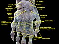 Wrist joint. Deep dissection. Posterior view.
 -
 Wrist joint. Deep dissection. Posterior view.
 -
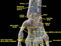 Wrist joint. Deep dissection.Anterior, palmar, view.
 -
 Wrist joint. Deep dissection.Anterior, palmar, view.


References
[edit]- ^Behnke 2006,p. 76 "The wrist contains eight bones, roughly aligned in two rows, known as the carpal bones."
- ^abMoore KL, Agur AM (2006).Essential clinical anatomy.Lippincott Williams & Wilkins. p. 485.ISBN0-7817-6274-X.
The wrist (carpus), the proximal segment of the hand, is a complex of eight carpal bones. The carpus articulates proximally with the forearm at the wrist joint and distally with the five metacarpals. The joints formed by the carpus include the wrist (the radiocarpal joint), intercarpal, carpometacarpal, and intermetacarpal joints. Augmenting movement at the wrist joint, the rows of carpals glide on each other [...]
- ^Behnke 2006,p. 77 "With the large number of bones composing the wrist (ulna, radius, eight carpas, and five metacarpals), it makes sense that there are many, many joints that make up the structure known as the wrist."
- ^Baratz M, Watson AD, Imbriglia JE (1999).Orthopaedic surgery: the essentials.Thieme. p. 391.ISBN0-86577-779-9.
The wrist joint is composed of not only the radiocarpal and distal radioulnar joints but also the intercarpal articulations.
- ^Platzer 2004,p. 122
- ^abPlatzer 2004,p. 130
- ^"Wrist Joint".The Lecturio Medical Concept Library.Retrieved2021-06-23.
- ^Platzer 2004,pp. 126–129
- ^Al-Khater KM, Hegazi TM, Al-Thani HF, Al-Muhanna HT, Al-Hamad BW, Alhuraysi SM, et al. (September 2020)."Time of appearance of ossification centers in carpal bones. A radiological retrospective study on Saudi children".Saudi Medical Journal.41(9): 938–946.doi:10.15537/smj.2020.9.25348.PMC7557557.PMID32893275.
- ^Isenberg DA, Maddison P, Woo P (2004).Oxford textbook of rheumatology.Oxford University Press. p. 87.ISBN0-19-850948-0.
- ^"Wrist Joint".Earth's Lab.
- ^Rea P (2016-01-01). "Chapter 3 - Neck". In Rea P (ed.).Essential Clinically Applied Anatomy of the Peripheral Nervous System in the Head and Neck.Academic Press. pp. 131–183.doi:10.1016/b978-0-12-803633-4.00003-x.ISBN978-0-12-803633-4.
- ^Saladin KS (2003).Anatomy & Physiology: The Unity of Form and Function(3rd ed.). McGraw-Hill. pp. 361, 365.
- ^Platzer 2004,p. 132
- ^Platzer 2004,p. 172
- ^abcdeLalani I, Argoff CE (2008-01-01). "Chapter 10 - History and Physical Examination of the Pain Patient". In Benzon HT, Rathmell JP, Wu CL, Turk DC (eds.).Raj's Practical Management of Pain(Fourth ed.). Philadelphia: Mosby. pp. 177–188.doi:10.1016/B978-032304184-3.50013-3.ISBN978-0-323-04184-3.
- ^Kingston B (2000).Understanding joints: a practical guide to their structure and function.Nelson Thornes. pp. 126–127.ISBN0-7487-5399-0.
- ^Döring AC, Overbeek CL, Teunis T, Becker SJ, Ring D (October 2016)."A Slightly Dorsally Tilted Lunate on MRI can be Considered Normal".The Archives of Bone and Joint Surgery.4(4): 348–352.PMC5100451.PMID27847848.
- ^Stretanski MF (2020-01-01). "Chapter 32 - Hand and Wrist Ganglia". In Frontera WR, Silver JK, Rizzo TD (eds.).Essentials of Physical Medicine and Rehabilitation(Fourth ed.). Philadelphia: Content Repository Only!. pp. 169–173.doi:10.1016/B978-0-323-54947-9.00032-8.ISBN978-0-323-54947-9.S2CID229189365.
- ^Waldman SD (2014-01-01). "Chapter 58 - Flexor Carpi Radialis Tendinitis". In Waldman SD (ed.).Atlas of Uncommon Pain Syndromes(Third ed.). Philadelphia: W.B. Saunders. pp. 172–174.doi:10.1016/b978-1-4557-0999-1.00058-7.ISBN978-1-4557-0999-1.
- ^"Hand Etymology".American Society for Surgery of the Hand.
Sources
[edit]- Behnke RS (2006).Kinetic anatomy.Human Kinetics.ISBN0-7360-5909-1.
- Platzer W (2004).Color Atlas of Human Anatomy, Vol. 1: Locomotor System(5th ed.).Thieme.ISBN3-13-533305-1.