Aboneis arigidorgan[1]that constitutes part of theskeletonin mostvertebrateanimals. Bones protect the various other organs of the body, produceredandwhite blood cells,storeminerals,provide structure and support for the body, and enablemobility.Bones come in a variety of shapes and sizes and have complex internal and external structures.[2]They are lightweight yet strong and hard and serve multiplefunctions.
In thehuman bodyat birth, approximately 300 bones are present. Many of these fuse together during development, leaving a total of 206 separate bones in the adult, not counting numerous smallsesamoid bones.[3][4]The largest bone in the body is thefemuror thigh-bone, and the smallest is thestapesin themiddle ear.
Bone is not uniformly solid, but consists of a flexiblematrix(about 30%) and bound minerals (about 70%), which are intricately woven and continuously remodeled by a group of specialized bone cells. Their unique composition and design allows bones to be relativelyhardand strong, while remaining lightweight.
Bone matrix is 90 to 95% composed of elasticcollagenfibers, also known as ossein,[5]and the remainder isground substance.[6]The elasticity ofcollagenimproves fracture resistance.[7]The matrix is hardened by the binding of inorganic mineral salt,calcium phosphate,in a chemical arrangement known asbone mineral,a form of calciumapatite.[8][9]It is the mineralization that gives bones rigidity.
Bone is actively constructed and remodeled throughout life by special bone cells known as osteoblasts and osteoclasts. Within any single bone, the tissue is woven into two main patterns, known as cortical and cancellous bone, each with a different appearance and characteristics.
The hard outer layer of bones is composed ofcortical bone,which is also calledcompact boneas it is much denser than cancellous bone. It forms the hard exterior (cortex) of bones. The cortical bone gives bone its smooth, white, and solid appearance, and accounts for 80% of the total bone mass of an adulthuman skeleton.[10]It facilitates bone's main functions—to support the whole body, to protect organs, to provideleversfor movement, and to store and release chemical elements, mainly calcium. It consists of multiple microscopic columns, each called anosteonor Haversian system. Each column is multiple layers ofosteoblastsandosteocytesaround a central canal called theosteonic canal.Volkmann's canalsat right angles connect the osteons together. The columns are metabolically active, and as bone is reabsorbed and created the nature and location of the cells within the osteon will change. Cortical bone is covered by aperiosteumon its outer surface, and anendosteumon its inner surface. The endosteum is the boundary between the cortical bone and the cancellous bone.[11]The primary anatomical and functional unit of cortical bone is theosteon.
Cancellous boneorspongy bone,[12][11]also known astrabecular bone,is the internal tissue of the skeletal bone and is an open cellporousnetwork that follows the material properties ofbiofoams.[13][14]Cancellous bone has a highersurface-area-to-volume ratiothan cortical bone and it is lessdense.This makes it weaker and more flexible. The greater surface area also makes it suitable for metabolic activities such as the exchange of calcium ions. Cancellous bone is typically found at the ends of long bones, near joints, and in the interior of vertebrae. Cancellous bone is highlyvascularand often contains redbone marrowwherehematopoiesis,the production of blood cells, occurs. The primary anatomical and functional unit of cancellous bone is thetrabecula.The trabeculae are aligned towards the mechanical load distribution that a bone experiences within long bones such as thefemur.As far as short bones are concerned, trabecular alignment has been studied in thevertebralpedicle.[15]Thin formations ofosteoblastscovered in endosteum create an irregular network of spaces,[16]known as trabeculae. Within these spaces arebone marrowandhematopoietic stem cellsthat give rise toplatelets,red blood cellsandwhite blood cells.[16]Trabecular marrow is composed of a network of rod- and plate-like elements that make the overall organ lighter and allow room for blood vessels and marrow. Trabecular bone accounts for the remaining 20% of total bone mass but has nearly ten times the surface area of compact bone.[17]
The wordscancellousandtrabecularrefer to the tiny lattice-shaped units (trabeculae) that form the tissue. It was first illustrated accurately in the engravings ofCrisóstomo Martinez.[18]
Bone marrow,also known asmyeloid tissuein red bone marrow, can be found in almost any bone that holdscancellous tissue.Innewborns,all such bones are filled exclusively with red marrow orhematopoieticmarrow, but as the child ages the hematopoietic fraction decreases in quantity and the fatty/ yellow fraction calledmarrow adipose tissue(MAT) increases in quantity. In adults, red marrow is mostly found in the bone marrow of the femur, the ribs, the vertebrae andpelvic bones.[19]
Bone receives about 10% of cardiac output.[20]Blood enters theendosteum,flows through the marrow, and exits through small vessels in the cortex.[20]In humans,blood oxygen tensionin bone marrow is about 6.6%, compared to about 12% in arterial blood, and 5% in venous and capillary blood.[20]
Light micrographofdecalcifiedcancellous bone tissue displaying osteoblasts actively synthesizing osteoid, containing two osteocytes.
Osteoblastsare mononucleate bone-forming cells. They are located on the surface of osteon seams and make aproteinmixture known asosteoid,which mineralizes to become bone.[23]The osteoid seam is a narrow region of a newly formed organic matrix, not yet mineralized, located on the surface of a bone. Osteoid is primarily composed of Type Icollagen.Osteoblasts also manufacturehormones,such asprostaglandins,to act on the bone itself. The osteoblast creates and repairs new bone by actually building around itself. First, the osteoblast puts up collagen fibers. These collagen fibers are used as a framework for the osteoblasts' work. The osteoblast then deposits calcium phosphate which is hardened byhydroxideandbicarbonateions. The brand-new bone created by the osteoblast is calledosteoid.[24]Once the osteoblast is finished working it is actually trapped inside the bone once it hardens. When the osteoblast becomes trapped, it becomes known as an osteocyte. Other osteoblasts remain on the top of the new bone and are used to protect the underlying bone, these become known as bone lining cells.[25]
Osteocytesare cells of mesenchymal origin and originate from osteoblasts that have migrated into and become trapped and surrounded by a bone matrix that they themselves produced.[11]The spaces the cell body of osteocytes occupy within the mineralized collagen type I matrix are known aslacunae,while the osteocyte cell processes occupy channels called canaliculi. The many processes of osteocytes reach out to meet osteoblasts, osteoclasts, bone lining cells, and other osteocytes probably for the purposes of communication.[26]Osteocytes remain in contact with other osteocytes in the bone through gap junctions—coupled cell processes which pass through the canalicular channels.
Osteoclastsare very largemultinucleatecells that are responsible for the breakdown of bones by the process ofbone resorption.New bone is then formed by the osteoblasts. Bone is constantlyremodeledby the resorption of osteoclasts and created by osteoblasts.[21]Osteoclasts are large cells with multiplenucleilocated on bone surfaces in what are calledHowship's lacunae(orresorption pits). These lacunae are the result of surrounding bone tissue that has been reabsorbed.[27]Because the osteoclasts are derived from amonocytestem-celllineage, they are equipped withphagocytic-like mechanisms similar to circulatingmacrophages.[21]Osteoclasts mature and/or migrate to discrete bone surfaces. Upon arrival, active enzymes, such astartrate-resistant acid phosphatase,aresecretedagainst the mineral substrate.[citation needed]The reabsorption of bone by osteoclasts also plays a role incalciumhomeostasis.[27]
Bones consist of living cells (osteoblasts and osteocytes) embedded in a mineralized organic matrix. The primary inorganic component of human bone ishydroxyapatite,the dominantbone mineral,having the nominal composition of Ca10(PO4)6(OH)2.[28]The organic components of this matrix consist mainly oftype I collagen— "organic" referring to materials produced as a result of the human body—and inorganic components, which alongside the dominanthydroxyapatitephase, include other compounds ofcalciumandphosphateincluding salts. Approximately 30% of the acellular component of bone consists of organic matter, while roughly 70% by mass is attributed to the inorganic phase.[29]Thecollagenfibers give bone itstensile strength,and the interspersed crystals ofhydroxyapatitegive bone itscompressive strength.These effects aresynergistic.[29]The exact composition of the matrix may be subject to change over time due to nutrition andbiomineralization,with the ratio ofcalciumtophosphatevarying between 1.3 and 2.0 (per weight), and trace minerals such asmagnesium,sodium,potassiumandcarbonatealso be found.[29]
Type I collagen composes 90–95% of the organic matrix, with the remainder of the matrix being a homogenous liquid calledground substanceconsisting ofproteoglycanssuch ashyaluronic acidandchondroitin sulfate,[29]as well as non-collagenous proteins such asosteocalcin,osteopontinorbone sialoprotein.Collagen consists of strands of repeating units, which give bone tensile strength, and are arranged in an overlapping fashion that prevents shear stress. The function of ground substance is not fully known.[29]Two types of bone can be identified microscopically according to the arrangement of collagen: woven and lamellar.
Woven bone (also known asfibrous bone), which is characterized by a haphazard organization of collagen fibers and is mechanically weak.[30]
Lamellar bone, which has a regular parallel alignment of collagen into sheets ( "lamellae" ) and is mechanically strong.[14][30]
Transmissionelectron micrographof decalcified woven bone matrix displaying characteristic irregular orientation of collagen fibers
Woven bone is produced when osteoblasts produce osteoid rapidly, which occurs initially in allfetalbones, but is later replaced by more resilient lamellar bone. In adults, woven bone is created afterfracturesor inPaget's disease.Woven bone is weaker, with a smaller number of randomly oriented collagen fibers, but forms quickly; it is for this appearance of the fibrous matrix that the bone is termedwoven.It is soon replaced by lamellar bone, which is highly organized inconcentricsheets with a much lower proportion of osteocytes to surrounding tissue. Lamellar bone, which makes its first appearance in humans in thefetusduring the third trimester,[31]is stronger and filled with many collagen fibers parallel to other fibers in the same layer (these parallel columns are called osteons). Incross-section,the fibers run in opposite directions in alternating layers, much like inplywood,assisting in the bone's ability to resisttorsionforces. After a fracture, woven bone forms initially and is gradually replaced by lamellar bone during a process known as "bony substitution". Compared to woven bone, lamellar bone formation takes place more slowly. The orderly deposition of collagen fibers restricts the formation of osteoid to about 1 to 2μmper day. Lamellar bone also requires a relatively flat surface to lay the collagen fibers in parallel or concentric layers.[32]
The extracellular matrix of bone is laid down byosteoblasts,which secrete both collagen and ground substance. These cells synthesise collagen Alpha polypetpide chains and then secrete collagen molecules. The collagen molecules associate with their neighbors and crosslink via lysyl oxidase to form collagen fibrils. At this stage, they are not yet mineralized, and this zone of unmineralized collagen fibrils is called "osteoid". Around and inside collagen fibrils calcium and phosphate eventuallyprecipitatewithin days to weeks becoming then fully mineralized bone with an overall carbonate substituted hydroxyapatite inorganic phase.[33][29]
In order to mineralise the bone, the osteoblasts secrete alkaline phosphatase, some of which is carried byvesicles.This cleaves the inhibitory pyrophosphate and simultaneously generates free phosphate ions for mineralization, acting as the foci for calcium and phosphate deposition. Vesicles may initiate some of the early mineralization events by rupturing and acting as a centre for crystals to grow on. Bone mineral may be formed from globular and plate structures, and via initially amorphous phases.[34][35]
Structure of a long boneOne way to classify bones is by their shape or appearance.
Five types of bones are found in the human body: long, short, flat, irregular, and sesamoid.[36]
Skeletal System of Human BodyLong bonesare characterized by a shaft, thediaphysis,that is much longer than its width; and by anepiphysis,a rounded head at each end of the shaft. They are made up mostly ofcompact bone,with lesser amounts ofmarrow,located within themedullary cavity,and areas of spongy, cancellous bone at the ends of the bones.[37]Most bones of thelimbs,including those of thefingersandtoes,are long bones. The exceptions are the eightcarpal bonesof thewrist,the seven articulatingtarsal bonesof theankleand the sesamoid bone of thekneecap.Long bones such as the clavicle, that have a differently shaped shaft or ends are also calledmodified long bones.
Short bonesare roughlycube-shaped, and have only a thin layer of compact bone surrounding a spongy interior. Short bones provide stability and support as well as some limited motion.[38]The bones of the wrist and ankle are short bones.
Flat bonesare thin and generally curved, with two parallel layers of compact bone sandwiching a layer of spongy bone. Most of the bones of theskullare flat bones, as is thesternum.[39]
Sesamoid bonesare bones embedded in tendons. Since they act to hold the tendon further away from the joint, the angle of the tendon is increased and thus the leverage of the muscle is increased. Examples of sesamoid bones are thepatellaand thepisiform.[40]
Irregular bonesdo not fit into the above categories. They consist of thin layers of compact bone surrounding a spongy interior. As implied by the name, their shapes are irregular and complicated. Often this irregular shape is due to their many centers of ossification or because they contain bony sinuses. The bones of thespine,pelvis,and some bones of the skull are irregular bones. Examples include theethmoidandsphenoidbones.[41]
In the study ofanatomy,anatomists use a number ofanatomical termsto describe the appearance, shape and function of bones. Other anatomical terms are also used to describe thelocation of bones.Like other anatomical terms, many of these derive fromLatinandGreek.Some anatomists still use Latin to refer to bones. The term "osseous", and the prefix "osteo-", referring to things related to bone, are still used commonly today.
Some examples of terms used to describe bones include the term "foramen" to describe a hole through which something passes, and a "canal" or "meatus" to describe a tunnel-like structure. A protrusion from a bone can be called a number of terms, including a "condyle", "crest", "spine", "eminence", "tubercle" or "tuberosity", depending on the protrusion's shape and location. In general,long bonesare said to have a "head", "neck", and "body".
When two bones join, they are said to "articulate". If the two bones have a fibrous connection and are relatively immobile, then the joint is called a "suture".
Intramembranous ossificationmainly occurs during formation of the flat bones of theskullbut also the mandible, maxilla, and clavicles; the bone is formed from connective tissue such asmesenchymetissue rather than from cartilage. The process includes: the development of theossification center,calcification,trabeculae formation and the development of the periosteum.[43]
Endochondral ossificationoccurs in long bones and most other bones in the body; it involves the development of bone from cartilage. This process includes the development of a cartilage model, its growth and development, development of the primary and secondaryossification centers,and the formation of articular cartilage and theepiphyseal plates.[44]
Endochondral ossification begins with points in the cartilage called "primary ossification centers". They mostly appear during fetal development, though a few short bones begin their primary ossification afterbirth.They are responsible for the formation of the diaphyses of long bones, short bones and certain parts of irregular bones. Secondary ossification occurs after birth and forms theepiphysesof long bones and the extremities of irregular and flat bones. The diaphysis and both epiphyses of a long bone are separated by a growing zone of cartilage (theepiphyseal plate). At skeletal maturity (18 to 25 years of age), all of the cartilage is replaced by bone, fusing the diaphysis and both epiphyses together (epiphyseal closure).[45]In the upper limbs, only the diaphyses of the long bones and scapula are ossified. The epiphyses, carpal bones, coracoid process, medial border of the scapula, and acromion are still cartilaginous.[46]
The following steps are followed in the conversion of cartilage to bone:
Zone of reserve cartilage. This region, farthest from the marrow cavity, consists of typical hyaline cartilage that as yet shows no sign of transforming into bone.[47]
Zone of cell proliferation. A little closer to the marrow cavity, chondrocytes multiply and arrange themselves into longitudinal columns of flattened lacunae.[47]
Zone of cell hypertrophy. Next, the chondrocytes cease to divide and begin to hypertrophy (enlarge), much like they do in the primary ossification center of the fetus. The walls of the matrix between lacunae become very thin.[47]
Zone of calcification. Minerals are deposited in the matrix between the columns of lacunae and calcify the cartilage. These are not the permanent mineral deposits of bone, but only a temporary support for the cartilage that would otherwise soon be weakened by the breakdown of the enlarged lacunae.[47]
Zone of bone deposition. Within each column, the walls between the lacunae break down and the chondrocytes die. This converts each column into a longitudinal channel, which is immediately invaded by blood vessels and marrow from the marrow cavity. Osteoblasts line up along the walls of these channels and begin depositing concentric lamellae of matrix, while osteoclasts dissolve the temporarily calcified cartilage.[47]
Bones serve a variety of mechanical functions. Together the bones in the body form theskeleton.They provide a frame to keep the body supported, and an attachment point forskeletal muscles,tendons,ligamentsandjoints,which function together to generate and transfer forces so that individual body parts or the whole body can be manipulated in three-dimensional space (the interaction between bone and muscle is studied inbiomechanics).
Bones protect internal organs, such as theskullprotecting thebrainor theribsprotecting theheartandlungs.Because of the way that bone is formed, bone has a highcompressive strengthof about 170MPa(1,700kgf/cm2),[7]poortensile strengthof 104–121 MPa, and a very lowshear stressstrength (51.6 MPa).[48][49]This means that bone resists pushing (compressional) stress well, resist pulling (tensional) stress less well, but only poorly resists shear stress (such as due to torsional loads). While bone is essentiallybrittle,bone does have a significant degree ofelasticity,contributed chiefly bycollagen.
Mechanically, bones also have a special role inhearing.Theossiclesare three small bones in themiddle earwhich are involved in sound transduction.
The cancellous part of bones containbone marrow.Bone marrow produces blood cells in a process calledhematopoiesis.[50]Blood cells that are created in bone marrow includered blood cells,plateletsandwhite blood cells.[51]Progenitor cells such as thehematopoietic stem celldivide in a process calledmitosisto produce precursor cells. These include precursors which eventually give rise towhite blood cells,anderythroblastswhich give rise to red blood cells.[52]Unlike red and white blood cells, created by mitosis, platelets are shed from very large cells calledmegakaryocytes.[53]This process of progressive differentiation occurs within the bone marrow. After the cells are matured, they enter thecirculation.[54]Every day, over 2.5 billion red blood cells and platelets, and 50–100 billiongranulocytesare produced in this way.[22]
As well as creating cells, bone marrow is also one of the major sites where defective or aged red blood cells are destroyed.[22]
Mineral storage – bones act as reserves of minerals important for the body, most notablycalciumandphosphorus.[55][56][57]
Determined by the species, age, and the type of bone, bone cells make up to 15 percent of the bone.Growth factorstorage—mineralized bone matrix stores important growth factors such asinsulin-like growth factors, transforming growth factor,bone morphogenetic proteinsand others.[58]
Acid-basebalance – bone buffers the blood against excessivepHchanges by absorbing or releasingalkaline salts.[60]
Detoxification – bone tissues can also storeheavy metalsand other foreign elements, removing them from the blood and reducing their effects on other tissues. These can later be gradually released forexcretion.[61]
Calcium balance – the process of bone resorption by the osteoclasts releases stored calcium into the systemic circulation and is an important process in regulating calcium balance. As bone formation activelyfixescirculating calcium in its mineral form, removing it from the bloodstream, resorption activelyunfixesit thereby increasing circulating calcium levels. These processes occur in tandem at site-specific locations.[63]
Bone is constantly being created and replaced in a process known asremodeling.This ongoing turnover of bone is a process of resorption followed by replacement of bone with little change in shape. This is accomplished through osteoblasts and osteoclasts. Cells are stimulated by a variety ofsignals,and together referred to as a remodeling unit. Approximately 10% of the skeletal mass of an adult is remodelled each year.[64]The purpose of remodeling is to regulatecalcium homeostasis,repairmicrodamaged bonesfrom everyday stress, and to shape the skeleton during growth.[65]Repeated stress, such as weight-bearingexerciseor bone healing, results in the bone thickening at the points of maximum stress (Wolff's law). It has been hypothesized that this is a result of bone'spiezoelectricproperties, which cause bone to generate small electrical potentials under stress.[66]
The action of osteoblasts and osteoclasts are controlled by a number of chemicalenzymesthat either promote or inhibit the activity of the bone remodeling cells, controlling the rate at which bone is made, destroyed, or changed in shape. The cells also useparacrine signallingto control the activity of each other.[67][68]For example, the rate at which osteoclasts resorb bone is inhibited bycalcitoninandosteoprotegerin.Calcitonin is produced byparafollicular cellsin thethyroid gland,and can bind to receptors on osteoclasts to directly inhibit osteoclast activity. Osteoprotegerin is secreted by osteoblasts and is able to bind RANK-L, inhibiting osteoclast stimulation.[69]
Osteoblasts can also be stimulated to increase bone mass through increased secretion ofosteoidand byinhibitingthe ability of osteoclasts to break downosseous tissue.[citation needed]Increased secretion of osteoid is stimulated by the secretion ofgrowth hormoneby thepituitary,thyroid hormoneand the sex hormones (estrogensandandrogens). These hormones also promote increased secretion of osteoprotegerin.[69]Osteoblasts can also be induced to secrete a number ofcytokinesthat promote reabsorption of bone by stimulating osteoclast activity and differentiation from progenitor cells.Vitamin D,parathyroid hormoneand stimulation from osteocytes induce osteoblasts to increase secretion of RANK-ligandandinterleukin 6,which cytokines then stimulate increased reabsorption of bone by osteoclasts. These same compounds also increase secretion ofmacrophage colony-stimulating factorby osteoblasts, which promotes the differentiation of progenitor cells into osteoclasts, and decrease secretion of osteoprotegerin.[citation needed]
Bone volume is determined by the rates of bone formation and bone resorption. Certain growth factors may work to locally alter bone formation by increasing osteoblast activity. Numerous bone-derived growth factors have been isolated and classified via bone cultures. These factors include insulin-like growth factors I and II, transforming growth factor-beta, fibroblast growth factor, platelet-derived growth factor, and bone morphogenetic proteins.[70]Evidence suggests that bone cells produce growth factors for extracellular storage in the bone matrix. The release of these growth factors from the bone matrix could cause the proliferation of osteoblast precursors. Essentially, bone growth factors may act as potential determinants of local bone formation.[70]Cancellous bone volume in postmenopausal osteoporosis may be determined by the relationship between the total bone forming surface and the percent of surface resorption.[71]
A number of diseases can affect bone, including arthritis, fractures, infections, osteoporosis and tumors. Conditions relating to bone can be managed by a variety of doctors, includingrheumatologistsfor joints, andorthopedicsurgeons, who may conduct surgery to fix broken bones. Other doctors, such asrehabilitation specialistsmay be involved in recovery,radiologistsin interpreting the findings on imaging, andpathologistsin investigating the cause of the disease, andfamily doctorsmay play a role in preventing complications of bone disease such as osteoporosis.
When a doctor sees a patient, a history and exam will be taken. Bones are then often imaged, calledradiography.This might includeultrasoundX-ray,CT scan,MRI scanand other imaging such as aBone scan,which may be used to investigate cancer.[72]Other tests such as a blood test for autoimmune markers may be taken, or asynovial fluidaspirate may be taken.[72]
In normal bone,fracturesoccur when there is significant force applied or repetitive trauma over a long time. Fractures can also occur when a bone is weakened, such as with osteoporosis, or when there is a structural problem, such as when the bone remodels excessively (such asPaget's disease) or is the site of the growth of cancer.[73]Common fractures includewrist fracturesandhip fractures,associated withosteoporosis,vertebral fracturesassociated with high-energy trauma and cancer, and fractures of long-bones. Not all fractures are painful.[73]When serious, depending on the fractures type and location, complications may includeflail chest,compartment syndromesorfat embolism.
Compound fracturesinvolve the bone's penetration through the skin. Some complex fractures can be treated by the use ofbone graftingprocedures that replace missing bone portions.
Fractures and their underlying causes can be investigated byX-rays,CT scansandMRIs.[73]Fractures are described by their location and shape, and several classification systems exist, depending on the location of the fracture. A common long bone fracture in children is aSalter–Harris fracture.[74]When fractures are managed, pain relief is often given, and the fractured area is often immobilised. This is to promotebone healing.In addition, surgical measures such asinternal fixationmay be used. Because of the immobilisation, people with fractures are often advised to undergorehabilitation.[73]
Cancercan arise in bone tissue, and bones are also a common site for other cancers to spread (metastasise) to.[76]Cancers that arise in bone are called "primary" cancers, although such cancers are rare.[76]Metastases within bone are "secondary" cancers, with the most common beingbreast cancer,lung cancer,prostate cancer,thyroid cancer,andkidney cancer.[76]Secondary cancers that affect bone can either destroy bone (called a "lytic"cancer) or create bone (a"sclerotic"cancer). Cancers of the bone marrow inside the bone can also affect bone tissue, examples includingleukemiaandmultiple myeloma.Bone may also be affected by cancers in other parts of the body. Cancers in other parts of the body may releaseparathyroid hormoneorparathyroid hormone-related peptide.This increases bone reabsorption, and can lead to bone fractures.
Bone tissue that is destroyed or altered as a result of cancers is distorted, weakened, and more prone to fracture. This may lead to compression of thespinal cord,destruction of the marrow resulting inbruising,bleedingandimmunosuppression,and is one cause of bone pain. If the cancer is metastatic, then there might be other symptoms depending on the site of the original cancer. Some bone cancers can also be felt.
Cancers of the bone are managed according to their type, theirstage,prognosis, and what symptoms they cause. Many primary cancers of bone are treated withradiotherapy.Cancers of bone marrow may be treated withchemotherapy,and other forms of targeted therapy such asimmunotherapymay be used.[77]Palliative care,which focuses on maximising a person'squality of life,may play a role in management, particularly if the likelihood ofsurvival within five yearsis poor.
Skeletal fluorosisis a bone disease caused by an excessive accumulation offluoridein the bones. In advanced cases, skeletal fluorosis damages bones and joints and is painful.[83]
Reduced bone mineral density in Osteoporosis (R), increasing the likelihood of fractures
Osteoporosis is a disease of bone where there is reducedbone mineral density,increasing the likelihood offractures.[84]Osteoporosis is defined in women by theWorld Health Organizationas a bone mineral density of 2.5standard deviationsbelow peak bone mass, relative to the age and sex-matched average. This density is measured usingdual energy X-ray absorptiometry(DEXA), with the term "established osteoporosis" including the presence of afragility fracture.[85]Osteoporosis is most common in women aftermenopause,when it is called "postmenopausal osteoporosis", but may develop in men and premenopausal women in the presence of particular hormonal disorders and otherchronicdiseases or as a result ofsmokingandmedications,specificallyglucocorticoids.[84]Osteoporosis usually has no symptoms until a fracture occurs.[84]For this reason, DEXA scans are often done in people with one or more risk factors, who have developed osteoporosis and are at risk of fracture.[84]
Osteopathic medicineis a school of medical thought that links the musculoskeletal system to overall health. As of 2012[update],over 77,000 physiciansin the United Statesare trained in osteopathic medical schools.[88]
Human femurs and humerus from Roman period, with evidence of healedfractures
The study of bones and teeth is referred to asosteology.It is frequently used inanthropology,archeologyandforensic sciencefor a variety of tasks. This can include determining the nutritional, health, age or injury status of the individual the bones were taken from. Preparing fleshed bones for these types of studies can involve the process ofmaceration.
Typically anthropologists and archeologists studybone toolsmade byHomo sapiensandHomo neanderthalensis.Bones can serve a number of uses such as projectile points or artistic pigments, and can also be made from external bones such asantlers.
Skeletal fluorosisin a cow's leg, due to industrial contaminationLeg and pelvic girdle bones of bird
Birdskeletons are very lightweight. Their bones are smaller and thinner, to aid flight. Among mammals,batscome closest to birds in terms of bone density, suggesting that small dense bones are a flight adaptation. Many bird bones have little marrow due to them being hollow.[89]
A bird'sbeakis primarily made of bone as projections of themandibleswhich are covered inkeratin.
Some bones, primarily formed separately in subcutaneous tissues, include headgears (such as bony core of horns, antlers, ossicones), osteoderm, andos penis/os clitoris.[90]Adeer'santlersare composed of bone which is an unusual example of bone being outside the skin of the animal once the velvet is shed.[91]
The extinct predatory fishDunkleosteushad sharp edges of hard exposed bone along its jaws.[92][93]
The proportion of cortical bone that is 80% in the human skeleton may be much lower in other animals, especially inmarine mammalsandmarine turtles,or in variousMesozoicmarine reptiles,such asichthyosaurs,[94]among others.[95]This proportion can vary quickly in evolution; it often increases in early stages of returns to an aquatic lifestyle, as seen in earlywhalesandpinnipeds,among others. It subsequently decreases in pelagic taxa, which typically acquire spongy bone, but aquatic taxa that live in shallow water can retain very thick,pachyostotic,[96]osteosclerotic,or pachyosteosclerotic[97]bones, especially if they move slowly, likesea cows.In some cases, even marine taxa that had acquired spongy bone can revert to thicker, compact bones if they become adapted to live in shallow water, or inhypersaline(denser) water.[98][99][100]
Many animals, particularlyherbivores,practiceosteophagy—the eating of bones. This is presumably carried out in order to replenish lackingphosphate.
Many bone diseases that affect humans also affect other vertebrates—an example of one disorder is skeletal fluorosis.
Bone gluecan be made by prolonged boiling of ground or cracked bones, followed by filtering and evaporation to thicken the resulting fluid. Historically once important, bone glue and other animal glues today have only a few specialized uses, such as inantiques restoration.Essentially the same process, with further refinement, thickening and drying, is used to makegelatin.
Brothis made by simmering several ingredients for a long time, traditionally including bones.
Oracle bone scriptwas a writing system used inAncient Chinabased on inscriptions in bones. Its name originates from oracle bones, which were mainly ox clavicle. The Ancient Chinese (mainly in theShang dynasty), would write their questions on theoracle bone,and burn the bone, and where the bone cracked would be the answer for the questions.
Thewishbonesof fowl have been used fordivination,and are still customarily used in a tradition to determine which one of two people pulling on either prong of the bone may make a wish.
Various cultures throughout history have adopted the custom of shaping an infant's head by the practice ofartificial cranial deformation.A widely practised custom in China was that offoot bindingto limit the normal growth of the foot.
^Barnes-Svarney, Patricia L.; Svarney, Thomas E. (2016).The Handy Anatomy Answer Book: Includes Physiology.Detroit: Visible Ink Press. pp. 90–91.ISBN9781578595426.
^Sims, Natalie A.; Vrahnas, Christina (2014). "Regulation of cortical and trabecular bone mass by communication between osteoblasts, osteocytes and osteoclasts".Archives of Biochemistry and Biophysics.561:22–28.doi:10.1016/j.abb.2014.05.015.PMID24875146.
^Bertazzo, S.; Bertran, C.A.; Camilli, J.A. (2006). "Morphological Characterization of Femur and Parietal Bone Mineral of Rats at Different Ages".Key Engineering Materials.309–311: 11–14.doi:10.4028/ scientific.net/kem.309-311.11.S2CID135813389.
^Adriana Jerez; Susana Mangione; Virginia Abdala (2010), "Occurrence and distribution of sesamoid bones in squamates: a comparative approach",Acta Zoologica,91(3): 295–305,doi:10.1111/j.1463-6395.2009.00408.x,hdl:11336/74304
^Pratt, Rebecca."Bone as an Organ".AnatomyOne.Amirsys, Inc. Archived fromthe originalon 30 October 2019.Retrieved28 September2012.
^Turner, C.H.; Wang, T.; Burr, D.B. (2001). "Shear Strength and Fatigue Properties of Human Cortical Bone Determined from Pure Shear Tests".Calcified Tissue International.69(6): 373–378.doi:10.1007/s00223-001-1006-1.PMID11800235.S2CID30348345.
^Fernández, KS; de Alarcón, PA (December 2013). "Development of the hematopoietic system and disorders of hematopoiesis that present during infancy and early childhood".Pediatric Clinics of North America.60(6): 1273–1289.doi:10.1016/j.pcl.2013.08.002.PMID24237971.
^abBoulpaep, Emile L.; Boron, Walter F. (2005).Medical physiology: a cellular and molecular approach.Philadelphia: Saunders. pp. 1089–1091.ISBN978-1-4160-2328-9.
^Nordin, BE; Aaron, J; Speed, R; Crilly, RG (8 August 1981). "Bone formation and resorption as the determinants of trabecular bone volume in postmenopausal osteoporosis".Lancet.2(8241): 277–279.doi:10.1016/S0140-6736(81)90526-2.PMID6114324.S2CID29646037.
^WHO (1994). "Assessment of fracture risk and its application to screening for postmenopausal osteoporosis. Report of a WHO Study Group".World Health Organization Technical Report Series.843:1–129.PMID7941614.
^Chen Q, Liu K, Robinson AR, et al. DNA damage drives accelerated bone aging via an NF-κB-dependent mechanism. J Bone Miner Res. 2013;28(5):1214-1228.doi:10.1002/jbmr.1851
^Laurin, M.; Canoville, A.; Germain, D. (2011). "Bone microanatomy and lifestyle: a descriptive approach".Comptes Rendus Palevol.10(5–6): 381–402.doi:10.1016/j.crpv.2011.02.003.
^de Buffrénil, Vivian; Canoville, Aurore; D'Anastasio, Ruggero; Domning, Daryl P. (June 2010). "Evolution of Sirenian Pachyosteosclerosis, a Model-case for the Study of Bone Structure in Aquatic Tetrapods".Journal of Mammalian Evolution.17(2): 101–120.doi:10.1007/s10914-010-9130-1.S2CID39169019.
Katja Hoehn; Marieb, Elaine Nicpon (2007).Human Anatomy & Physiology(7th ed.). San Francisco: Benjamin Cummings.ISBN978-0-8053-5909-1.
Bryan H. Derrickson; Tortora, Gerard J. (2005).Principles of anatomy and physiology.New York: Wiley.ISBN978-0-471-68934-8.
Davidson, Stanley (2010). Colledge, Nicki R.; Walker, Brian R.; Ralston, Stuart H. (eds.).Davidson's principles and practice of medicine.Illustrated by Robert Britton (21st ed.). Edinburgh: Churchill Livingstone/Elsevier.ISBN978-0-7020-3085-7.
Deakin, Barbara Young; et al. (2006).Wheater's functional histology: a text and colour atlas(5th ed.). London: Churchill Livingstone/Elsevier.ISBN978-0-443-068-508.–drawings by Philip J.
Hall, Arthur C.; Guyton, John E. (2005).Textbook of medical physiology(11th ed.). Philadelphia: W.B. Saunders.ISBN978-0-7216-0240-0.
Anthony, S. Fauci; Harrison, T.R.; et al. (2008).Harrison's principles of internal medicine(17th ed.). New York [etc.]: McGraw-Hill Medical.ISBN978-0-07-147692-8.–Anthony edits the current version; Harrison edited previous versions.


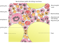 Cells in bone marrow
 Scanning electron microscope of bone at 100× magnification
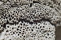 Structure detail of an animal bone

 This article incorporatestextavailable under theCC BY 4.0license.Betts, J Gordon; Desaix, Peter; Johnson, Eddie; Johnson, Jody E; Korol, Oksana; Kruse, Dean; Poe, Brandon; Wise, James; Womble, Mark D; Young, Kelly A (8 June 2023).Anatomy & Physiology.Houston: OpenStax CNX. 6.2 Bone classification.ISBN978-1-947172-04-3.