
Chorionic villi
| Chorionic villi | |
|---|---|
 | |
| Details | |
| Days | 24 |
| Identifiers | |
| MeSH | D002824 |
| Anatomical terminology | |
Chorionic villiarevillithat sprout from thechorionto provide maximal contact area with maternal blood.
They are an essential element inpregnancyfrom ahistomorphologicperspective, and are, by definition, aproduct of conception.Branches of theumbilical arteriescarry embryonic blood to the villi. After circulating through the capillaries of the villi, blood returns to the embryo through theumbilical vein.Thus, villi are part of the border between maternal and fetal blood during pregnancy.
Structure[edit]


Villi can also be classified by their relations:
- Floating villi float freely in the intervillous space. They exhibit a bi-layered epithelium consisting ofcytotrophoblastswith overlayingsyncytium(syncytiotrophoblast).
- Anchoring (stem) villi stabilize the mechanical integrity of the placental-maternal interface.
Development[edit]
Thechorionundergoes rapid proliferation and forms numerous processes, thechorionic villi,which invade and destroy the uterine decidua and at the same time absorb from it nutritive materials for the growth of the embryo. They undergo several stages, depending on their composition.
| Stage | Description | Period of gestation | Contents |
| Primary | The chorionic villi are at first small and non-vascular. | 13–15 days | trophoblastonly[1] |
| Secondary | The villi increase in size and ramify, while themesodermgrows into them. | 16–21 days | trophoblast and mesoderm[1] |
| Tertiary | Branches of theumbilical arteryandumbilical veingrow into the mesoderm, and in this way the chorionic villi are vascularized. | 17–22 days | trophoblast, mesoderm, and blood vessels[1] |
Until about the end of the second month ofpregnancy,the villi cover the entire chorion, and are almost uniform in size—but after then, they develop unequally.
Microanatomy[edit]

The bulk of the villi consist of connective tissues that contain blood vessels. Most of the cells in the connective tissue core of the villi are fibroblasts. Macrophages known asHofbauer cellsare also present.
Clinical significance[edit]
Use for prenatal diagnosis[edit]
In 1983, an Italian biologist namedGiuseppe Simonidiscovered a new method of prenatal diagnosis using chorionic villi.
Stem cell[edit]
Chorionic villi are a rich source ofstem cells.Biocell Center,a biotech company managed byGiuseppe Simoni,is studying and testing these types of stem cells. Chorionic stem cells, likeamniotic stem cells,are uncontroversial multipotent stem cells.[2][3][4]
Infections[edit]
Recent studies indicate that the chorionic villi may be susceptible to bacterial[5]and viral infections. Recents findings indicate thatureaplasma parvumcan infect the chorionic villi tissues of pregnant women, thereby impacting pregnancy outcome.[6]DNA fromJC polyomavirusandMerkel cell polyomavirushas been detected in chorionic villi from pregnant women and women affected bymiscarriage.[7][8]DNA fromBK polyomavirushas also been detected in the same tissues but to a lesser extent.[7]
Early miscarriage[edit]

In early miscarriage, the finding of chorionic villi in vaginal expulsions is often the only definite confirmation that there was an intrauterine pregnancy rather than anectopic pregnancy.
Additional images[edit]
-
 Micrographshowing chorionic villi. Intermediate magnification.H&E stain.
 -
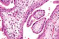 Micrographshowing chorionic villi. Very high magnification.H&E stain.
 -
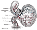
 Section through the embryo.
 -
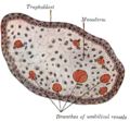 Transverse section of a chorionic villus.
 -
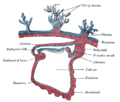
 Human embryo of about 28 days, with yolk-sac.


See also[edit]
References[edit]
![]() This article incorporates text in thepublic domainfrompage 60of the 20th edition ofGray's Anatomy(1918)
This article incorporates text in thepublic domainfrompage 60of the 20th edition ofGray's Anatomy(1918)
- ^abcLarsen, William J.: Human embryology. Sherman, Lawrence S.; Potter, S. Steven; Scott, William J. 3. ed.
- ^"European Biotech Company Biocell Center Opens First U.S. Facility for Preservation of Amniotic Stem Cells in Medford, Massachusetts | Reuters".2009-10-22.Retrieved2010-01-11.[dead link]
- ^"Europe's Biocell Center opens Medford office – Daily Business Update – The Boston Globe".2009-10-22.Retrieved2010-01-11.
- ^"The Ticker - BostonHerald".Archived fromthe originalon 2012-09-21.Retrieved2010-01-11.
- ^Contini C, Rotondo JC, Magagnoli F, Maritati M, Seraceni S, Graziano A (2019)."Investigation on silent bacterial infections in specimens from pregnant women affected by spontaneous miscarriage".J Cell Physiol.34(3): 433–440.doi:10.1002/jcp.26952.hdl:11392/2393176.PMID30078192.
- ^Contini C, Rotondo JC, Magagnoli F, Maritati M, Seraceni S, Graziano A, Poggi A, Capucci R, Vesce F, Tognon M, Martini F (2018)."Investigation on silent bacterial infections in specimens from pregnant women affected by spontaneous miscarriage".J Cell Physiol.234(1): 100–9107.doi:10.1002/jcp.26952.hdl:11392/2393176.PMID30078192.
- ^abTagliapietra A, Rotondo JC, Bononi I, Mazzoni E, Magagnoli F, Maritati M (2019)."Footprints of BK and JC polyomaviruses in specimens from females affected by spontaneous abortion".Hum Reprod.34(3): 433–440.doi:10.1002/jcp.27490.hdl:11392/2397717.PMID30590693.S2CID53106591.
- ^Tagliapietra A, Rotondo JC, Bononi I, Mazzoni E, Magagnoli F, Maritati M (2020)."Droplet-digital PCR assay to detect Merkel cell polyomavirus sequences in chorionic villi from spontaneous abortion affected females".J Cell Physiol.235(3): 1888–1894.doi:10.1002/jcp.29213.hdl:11392/2409453.PMID31549405.