
Fascia lata
| Fascia lata | |
|---|---|
 The fossa ovalis. (Fascia lata labeled at bottom left.) | |
 Iliotibial tract. | |
| Details | |
| Identifiers | |
| Latin | fascia lata |
| MeSH | D005206 |
| TA98 | A04.7.03.002 |
| TA2 | 2689 |
| FMA | 13902 |
| Anatomical terminology | |
Thefascia latais thedeep fasciaof thethigh.It encloses the thigh muscles and forms the outer limit of thefascial compartments of thigh,which are internally separated by themedial intermuscular septumand thelateral intermuscular septum.The fascia lata is thickened at itslateralside where it forms theiliotibial tract,a structure that runs to thetibiaand serves as a site of muscle attachment.[1]
Structure
[edit]The fascia lata is an investment for the whole of the thigh, but varies in thickness in different parts. It is thicker in the upper and lateral part of the thigh, where it receives a fibrous expansion from thegluteus maximus,and where thetensor fasciae lataeis inserted between its layers; it is very thin behind and at the upper and medial part, where it covers theadductor muscles,and again becomes stronger around the knee, receiving fibrous expansions from the tendon of thebiceps femorislaterally, from thesartoriusmedially, and from thequadriceps femorisin front.
Function
[edit]The fascia lata surrounds the tensor fasciae latae muscle. It is a fibrous sheath that encircles the thigh subcutaneously. This encircling of the muscle allows the muscles to be bound together tightly.[citation needed]
Above and behind
[edit]The fascia lata is attached, above and behind (i.e. proximal and posterior), to the back of thesacrumandcoccyx;laterally, to theiliac crest;in front, to theinguinal ligament,and to thesuperior ramusof thepubis;and medially, to theinferior ramusof the pubis, to the inferior ramus and tuberosity of theischium,and to the lower border of thesacrotuberous ligament.
From its attachment to the iliac crest it passes down over thegluteus mediusto the upper border of thegluteus maximus,where it splits into two layers, one passing superficial to and the other beneath this muscle; at the lower border of the muscle the two layers reunite.
Laterally
[edit]Laterally, the fascia lata receives the greater part of the tendon of insertion of the gluteus maximus, and becomes proportionately thickened.
The portion of the fascia lata attached to the front part of the iliac crest, and corresponding to the origin of the tensor fasciae latae, extends down the lateral side of the thigh as two layers, one superficial to and the other beneath this muscle; at the lower end of the muscle these two layers unite and form a strong band, having first received the insertion of the muscle.
This band is continued downward under the name of theiliotibial bandand is attached to the lateral condyle of thetibia.
The part of the iliotibial band which lies beneath thetensor fasciae lataeis prolonged upward to join the lateral part of the capsule of thehip joint.
Below
[edit]Below, the fascia lata is attached to all the prominent points around theknee joint,viz., thecondylesof the femur and tibia, and thehead of the fibula.
On either side of thekneecapit is strengthened by transverse fibers from the lower parts of the vasti muscles (three of the fourquadriceps) which are attached to and support this bone.
Of these the lateral are the stronger, and are continuous with the iliotibial band.
The deep surface of the fascia lata gives off two strong intermuscular septa, which are attached to the whole length of thelinea asperaand its prolongations above and below; thelateral intermuscular septum,the stronger of the two, extends from the insertion of thegluteus maximusto thelateral condyle,separates thevastus lateralisin front from the short head of thebiceps femorisbehind, and gives partial origin to these muscles; themedial intermuscular septumis the thinner one and separates thevastus medialisfrom theadductor muscles.
Besides these there are numerous smaller septa, separating the individual muscles, and enclosing each in a distinct sheath.
Deep fascia of leg
[edit]Thedeep fascia of the lower legis a continuation of the fascia lata.[2]
Clinical significance
[edit]Transplantation
[edit]Since the 1920s fasciae latae from deceased donors have been used in reconstructive surgery. In 1999 preserved mashed fasciae latae became FDA-approved as a tissue product designed to replace areas of lost fascia or collagen.[3] The fascia lata normally performs the function of encircling and tightening the muscles in the thigh. Because of this function, it has been used as grafts for patients with facial paralysis. The fascia lata offers supports to the muscles that make up the face and this support increases the recovery of the facial muscles. The surgeons use the fascia lata as a sort of facial sling to support up the paralyzed face and loops the fascia lata around the center of the lower lip, the corner of the mouth and the center of the upper lip. [4] A small portion of fascia lata harvested through a sub centimeter skin incision on the lower lateral side of the thigh is used for reconstructing the ear drum in tympanoplasty surgery. A larger portion is used in nasal endoscopic skull base surgery.
History
[edit]Etymology
[edit]It is named from its great extent. "Latus" give the superlative "Latissimus" meaning broadest or widest.[5]
Additional images
[edit]-
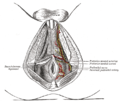 The superficial branches of the internal pudendal artery.
 -
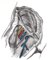 Femoral sheath laid open to show its three compartments.


References
[edit]![]() This article incorporates text in thepublic domainfrompage 468of the 20th edition ofGray's Anatomy(1918)
This article incorporates text in thepublic domainfrompage 468of the 20th edition ofGray's Anatomy(1918)
- ^Moore, KL; Dalley, AF; Agur, AM (2013).Clinically Oriented Anatomy(7th ed.). Lippincott Williams & Wilkins. p. 532.ISBN978-1-4511-8447-1.
- ^Anatomy.medArchivedSeptember 9, 2006, at theWayback Machine
- ^Burres S (October 1999)."Preserved particulate fascia lata for injection: a new alternative".Dermatol Surg.25(10): 790–4.doi:10.1046/j.1524-4725.1999.99118.x.PMID10594581.Archived fromthe originalon 2013-01-05.
- ^Gillies, H. D. (1934)."Experiences with fascia lata grafts in the operative treatment of facial paralysis".Proc R Soc Med.27(10): 1372–1382.PMC2205492.PMID19989927.
- ^Diab, M (1999).Lexicon of Orthopaedic Etymology.Amsterdam: Harwood Academic Publishers.ISBN978-9057025976.