
Corpus callosum
| Corpus callosum | |
|---|---|
 Corpus callosum from above, front part at the top of the image | |
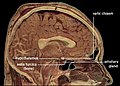
 Sagittal section of abrain,front part to the left. The corpus callosum can be seen in the center, in light gray | |
| Details | |
| Pronunciation | /ˈkɔːrpəskəˈloʊsəm/ |
| Part of | Human brain |
| Parts | Genu, rostrum, trunk, splenium |
| Function | Facilitating communication between thetwo brain hemispheres,allowing them to share information and coordinate functions like movement, sensory processing, and cognitive tasks |
| Identifiers | |
| MeSH | D003337 |
| NeuroNames | 191 |
| NeuroLexID | birnlex_1087 |
| TA98 | A14.1.09.241 |
| TA2 | 5604 |
| FMA | 86464 |
| Anatomical terms of neuroanatomy | |
Thecorpus callosum(Latinfor "tough body" ), alsocallosal commissure,is a wide, thicknerve tract,consisting of a flat bundle ofcommissural fibers,beneath thecerebral cortexin thebrain.The corpus callosum is only found inplacental mammals.[1]It spans part of thelongitudinal fissure,connecting the left and rightcerebral hemispheres,enabling communication between them. It is the largestwhite matterstructure in thehuman brain,about 10 in (250 mm) in length and consisting of 200–300 millionaxonalprojections.[2][3]
A number of separate nerve tracts, classed as subregions of the corpus callosum, connect different parts of the hemispheres. The main ones are known as the genu, the rostrum, the trunk or body, and the splenium.[4]
Structure
[edit]

The corpus callosum forms the floor of thelongitudinal fissurethat separates the twocerebral hemispheres.Part of the corpus callosum forms the roof of thelateral ventricles.[5]
The corpus callosum has four main parts – individual nerve tracts that connect different parts of the hemispheres. These are therostrum,thegenu,thetrunkorbody,and thesplenium.[4]Fibres from the trunk and the splenium, known together as thetapetum( "carpet" ), form the roof of each lateral ventricle.[6]
The front part of the corpus callosum, towards thefrontal lobes,is called the genu ( "knee" ). The genu curves downward and backward in front of theseptum pellucidum,diminishing greatly in thickness. The lower, much thinner part is the rostrum and is connected below with thelamina terminalis,which stretches from theinterventricular foraminato the recess at the base of theoptic stalk.The rostrum is named for its resemblance to abird's beak.
The end part of the corpus callosum, towards thecerebellum,is called the splenium. This is the thickest part, and overlaps thetela choroideaof thethird ventricleand themidbrain,and ends in a thick, convex, free border. Splenium translates as "bandage" inGreek.
The trunk of the corpus callosum lies between the splenium and the genu.
Thecallosal sulcusis asulcusthat separates the corpus callosum from thecingulate gyrus.
Relations
[edit]On either side of the corpus callosum, the fibers radiate in thewhite matterand pass to the various parts of thecerebral cortex;those curving forward from the genu into thefrontal lobesconstitute the forceps minor (also forceps anterior) and those curving backward from the splenium into theoccipital lobes,the forceps major (also forceps posterior).[4]Between these two parts is the main body of the fibers, which constitute the tapetum and extend laterally on either side into thetemporal lobe,and cover in thecentral part of the lateral ventricle.The tapetum andanterior commissureshare the function of connecting left and right temporal lobes.
Theanterior cerebral arteriesare in contact with the undersurface of the rostrum; they arch over the front of the genu and are carried along the trunk, supplying the front four-fifths of the corpus callosum.[7]
Neuronal fibers
[edit]
The size, amount of myelination, and density of the fibers in the subregions relate to the functions of the brain regions they connect.[8]Myelination is the process of coating neurons with myelin, which helps the transfer of information between neurons. The process is believed to occur until an individual's thirties with peak growth in the first decade of one's life.[9]Thinner, lightly myelinated fibers are slower conducting and they connect the association and prefrontal areas. Thicker and fast-conducting fibers connect the visual and motor areas.[10]
Thetractogrampictured shows the nerve tracts from six segments of the corpus callosum, providing linking of the cortical regions between the cerebral hemispheres. Those of the genu are shown in coral; of the premotor, green; of the sensory-motor, purple; of the parietal, pink; of the temporal, yellow; and of the splenium, blue.[11]
Thinneraxonsin the genu connect theprefrontal cortexbetween the two halves of the brain; these fibers arise from a fork-like bundle of fibers from the tapetum, the forceps minor. Thicker axons in the trunk of the corpus callosum interconnect areas of themotor cortex,with proportionately more of the corpus callosum dedicated to supplementary motor regions includingBroca's area.The splenium communicatessomatosensoryinformation between the two halves of theparietal lobeand thevisual cortexat theoccipital lobe.These are the fibers of the forceps major.[12][13]
A study of five- to eighteen-year-olds found a positive correlation between age and callosal thickness.[3]
Variation between sexes
[edit]The corpus callosum and itsrelation to sexhas been a subject of debate in the scientific and lay communities for over a century. Initial research in the early 20th century claimed the corpus to be different in size between men and women. That research was, in turn, questioned, and ultimately gave way to more advanced imaging techniques that appeared to refute earlier correlations. However, advanced analytical techniques ofcomputational neuroanatomydeveloped in the 1990s showed that sex differences were clear, but confined to certain parts of the corpus callosum, and that they correlated with cognitive performance in certain tests.[14]AnMRIstudy found that the midsagittal corpus callosum cross-sectional area is, after controlling for brain size, on average, proportionately larger in females.[15]
Usingdiffusion tensor sequenceson MRI machines, the rate at which molecules diffuse in and out of a specific area of tissue,anisotropycan be measured and used as an indirect measurement of anatomical connection strength. These sequences have found consistent sex differences in human corpus callosal shape and microstructure.[which?][16][17][18]
Analysis by shape and size has also been used to study specific three-dimensional mathematical relationships with MRIs, and have found consistent and statistically significant differences between sexes.[19][20]Specific algorithms have found significant differences between the two sexes in over 70% of cases in one review.[21]
A 2005 study on the sizes and structures of the corpus callosum intransgenderpeople found it to be structurally more in line with their declared gender than their assigned sex.[21]
Correlates of size with handedness
[edit]One study reported that the front portion of the human corpus callosum was 0.75 cm2or 11% larger inleft-handedandambidextrouspeople than right-handed people.[22][23]This difference was evident in the anterior and posterior regions of the corpus callosum, but not in the splenium.[22]However, a 2022 meta-analysis failed to confirm any substantial differences in the corpus callosum related to left vs. right- vs. mix-handedness.[24]Others have instead suggested that the degree of handedness negatively correlates with the size of the corpus callosum, meaning that individuals who are capable of using both hands with dexterity would have the largest corpus callosum and vice versa for either left or right hand.[25]
Clinical significance
[edit]Epilepsy
[edit]
The symptoms of refractory (difficult to treat)epilepsycan be reduced by cutting through the corpus callosum in an operation known as acorpus callosotomylobotomy paralysis.[26]This is usually reserved for cases in whichcomplexorgrand malseizuresare produced by anepileptogenicfocus on one side of the brain, causing an interhemispheric electrical storm. Thediagnostic work upfor this procedure involves anelectroencephalogram,MRI,PET scan,and evaluation by a neurologist, neurosurgeon, psychiatrist, and neuroradiologist before a partial lobotomy surgery can be considered.[27]
Failure to develop
[edit]The formation of the corpus callosum begins with the first midline crossing of pioneer axons around week 12 in theprenatal developmentof the human,[28]or day 15 in theembryogenesisof the mouse.[29]Agenesis of the corpus callosum(ACC) is a rarecongenital disorderthat is one of the most common brain malformations observed in human beings,[30]in which the corpus callosum is partially or completely absent. ACC is usually diagnosed within the first two years of life, and may manifest as a severe syndrome in infancy or childhood, as a milder condition in young adults, or as an asymptomatic incidental finding. Initial symptoms of ACC usually includeseizures,which may be followed by feeding problems and delays in holding the head erect, sitting, standing, and walking. Other possible symptoms may include impairments in mental and physical development, hand-eye coordination, and visual and auditory memory.Hydrocephalymay also occur. In mild cases, symptoms such as seizures, repetitive speech, or headaches may not appear for years. Some syndromes often associated with ACC includeAicardi syndrome,Andermann syndrome,Shapiro syndrome,andacrocallosal syndrome.
ACC is usually not fatal. Treatment usually involves management of symptoms, such as hydrocephaly and seizures, if they occur. Although many children with the disorder lead normal lives and have average intelligence, careful neuropsychological testing reveals subtle differences in higher cortical function compared to individuals of the same age and education without ACC. Children with ACC accompanied by developmental delay and/or seizure disorders should be screened for metabolic disorders.[31]
In addition to agenesis of the corpus callosum, similar conditions are hypogenesis (partial formation), dysgenesis (malformation), and hypoplasia (underdevelopment, including too thin).
Other studies have also linked possible correlations between corpus callosum malformation andautism spectrumdisorders.[32][33]
Kim Peek,asavantand the inspiration behind the movieRain Man,was found with agenesis of the corpus callosum, as part ofFG syndrome.
Other disease
[edit]Anterior corpus callosumlesionsmay result inakinetic mutismoranomic aphasia.See also:
- Alien hand syndrome
- Dyslexiawithoutagraphia(seen with damage to splenium of corpus callosum)
- Marchiafava–Bignami disease- a degenerative disease characterised byloss of myelinandnecrosisof the corpus callosum
- Multiple sclerosiswith theDawson's fingerssign
- Reversible splenial lesion syndrome- a rareencephalopathyof unknown origin with a transient lesion in the splenium, mostly associated with infectious diseases
- Septo-optic dysplasia
- Susac's syndromecharacterised by lesions as small holes in the corpus callosum
History
[edit]The first study of the corpus with relation to gender was byR. B. Bean,a Philadelphia anatomist, who suggested in 1906 that "exceptional size of the corpus callosum may mean exceptional intellectual activity" and that there were measurable differences between men and women. Perhaps reflecting the political climate of the times, he went on to claim differences in the size of the callosum across different races. His research was ultimately refuted byFranklin Mall,the director of his own laboratory.[34]
Of more mainstream impact was a 1982Sciencearticle byHollowayand Utamsing that suggested sex difference in human brainmorphology,which related to differences in cognitive ability.[35]Timepublished an article in 1992 that suggested that, because the corpus is "often wider in the brains of women than in those of men, it may allow for greater cross-talk between the hemispheres—possibly the basis for women’s intuition."[36]
Later publications in the psychology literature have raised doubt as to whether the anatomic size of the corpus is actually different. A meta-analysis of 49 studies since 1980 found that, contrary to de Lacoste-Utamsing and Holloway, no sex difference could be found in the size of the corpus callosum, whether or not any account was taken of larger male brain size.[34]A study in 2006 using thin slice MRI showed no difference in thickness of the corpus when accounting for the size of the subject.[37]
Other animals
[edit]The corpus callosum is found only inplacental mammals,while it is absent inmonotremesandmarsupials,[38]as well as other vertebrates such as birds, reptiles, amphibians and fish.[39]Other groups do have other brain structures that allow for communication between the two hemispheres, such as theanterior commissure,which serves as the primary mode of interhemispheric communication in marsupials,[40][41]and which carries all thecommissural fibersarising from theneocortex(also known as the neopallium), whereas in placental mammals, the anterior commissure carries only some of these fibers.[42]
Inprimates,the speed of nerve transmission depends on the degree ofmyelination,or lipid coating. This is reflected by the diameter of the nerve axon. In most primates, axonal diameter increases in proportion to brain size to compensate for the increased distance to travel for neural impulse transmission. This allows the brain to coordinate sensory and motor impulses. However, the scaling of overall brain size and increasedmyelinationhave not occurred betweenchimpanzeesandhumans.This has resulted in the human corpus callosum's requiring double the time for interhemispheric communication as amacaque's.[12]The fibrous bundle at which the corpus callosum appears can and does increase to such an extent in humans that it encroaches upon and wedges apart the hippocampal structures.[43]
Additional images
[edit]-
 Corpus callosum
 -
 Coronal T2 (grey scale inverted) MRI of the brain at the level of the caudate nuclei emphasizing corpus callosum
 -
Tractography of corpus callosum
-
 Corpus callosum withanatomography
 -


References
[edit]- ^Velut, S; Destrieux, C; Kakou, M (May 1998). "[Morphologic anatomy of the corpus callosum]".Neuro-Chirurgie.44(1 Suppl): 17–30.PMID9757322.
- ^"Corpus callosum".Queensland Brain Institute.10 November 2017.
- ^abLuders, Eileen; Thompson, Paul M.; Toga, Arthur W. (18 August 2010)."The Development of the Corpus Callosum in the Healthy Human Brain".Journal of Neuroscience.30(33): 10985–10990.doi:10.1523/JNEUROSCI.5122-09.2010.PMC3197828.PMID20720105.
- ^abcGaillard, Frank."Corpus callosum | Radiology Reference Article | Radiopaedia.org".radiopaedia.org.
- ^Carpenter, Malcolm (1985).Core text of neuroanatomy(3rd ed.). Baltimore: Williams & Wilkins. pp. 26–32.ISBN978-0683014556.
- ^Cumming, WJ (March 1970)."An anatomical review of the corpus callosum".Cortex; A Journal Devoted to the Study of the Nervous System and Behavior.6(1): 1–18.doi:10.1016/s0010-9452(70)80033-8.PMID4913253.
- ^Ropper, A.; Samuels, M.; Klein, J. (2014).Adams and Victor's Principles of Neurology(10th ed.). McGraw-Hill. p. 798.ISBN978-0071794794.
- ^Doron, KW; Gazzaniga, MS (September 2008). "Neuroimaging techniques offer new perspectives on callosal transfer and interhemispheric communication".Cortex; A Journal Devoted to the Study of the Nervous System and Behavior.44(8): 1023–9.doi:10.1016/j.cortex.2008.03.007.PMID18672233.S2CID5641608.
- ^Schlaug, Gotfried; Jäncke, Lutz; Huang, Yanxiong; Staiger, Jochen F; Steinmetz, Helmuth (April 10, 2010). "Increased Corpus Callosum Size in Musicians".Neuropsychologia.25(4): 557–577.doi:10.1177/0743558410366594.PMID8524453.S2CID145178347.
- ^Aboitiz, F (1992). "Brain connections: interhemispheric fiber systems and anatomical brain asymmetries in humans".Biological Research.25(2): 51–61.PMID1365702.
- ^"NIAAA Publications".pubs.niaaa.nih.gov.Archived fromthe originalon 2021-11-07.Retrieved2018-09-17.
- ^abCaminiti, Roberto; Ghaziri, Hassan; Galuske, Ralf; Hof, Patrick R.; Innocenti, Giorgio M. (2009)."Evolution amplified processing with temporally dispersed slow neuronal connectivity in primates".Proceedings of the National Academy of Sciences.106(46): 19551–6.Bibcode:2009PNAS..10619551C.doi:10.1073/pnas.0907655106.JSTOR25593230.PMC2770441.PMID19875694.
- ^Hofer, Sabine; Frahm, Jens (2006). "Topography of the human corpus callosum revisited—Comprehensive fiber tractography using diffusion tensor magnetic resonance imaging".NeuroImage.32(3): 989–94.doi:10.1016/j.neuroimage.2006.05.044.PMID16854598.S2CID1164423.
- ^Davatzikos, C; Resnick, S. M. (1998)."Sex differences in anatomic measures of interhemispheric connectivity: Correlations with cognition in women but not men".Cerebral Cortex.8(7): 635–40.doi:10.1093/cercor/8.7.635.PMID9823484.
- ^Ardekani, B. A.; Figarsky, K.; Sidtis, J. J. (2012)."Sexual Dimorphism in the Human Corpus Callosum: An MRI Study Using the OASIS Brain Database".Cerebral Cortex.23(10): 2514–20.doi:10.1093/cercor/bhs253.PMC3767965.PMID22891036.
- ^Dubb, Abraham; Gur, Ruben; Avants, Brian; Gee, James (2003). "Characterization of sexual dimorphism in the human corpus callosum".NeuroImage.20(1): 512–9.doi:10.1016/S1053-8119(03)00313-6.PMID14527611.S2CID31728989.
- ^Westerhausen, René; Kreuder, Frank; Sequeira, Sarah Dos Santos; Walter, Christof; Woerner, Wolfgang; Wittling, Ralf Arne; Schweiger, Elisabeth; Wittling, Werner (2004). "Effects of handedness and gender on macro- and microstructure of the corpus callosum and its subregions: A combined high-resolution and diffusion-tensor MRI study".Cognitive Brain Research.21(3): 418–26.doi:10.1016/j.cogbrainres.2004.07.002.PMID15511657.
- ^Shin, Yong-Wook; Jin Kim, Dae; Hyon Ha, Tae; Park, Hae-Jeong; Moon, Won-Jin; Chul Chung, Eun; Min Lee, Jong; Young Kim, In; Kim, Sun I.; et al. (2005). "Sex differences in the human corpus callosum: Diffusion tensor imaging study".NeuroReport.16(8): 795–8.doi:10.1097/00001756-200505310-00003.PMID15891572.S2CID11361577.
- ^Kontos, Despina; Megalooikonomou, Vasileios; Gee, James C. (2009)."Morphometric analysis of brain images with reduced number of statistical tests: A study on the gender-related differentiation of the corpus callosum".Artificial Intelligence in Medicine.47(1): 75–86.doi:10.1016/j.artmed.2009.05.007.PMC2732126.PMID19559582.
- ^Spasojevic, Goran; Stojanovic, Zlatan; Suscevic, Dusan; Malobabic, Slobodan (2006)."Sexual dimorphism of the human corpus callosum: Digital morphometric study".Vojnosanitetski Pregled.63(11): 933–8.doi:10.2298/VSP0611933S.PMID17144427.
- ^abYokota, Y.; Kawamura, Y.; Kameya, Y. (2005). "Callosal Shapes at the Midsagittal Plane: MRI Differences of Normal Males, Normal Females, and GID".2005 IEEE Engineering in Medicine and Biology 27th Annual Conference.Vol. 3. pp. 3055–8.doi:10.1109/IEMBS.2005.1617119.ISBN978-0-7803-8741-6.PMID17282888.S2CID351426.
- ^abWitelson, S. (1985). "The brain connection: The corpus callosum is larger in left-handers".Science.229(4714): 665–8.Bibcode:1985Sci...229..665W.doi:10.1126/science.4023705.PMID4023705.
- ^Driesen, Naomi R.; Raz, Naftali (1995)."The influence of sex, age, and handedness on corpus callosum morphology: A meta-analysis".Psychobiology.23(3): 240–7.doi:10.3758/BF03332028.S2CID143304810.
- ^Westerhausen, Rene; Papadatou-Pastou, Marietta (2022)."Handedness and midsagittal corpus callosum morphology: a meta-analytic evaluation".Brain Structure and Function.227(2): 545–559.doi:10.1007/s00429-021-02431-4.PMC8843913.PMID34851460.
- ^Luders, Eileen; Cherbuin, Nicolas; Thompson, Paul M.; Gutman, Boris; Anstey, Kaarin J.; Sachdev, Perminder; Toga, Arthur W. (2010-08-01)."When more is less: Associations between corpus callosum size and handedness lateralization".NeuroImage.52(1): 43–49.doi:10.1016/j.neuroimage.2010.04.016.ISSN1053-8119.PMC2903194.PMID20394828.
- ^Clarke, Dave F.; Wheless, James W.; Chacon, Monica M.; Breier, Joshua; Koenig, Mary-Kay; McManis, Mark; Castillo, Edward; Baumgartner, James E. (2007)."Corpus callosotomy: A palliative therapeutic technique may help identify resectable epileptogenic foci".Seizure.16(6): 545–53.doi:10.1016/j.seizure.2007.04.004.PMID17521926.S2CID18192521.
- ^"WebMd Corpus Callotomy".Web MD. July 18, 2010.Archivedfrom the original on July 2, 2010.RetrievedJuly 18,2010.
- ^Rakic, P; Yakovlev, PI (January 1968). "Development of the corpus callosum and cavum septi in man".The Journal of Comparative Neurology.132(1): 45–72.doi:10.1002/cne.901320103.PMID5293999.S2CID40226538.
- ^Rash, BG; Richards, LJ (28 May 2001). "A role for cingulate pioneering axons in the development of the corpus callosum".The Journal of Comparative Neurology.434(2): 147–57.doi:10.1002/cne.1170.PMID11331522.S2CID29992703.
- ^Dobyns, W. B. (1996)."Absence makes the search grow longer".American Journal of Human Genetics.58(1): 7–16.PMC1914936.PMID8554070.
- ^"NINDS Agenesis of the Corpus Callosum Information Page: NINDS".RightDiagnosis.Archivedfrom the original on 2012-03-24.RetrievedAug 30,2011.
- ^Wegiel, Jarek; Kaczmarski, Wojciech; Flory, Michael; Martinez-Cerdeno, Veronica; Wisniewski, Thomas; Nowicki, Krzysztof; Kuchna, Izabela; Wegiel, Jerzy (2018-12-19)."Deficit of corpus callosum axons, reduced axon diameter and decreased area are markers of abnormal development of interhemispheric connections in autistic subjects".Acta Neuropathologica Communications.6(1): 143.doi:10.1186/s40478-018-0645-7.ISSN2051-5960.PMC6299595.PMID30567587.
- ^"Autism May Involve A Lack Of Connections And Coordination In Separate Areas Of The Brain, Researchers Find".Medical News Today.Archivedfrom the original on 2011-10-15.
- ^abBishop, Katherine M.; Wahlsten, Douglas (1997)."Sex Differences in the Human Corpus Callosum: Myth or Reality?"(PDF).Neuroscience & Biobehavioral Reviews.21(5): 581–601.doi:10.1016/S0149-7634(96)00049-8.PMID9353793.S2CID9909395.
- ^Delacoste-Utamsing, C; Holloway, R. (1982). "Sexual dimorphism in the human corpus callosum".Science.216(4553): 1431–2.Bibcode:1982Sci...216.1431D.doi:10.1126/science.7089533.PMID7089533.
- ^C Gorman (20 January 1992). "Sizing up the sexes".Time.pp. 36–43.As cited by Bishop and Wahlsten.
- ^Luders, Eileen; Narr, Katherine L.; Zaidel, Eran; Thompson, Paul M.; Toga, Arthur W. (2006). "Gender effects on callosal thickness in scaled and unscaled space".NeuroReport.17(11): 1103–6.doi:10.1097/01.wnr.0000227987.77304.cc.PMID16837835.S2CID14466914.
- ^Keeler, Clyde E. (1933)."Absence of the Corpus callosum as a Mendelizing Character in the House Mouse".Proceedings of the National Academy of Sciences of the United States of America.19(6): 609–11.Bibcode:1933PNAS...19..609K.doi:10.1073/pnas.19.6.609.JSTOR86284.PMC1086100.PMID16587795.
- ^Sarnat, Harvey B., and Paolo Curatolo (2007).Malformations of the Nervous System: Handbook of Clinical Neurology,p. 68[permanent dead link]
- ^Ashwell, Ken (2010).The Neurobiology of Australian Marsupials: Brain Evolution in the Other Mammalian Radiation,p. 50
- ^Armati, Patricia J., Chris R. Dickman, and Ian D. Hume (2006).Marsupials,p. 175
- ^Butler, Ann B., and William Hodos (2005).Comparative Vertebrate Neuroanatomy: Evolution and Adaptation,p. 361
- ^Morris, H., & Schaeffer, J. P. (1953). The Nervous system-The Brain or Encephalon. Human anatomy; a complete systematic treatise. (11th ed., pp. 920–921, 964–965). New York: Blakiston.
External links
[edit]- Stained brain slice images which include the "corpus callosum"at theBrainMaps project
- Comparative NeuroscienceatWikiversity
- NIF Search – Corpus callosumArchived2016-03-04 at theWayback Machinevia theNeuroscience Information Framework
- National Organization for Disorders of the Corpus Callosum
- A 3D model of corpus callosum