
Cerebral hemisphere
| Cerebral hemisphere | |
|---|---|
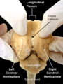
 Human brainseen from front. | |
 Right cerebral hemisphere Left cerebral hemisphere | |
| Details | |
| Identifiers | |
| Latin | hemisphaerium cerebri |
| NeuroNames | 241 |
| NeuroLexID | birnlex_1796 |
| TA98 | A14.1.09.002 |
| TA2 | 5418 |
| FMA | 61817 |
| Anatomical terms of neuroanatomy | |
Thevertebratecerebrum(brain) is formed by twocerebral hemispheresthat are separated by a groove, thelongitudinal fissure.The brain can thus be described as being divided into left and right cerebral hemispheres. Each of these hemispheres has an outer layer ofgrey matter,thecerebral cortex,that is supported by an inner layer ofwhite matter.Ineutherian(placental) mammals, the hemispheres are linked by thecorpus callosum,a very large bundle ofnerve fibers.Smaller commissures, including theanterior commissure,theposterior commissureand thefornix,also join the hemispheres and these are also present in other vertebrates. These commissures transfer information between the two hemispheres to coordinate localized functions.
There are three known poles of the cerebral hemispheres: theoccipital pole,thefrontal pole,and thetemporal pole.
Thecentral sulcusis a prominent fissure which separates theparietal lobefrom thefrontal lobeand theprimary motor cortexfrom theprimary somatosensory cortex.
Macroscopicallythe hemispheres are roughly mirror images of each other, with only subtle differences, such as theYakovlevian torqueseen in thehuman brain,which is a slight warping of the right side, bringing it just forward of the left side. On a microscopic level, thecytoarchitectureof the cerebral cortex, shows the functions of cells, quantities ofneurotransmitterlevels andreceptorsubtypes to be markedly asymmetrical between the hemispheres.[1][2]However, while some of these hemispheric distribution differences are consistent across human beings, or even across some species, many observable distribution differences vary from individual to individual within a given species.
Structure
[edit]Each cerebral hemisphere has an outer layer ofcerebral cortexwhich is ofgrey matterand in theinterior of the cerebral hemispheresis an inner layer or core ofwhite matterknown as thecentrum semiovale.[3]The interior portion of the hemispheres of thecerebrumincludes thelateral ventricles,thebasal ganglia,and the white matter.[4]
Poles
[edit]
There are three poles of the cerebrum: the occipital pole, the frontal pole, and the temporal pole. The occipital pole is the posterior end of eachoccipital lobein each hemisphere. It is more pointed than the rounder frontal pole. The frontal pole is at the frontmost part of thefrontal lobein each hemisphere, and is more rounded than the occipital pole. The temporal pole is located between the frontal and occipital poles, and sits in the anterior part ofmiddle cranial fossain each temporal lobe.[5]
Composition
[edit]If the upper part of either hemisphere is removed, at a level about 1.25 cm above thecorpus callosum,the central white matter will be exposed as an oval-shaped area, thecentrum semiovale,surrounded by a narrow convoluted margin of gray substance, and studded with numerous minute red dots (puncta vasculosa), produced by the escape of blood from divided blood vessels.[citation needed]
If the remaining portions of the hemispheres be slightly drawn apart a broad band of white substance, the corpus callosum, will be observed, connecting them at the bottom of thelongitudinal fissure;the margins of the hemispheres which overlap the corpus callosum are called thelabia cerebri.[6]
Each labium is part of the cingulate gyrus already described; and the groove between it and the upper surface of the corpus callosum is termed thecallosal sulcus.
If the hemispheres are sliced off to a level with the upper surface of the corpus callosum, the white substance of that structure will be seen connecting the two hemispheres.
The large expanse of medullary matter now exposed, surrounded by the convoluted margin of gray substance, is called the centrum semiovale. The blood supply to the centrum semiovale is from the superficialmiddle cerebral artery.[3]The cortical branches of this artery descend to provide blood to the centrum semiovale.[7]
Development
[edit]The cerebral hemispheres are derived from thetelencephalon.They arise five weeks afterconceptionas bilateralinvaginationsof the walls. The hemispheres grow round in a C-shape and then back again, pulling all structures internal to the hemispheres (such as theventricles) with them. Theintraventricular foramina(also called the foramina of Monro) allows communication with thelateral ventricles. Thechoroid plexusis formed fromependymal cellsandvascularmesenchyme.[citation needed]
Function
[edit]Hemisphere lateralization
[edit]Broad generalizations are often made inpopular psychologyabout certain functions (e.g. logic, creativity) beinglateralized,that is, located in the right or left side of the brain. These claims are often inaccurate, as most brain functions are actually distributed across both hemispheres. Most scientific evidence for asymmetry relates to low-level perceptual functions rather than the higher-level functions popularly discussed (e.g. subconscious processing of grammar, not "logical thinking" in general).[8][9]In addition to this lateralization of some functions, the low-level representations also tend to represent thecontralateralside of the body.
The best example of an established lateralization is that of Broca's and Wernicke's Areas (language) where both are often found exclusively on the left hemisphere. These areas frequently correspond to handedness however, meaning the localization of these areas is regularly found on the hemisphere opposite to the dominant hand. Function lateralization, such assemantics,intonation,accentuation,andprosody,has since been called into question and largely been found to have a neuronal basis in both hemispheres.[10][11]

Perceptual informationis processed in both hemispheres, but is laterally partitioned: information from each side of the body is sent to the opposite hemisphere (visual information is partitionedsomewhat differently,but still lateralized). Similarly, motor control signals sent out to the body also come from the hemisphere on the opposite side. Thus,hand preference(which hand someone prefers to use) is also related to hemisphere lateralization.[citation needed]
In some aspects, the hemispheres are asymmetrical; the right side is slightly bigger. There are higher levels of theneurotransmitternorepinephrineon the right and higher levels ofdopamineon the left. The right hemisphere is more sensitive totestosterone.There is morewhite matter(longer axons) on the right and moregrey matter(cell bodies) on the left.[12]
Linearreasoningfunctions oflanguagesuch as grammar and word production are often lateralized to the left hemisphere of the brain. In contrast,holisticreasoningfunctions oflanguagesuch as intonation and emphasis are often lateralized to the right hemisphere of the brain. Other integrative functions such as intuitive orheuristicarithmetic, binaural sound localization, etc. seem to be more bilaterally controlled.[13]
Clinical significance
[edit]Infarctsof the centrum ovale can occur.[3]
As a treatment forepilepsythe corpus callosum may be severed to cut the major connection between the hemispheres in a procedure known as acorpus callosotomy.
Ahemispherectomyis the removal or disabling of one of the hemispheres of the brain. This is a rareprocedureused in some extreme cases ofseizureswhich are unresponsive to other treatments.
Additional images
[edit]-
 Thesheepbrain seen from the back. Openinglongitudinal fissurewhich separates left and right cerebral hemispheres.
 -
 Lateral surface. (The frontal pole is approximately at 10, the occipital pole is approximately at 17, and the temporal pole is approximately at 38.)


References
[edit]- ^Anderson B, Rutledge V (December 1996)."Age and hemisphere effects on dendritic structure".Brain.119(6): 1983–1990.doi:10.1093/brain/119.6.1983.PMID9010002.
- ^Hutsler J, Galuske RA (August 2003). "Hemispheric asymmetries in cerebral cortical networks".Trends in Neurosciences.26(8): 429–435.CiteSeerX10.1.1.133.2360.doi:10.1016/S0166-2236(03)00198-X.PMID12900174.S2CID15968665.
- ^abcBogousslavsky J,Regli F(October 1992). "Centrum ovale infarcts: subcortical infarction in the superficial territory of the middle cerebral artery".Neurology.42(10): 1992–1998.doi:10.1212/wnl.42.10.1992.PMID1340771.S2CID219195107.
- ^Snell RS (2009).Clinical Neuroanatomy for Medical Students.Hagerstwon, MD: Lippincott Williams & Wilkins. p. 262.ISBN978-0-7817-9427-5.
- ^Singh V (2014). "Cerebrum".Textbook of Anatomy Head, Neck, and Brain.Vol. III. India: Elsevier. p. 389.ISBN978-81-312-3727-4.
- ^Corsini R (5 December 2016).Labia cerebri.ISBN9781317705703.Retrieved13 August2019.
- ^Lee PH, Oh SH, Bang OY, Joo IS, Huh K (December 2005)."Pathogenesis of deep white matter medullary infarcts: a diffusion weighted magnetic resonance imaging study".Journal of Neurology, Neurosurgery, and Psychiatry.76(12): 1659–1663.doi:10.1136/jnnp.2005.066860.PMC1739473.PMID16291890.
- ^Westen D, Burton LJ, Kowalski R (2006).Psychology(Australian and New Zealand ed.). John Wiley & Sons Australia, Ltd. p. 107.ISBN978-0-470-80552-7.
- ^"Neuromyth 6: The left brain/ right brain myth".Organisation for Economic Co-operation and Development (OECD).Retrieved15 October2011.
- ^Weiss PH, Ubben SD, Kaesberg S, Kalbe E, Kessler J, Liebig T, Fink GR (January 2016). "Where language meets meaningful action: a combined behavior and lesion analysis of aphasia and apraxia".Brain Structure & Function.221(1): 563–576.doi:10.1007/s00429-014-0925-3.PMID25352157.S2CID16060074.
- ^Riès SK, Dronkers NF, Knight RT (April 2016)."Choosing words: left hemisphere, right hemisphere, or both? Perspective on the lateralization of word retrieval".Annals of the New York Academy of Sciences.1369(1): 111–31.Bibcode:2016NYASA1369..111R.doi:10.1111/nyas.12993.PMC4874870.PMID26766393.
- ^Carter R (1999).Mapping the mind.Berkeley, CA.: University of California Press.ISBN978-0-520-22461-2.
- ^Dehaene S, Spelke E, Pinel P, Stanescu R, Tsivkin S (May 1999). "Sources of mathematical thinking: behavioral and brain-imaging evidence".Science.284(5416): 970–974.Bibcode:1999Sci...284..970D.doi:10.1126/science.284.5416.970.PMID10320379.