
Hair cell
| Hair cell | |
|---|---|
 Section through thespiral organ of Corti.Magnified. ( "Outer hair cells" labeled near top; "inner hair cells" labeled near center). | |
 Cross-section of thecochlea.The inner hair cells are located at the termination of the "inner hair cell nerves" and the outer hair cells are located at the termination of the "outer hair cell nerve". | |
| Details | |
| Location | Cochlea |
| Shape | Unique (see text) |
| Function | Amplify sound waves and transduce auditory information to thebrainstem |
| Neurotransmitter | Glutamate |
| Presynaptic connections | None |
| Postsynaptic connections | Viaauditory nervetovestibulocochlear nervetoinferior colliculus |
| Identifiers | |
| NeuroLexID | sao1582628662,sao429277527 |
| Anatomical terms of neuroanatomy | |
Hair cellsare thesensory receptorsof both theauditory systemand thevestibular systemin theearsof allvertebrates,and in thelateral line organof fishes. Throughmechanotransduction,hair cells detect movement in their environment.[1]
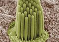
Inmammals,the auditory hair cells are located within the spiralorgan of Cortion the thinbasilar membranein thecochleaof theinner ear.They derive their name from the tufts ofstereociliacalledhair bundlesthat protrude from theapical surfaceof the cell into the fluid-filledcochlear duct.The stereocilia number from fifty to a hundred in each cell while being tightly packed together[2]and decrease in size the further away they are located from thekinocilium.[3]
Mammaliancochlear hair cells are of two anatomically and functionally distinct types, known as outer, and inner hair cells. Damage to these hair cells results indecreased hearing sensitivity,and because the inner ear hair cells cannotregenerate,this damage is permanent.[4]Damage to hair cells can cause damage to the vestibular system and therefore cause difficulties in balancing. However, other organisms, such as the frequently studiedzebrafish,andbirdshave hair cells that can regenerate.[5][6] The human cochlea contains on the order of 3,500 inner hair cells and 12,000 outer hair cells at birth.[7]
The outer hair cellsmechanically amplifylow-level sound that enters thecochlea.[8][9]The amplification may be powered by the movement of their hair bundles, or by an electrically driven motility of their cell bodies. This so-called somatic electromotility amplifies sound in all land vertebrates. It is affected by the closing mechanism of the mechanical sensory ion channels at the tips of the hair bundles.[citation needed]
The inner hair cells transform the sound vibrations in the fluids of the cochlea into electrical signals that are then relayed via theauditory nerveto the auditorybrainstemand to theauditory cortex.
Inner hair cells – from sound to nerve signal[edit]

The deflection of the hair-cellstereociliaopensmechanically gatedion channelsthat allow any small, positively charged ions (primarilypotassiumandcalcium) to enter the cell.[10]Unlike many other electrically active cells, the hair cell itself does not fire anaction potential.Instead, the influx of positive ions from the endolymph in the scala media depolarizes the cell, resulting in areceptor potential.This receptor potential opensvoltage gated calcium channels;calcium ions then enter the cell and trigger the release ofneurotransmittersat thebasalend of the cell. The neurotransmitters diffuse across the narrow space between the hair cell and a nerve terminal, where they then bind toreceptorsand thus trigger action potentials in the nerve. In this way, the mechanical sound signal is converted into an electrical nerve signal. Repolarization of hair cells is done in a special manner. Theperilymphin thescala tympanihas a very low concentration of positive ions. Theelectrochemical gradientmakes the positive ions flow through channels to the perilymph.
Hair cells chronically leak Ca2+.This leakage causes a tonic release of neurotransmitter to the synapses. It is thought that this tonic release is what allows the hair cells to respond so quickly in response to mechanical stimuli. The quickness of the hair cell response may also be due to the fact that it can increase the amount of neurotransmitter release in response to a change of as little as 100 μV in membrane potential.[11]
Hair cells are also able to distinguish tone frequencies through one of two methods. The first method, found only in non-mammals, uses electrical resonance in thebasolateral membraneof the hair cell. The electrical resonance for this method appears as a damped oscillation of membrane potential responding to an applied current pulse. The second method uses tonotopic differences of the basilar membrane. This difference comes from the different locations of the hair cells. Hair cells that have high-frequency resonance are located at the basal end while hair cells that have significantly lower frequency resonance are found at the apical end of theepithelium.[12]
Outer hair cells – acoustical pre-amplifiers[edit]
In mammalian outer hair cells, the varying receptor potential is converted to active vibrations of the cell body. This mechanical response to electrical signals is termed somatic electromotility;[13] it drives variations in the cell's length, synchronized to the incoming sound signal, and provides mechanical amplification by feedback to the traveling wave.[14]
Outer hair cells are found only in mammals. While hearing sensitivity of mammals is similar to that of other classes of vertebrates, without functioning outer hair cells, the sensitivity decreases by approximately 50 dB.[15]Outer hair cells extend the hearing range to about 200 kHz in some marine mammals.[16]They have also improved frequency selectivity (frequency discrimination), which is of particular benefit for humans, because it enabled sophisticated speech and music. Outer hair cells are functional even after cellular stores of ATP are depleted.[13]
The effect of this system is tononlinearly amplifyquiet sounds more than large ones so that a wide range of sound pressures can be reduced to a much smaller range of hair displacements.[17]This property of amplification is called thecochlear amplifier.
The molecular biology of hair cells has seen considerable progress in recent years, with the identification of themotor protein(prestin) that underlies somatic electromotility in the outer hair cells. Prestin's function has been shown to be dependent onchloride channelsignaling and that it is compromised by the common marine pesticidetributyltin.Because this class of pollutantbioconcentratesup the food chain, the effect is pronounced in top marine predators such asorcasandtoothed whales.[18]
Hair cell signal adaptation[edit]
Calcium ion influx plays an important role for the hair cells to adapt to the amplification of the signal. This allows humans to ignore constant sounds that are no longer new and allow us to be acute to other changes in our surrounding. The key adaptation mechanism comes from a motor protein myosin-1c that allows slow adaptation, provides tension to sensitize transduction channels, and also participate in signal transduction apparatus.[19][20]More recent research now shows that the calcium-sensitive binding ofcalmodulinto myosin-1c could actually modulate the interaction of the adaptation motor with other components of the transduction apparatus as well.[21][22]
Fast Adaptation: During fast adaptation, Ca2+ions that enter a stereocilium through an open MET channel bind rapidly to a site on or near the channel and induce channel closure. When channels close, tension increases in thetip link,pulling the bundle in the opposite direction.[19]Fast adaptation is more prominent in sound and auditory detecting hair cells, rather in vestibular cells.
Slow Adaption: The dominating model suggests that slow adaptation occurs when myosin-1c slides down the stereocilium in response to elevated tension during bundle displacement.[19]The resultant decreased tension in the tip link permits the bundle to move farther in the opposite direction. As tension decreases, channels close, producing the decline in transduction current.[19]Slow adaptation is most prominent in vestibular hair cells that sense spatial movement and less in cochlear hair cells that detect auditory signals.[20]
Neural connection[edit]
This sectionneeds additional citations forverification.(September 2016) |
Neurons of the auditory orvestibulocochlear nerve(the eighthcranial nerve) innervate cochlear and vestibular hair cells.[23]The neurotransmitter released by hair cells that stimulates the terminal neurites of peripheral axons of theafferent(towards the brain) neurons is thought to beglutamate.At the presynaptic juncture, there is a distinctpresynaptic dense bodyorribbon.This dense body is surrounded by synaptic vesicles and is thought to aid in the fast release of neurotransmitter.
Nerve fiber innervation is much denser for inner hair cells than for outer hair cells. A single inner hair cell is innervated by numerous nerve fibers, whereas a single nerve fiber innervates many outer hair cells. Inner hair cell nerve fibers are also very heavily myelinated, which is in contrast to the unmyelinated outer hair cell nerve fibers. The region of the basilar membrane supplying the inputs to a particular afferent nerve fibre can be considered to be itsreceptive field.
Efferent projections from the brain to the cochlea also play a role in the perception of sound. Efferent synapses occur on outer hair cells and on afferent axons under inner hair cells. The presynaptic terminal bouton is filled with vesicles containingacetylcholineand aneuropeptidecalledcalcitonin gene-related peptide.The effects of these compounds vary; in some hair cells the acetylcholine hyperpolarizes the cell, which reduces the sensitivity of the cochlea locally.
Regrowth[edit]
Research on the regrowth of cochlear cells may lead to medical treatments that restore hearing. Unlike birds and fish, humans and other mammals are generally incapable of regrowing the cells of the inner ear that convert sound into neural signals when those cells are damaged by age or disease.[6][24]Researchers are making progress ingene therapyandstem-cell therapythat may allow the damaged cells to be regenerated. Because hair cells ofauditoryandvestibular systemsin birds and fish have been found to regenerate, their ability has been studied at length.[6][25]In addition,lateral linehair cells, which have amechanotransductionfunction, have been shown to regrow in organisms, such as thezebrafish.[26]
Researchers have identified a mammalian gene that normally acts as amolecular switchto block the regrowth of cochlear hair cells in adults.[27]The Rb1 gene encodes theretinoblastoma protein,which is atumor suppressor.Rb stops cells from dividing by encouraging their exit from the cell cycle.[28][29]Not only do hair cells in a culture dish regenerate when the Rb1 gene is deleted, but mice bred to be missing the gene grow more hair cells than control mice that have the gene. Additionally, thesonic hedgehogprotein has been shown to block activity of theretinoblastoma protein,thereby inducing cell cycle re-entry and the regrowth of new cells.[30]
SeveralNotch signaling pathwayinhibitors, including thegamma secretaseinhibitor LY3056480, are being studied for their potential ability to regenerate hair cells in the cochlea.[31][32]
TBX2(T-box transcription factor 2) has been shown to be amaster regulatorin the differentiation of inner and outer hair cells.[33]This discovery has allowed researchers to direct hair cells to develop into either inner or outer hair cells, which could help in replacing hair cells that have died and prevent or reverse hearing loss.[34][35]
The cell cycle inhibitor p27kip1 (CDKN1B) has also been found to encourage regrowth of cochlear hair cells in mice following genetic deletion or knock down with siRNA targeting p27.[36][37]Research on hair cell regeneration may bring us closer to clinical treatment for humanhearing losscaused by hair cell damage or death.
See also[edit]
Additional images[edit]
-
 Thelamina reticularisand subjacent structures.
 -
 Stereocilia of frog inner ear


References[edit]
- ^Lumpkin, Ellen A.; Marshall, Kara L.; Nelson, Aislyn M. (2010)."The cell biology of touch".The Journal of Cell Biology.191(2): 237–248.doi:10.1083/jcb.201006074.PMC2958478.PMID20956378.
- ^McPherson, Duane (June 18, 2018)."Sensory Hair Cells: An Introduction to Structure and Physiology".Integrative and Comparative Biology.58(2): 282–300.doi:10.1093/icb/icy064.PMC6104712.PMID29917041.
- ^Schlosser, Gerhard (June 1, 2018)."A Short History of Nearly Every Sense – The Evolutionary History of Vertebrate Sensory Cell Types".Integrative and Comparative Biology.58(2): 301–316.doi:10.1093/icb/icy024.PMID29741623.
- ^Nadol, Joseph B. (1993). "Hearing loss".New England Journal of Medicine.329(15): 1092–1102.doi:10.1056/nejm199310073291507.PMID8371732.
- ^Lush, Mark E.; Piotrowski, Tatjana (2013)."Sensory hair cell regeneration in the zebrafish lateral line".Developmental Dynamics.243(10): 1187–1202.doi:10.1002/dvdy.24167.PMC4177345.PMID25045019.
- ^abcCotanche, Douglas A. (1994). "Hair cell regeneration in the bird cochlea following noise damage or ototoxic drug damage".Anatomy and Embryology.189(1): 1–18.doi:10.1007/bf00193125.PMID8192233.S2CID25619337.
- ^Rémy Pujol, Régis Nouvian, Marc Lenoir, "Hair cells(cochlea.eu)
- ^Ashmore, Jonathan Felix(1987)."A fast motile response in guinea-pig outer hair cells: the cellular basis of the cochlear amplifier".The Journal of Physiology.388(1): 323–347.doi:10.1113/jphysiol.1987.sp016617.ISSN1469-7793.PMC1192551.PMID3656195.
- ^Ashmore, Jonathan(2008). "Cochlear Outer Hair Cell Motility".Physiological Reviews.88(1): 173–210.doi:10.1152/physrev.00044.2006.ISSN0031-9333.PMID18195086.S2CID17722638.
- ^Müller, U (October 2008)."Cadherins and mechanotransduction by hair cells".Current Opinion in Cell Biology.20(5): 557–566.doi:10.1016/j.ceb.2008.06.004.PMC2692626.PMID18619539.
- ^Chan DK, Hudspeth AJ (February 2005)."Ca2+ current-driven nonlinear amplification by the mammalian cochlea in vitro".Nature Neuroscience.8(2): 149–155.doi:10.1038/nn1385.PMC2151387.PMID15643426.
- ^McPherson, Duane R (2018-08-01)."Sensory Hair Cells: An Introduction to Structure and Physiology".Integrative and Comparative Biology.58(2): 282–300.doi:10.1093/icb/icy064.ISSN1540-7063.PMC6104712.PMID29917041.
- ^abBrownell WE, Bader CR, Bertrand D, de Ribaupierre Y (1985-01-11). "Evoked mechanical responses of isolated cochlear outer hair cells".Science.227(4683): 194–196.Bibcode:1985Sci...227..194B.doi:10.1126/science.3966153.PMID3966153.
- ^A movie clip showing an isolated outer hair cell moving in response to electrical stimulation can be seenhere (physiol.ox.ac.uk).Archived2012-03-07 at theWayback Machine
- ^Géléoc GS, Holt JR (2003)."Auditory amplification: outer hair cells pres the issue".Trends Neurosci.26(3): 115–117.doi:10.1016/S0166-2236(03)00030-4.PMC2724262.PMID12591210.
- ^Wartzog D, Ketten DR (1999)."Marine Mammal Sensory Systems"(PDF).In Reynolds J, Rommel S (eds.).Biology of Marine Mammals.Smithsonian Institution Press.p. 132.S2CID48867300.Archived fromthe original(PDF)on 2018-09-19.
- ^Hudspeth AJ (2008-08-28)."Making an effort to listen: mechanical amplification in the ear".Neuron.59(4): 530–545.doi:10.1016/j.neuron.2008.07.012.PMC2724262.PMID18760690.
- ^Santos-Sacchi Joseph; Song Lei; Zheng Jiefu; Nuttall Alfred L (2006-04-12)."Control of mammalian cochlear amplification by chloride anions".Journal of Neuroscience.26(15): 3992–3998.doi:10.1523/JNEUROSCI.4548-05.2006.PMC6673883.PMID16611815.
- ^abcdGillespie, P. G.; Cyr, J. L. (2004). "Myosin-1c, the hair cell's adaptation motor".Annual Review of Physiology.66:521–545.doi:10.1146/annurev.physiol.66.032102.112842.PMID14977412.
- ^abStauffer, E. A.; Holt, J. R. (2007)."Sensory transduction and adaptation in inner and outer hair cells of the mouse auditory system".Journal of Neurophysiology.98(6): 3360–3369.doi:10.1152/jn.00914.2007.PMC2647849.PMID17942617.
- ^Cyr, J. L.; Dumont, R. A.; Gillespie, P. G. (2002)."Myosin-1c interacts with hair-cell receptors through its calmodulin-binding IQ domains".The Journal of Neuroscience.22(7): 2487–2495.doi:10.1523/JNEUROSCI.22-07-02487.2002.PMC6758312.PMID11923413.
- ^Housley, G D;Ashmore, J F(1992)."Ionic currents of outer hair cells isolated from the guinea-pig cochlea".The Journal of Physiology.448(1): 73–98.doi:10.1113/jphysiol.1992.sp019030.ISSN1469-7793.PMC1176188.PMID1593487.
- ^"Cranial Nerve VIII. Vestibulocochlear Nerve".Meddean.Loyola University Chicago.Retrieved2008-06-04.
- ^Edge AS, Chen ZY (2008)."Hair cell regeneration".Current Opinion in Neurobiology.18(4): 377–382.doi:10.1016/j.conb.2008.10.001.PMC5653255.PMID18929656.
- ^Lombarte A, Yan HY, Popper AN, Chang JS, Platt C (January 1993). "Damage and regeneration of hair cell ciliary bundles in a fish ear following treatment with gentamicin".Hear. Res.64(2): 166–174.doi:10.1016/0378-5955(93)90002-i.PMID8432687.S2CID4766481.
- ^Whitfield, T.T (2002)."Zebrafish as a model for hearing and deafness".Journal of Neurobiology.53(2): 157–171.doi:10.1002/neu.10123.PMID12382273.
- ^Henderson M (2005-01-15). "Gene that may no longer turn a deaf ear to old age".Times Online.
- ^Sage, Cyrille; Huang, Mingqian; Vollrath, Melissa A.; Brown, M. Christian; Hinds, Philip W.; Corey, David P.; Vetter, Douglas E.; Zheng-Yi, Chen (2005)."Essential role of retinoblastoma protein in mammalian hair cell development and hearing".Proceedings of the National Academy of Sciences of the United States of America.103(19): 7345–7350.Bibcode:2006PNAS..103.7345S.doi:10.1073/pnas.0510631103.PMC1450112.PMID16648263.
- ^Raphael Y, Martin DM (July 2005)."Deafness: lack of regulation encourages hair cell growth".Gene Ther.12(13): 1021–1022.doi:10.1038/sj.gt.3302523.PMID19202631.S2CID28974038.
- ^Lu, Na; Chen, Yan; Wang, Zhengmin; Chen, Guoling; Lin, Qin; Chen, Zheng-Yi; Li, Huawei (2013)."Sonic hedgehog initiates cochlear hair cell regeneration through downregulation of retinoblastoma protein".Biochemical and Biophysical Research Communications.430(2). Elsevier: 700–705.doi:10.1016/j.bbrc.2012.11.088.PMC3579567.PMID23211596.
- ^Erni, Silvia T.; Gill, John C.; Palaferri, Carlotta; Fernandes, Gabriella; Buri, Michelle; Lazarides, Katherine; Grandgirard, Denis; Edge, Albert S. B.; Leib, Stephen L.; Roccio, Marta (13 August 2021)."Hair Cell Generation in Cochlear Culture Models Mediated by Novel γ-Secretase Inhibitors".Frontiers in Cell and Developmental Biology.9.Frontiers Media SA: 710159.doi:10.3389/fcell.2021.710159.ISSN2296-634X.PMC8414802.PMID34485296.
- ^Samarajeewa, Anshula; Jacques, Bonnie E.; Dabdoub, Alain (8 May 2019)."Therapeutic Potential of Wnt and Notch Signaling and Epigenetic Regulation in Mammalian Sensory Hair Cell Regeneration".Molecular Therapy.27(5). Elsevier BV: 904–911.doi:10.1016/j.ymthe.2019.03.017.ISSN1525-0016.PMC6520458.PMID30982678.
- ^García-Añoveros, Jaime; Clancy, John C.; Foo, Chuan Zhi; García-Gómez, Ignacio; Zhou, Yingjie; Homma, Kazuaki; Cheatham, Mary Ann; Duggan, Anne (2022-05-04)."Tbx2 is a master regulator of inner versus outer hair cell differentiation".Nature.605(7909): 298–303.Bibcode:2022Natur.605..298G.doi:10.1038/s41586-022-04668-3.ISSN1476-4687.PMC9803360.PMID35508658.
- ^Paul, Marla (2022-05-04)."New Tool to Create Hearing Cells Lost in Aging".Northwestern Medicine News Center.Retrieved2022-05-11.
- ^Handsley-Davis, Matilda (2022-05-05)."Genetic discovery may help scientists reverse hearing loss".Cosmos.Royal Institution of Australia.Retrieved2022-05-11.
- ^Löwenheim H, Furness DN, Kil J, Zinn C, Gültig K, Fero ML, Frost D, Gummer AW, Roberts JM, Rubel EW, Hackney CM, Zenner HP (1999-03-30)."Gene disruption of p27(Kip1) allows cell proliferation in the postnatal and adult organ of corti".Proc Natl Acad Sci U S A.96(7): 4084–4088.Bibcode:1999PNAS...96.4084L.doi:10.1073/pnas.96.7.4084.PMC22424.PMID10097167.(primary source)
- ^Ono K, Nakagawa T, Kojima K, Matsumoto M, Kawauchi T, Hoshino M, Ito J (Dec 2009)."Silencing p27 reverses post-mitotic state of supporting cells in neonatal mouse cochleae"(PDF).Mol Cell Neurosci.42(4): 391–398.doi:10.1016/j.mcn.2009.08.011.hdl:2433/87734.PMID19733668.S2CID206831997.
Bibliography[edit]
- Coffin A, Kelley M, Manley GA, Popper AN (2004). "Evolution of sensory hair cells". In Manley, et al. (eds.).Evolution of the Vertebrate Auditory System.pp. 55–94.
- Fettiplace R, Hackney CM (2006). "The sensory and motor roles of auditory hair cells".Nature Reviews. Neuroscience.7(1): 19–29.doi:10.1038/nrn1828.PMID16371947.S2CID10155096.
- Kandel ER,Schwartz JH, Jessell TM (2000).Principles of Neural Science(4th ed.). New York: McGraw-Hill. pp.590–594.ISBN0-8385-7701-6.
- Manley GA, Popper AN, Fay RR (2004).Evolution of the Vertebrate Auditory System.New York: Springer-Verlag.ISBN0-387-21093-8.
- Manley GA (2004). "Advances and perspectives in the study of the evolution of the vertebrate auditory system". In Manley, et al. (eds.).Evolution of the Vertebrate Auditory System.pp. 360–368.
- Rabbitt RD, Boyle R, Highstein SM (1–5 February 2010)."Mechanical amplification by hair cells in the semicircular canals".Proceedings of the National Academy of Sciences.107(8): 3864–3869.Bibcode:2010PNAS..107.3864R.doi:10.1073/pnas.0906765107.PMC2840494.PMID20133682.
- "Built-in amps: How subtle head motions, quiet sounds are reported to the brain".Medical Xpress.February 9, 2010.
- Breneman KD, Brownell WE, Rabbitt RD (22 April 2009). Brezina V (ed.)."Hair cell bundles: flexoelectric motors of the inner ear".PLOS ONE.4(4): e5201.Bibcode:2009PLoSO...4.5201B.doi:10.1371/journal.pone.0005201.PMC2668172.PMID19384413.
- "Power steering for your hearing: Ears have tiny 'flexoelectric' motors to amplify sound".Phys.org(Press release). April 22, 2009.
External links[edit]
- Molecular Basis of Hearing
- Outer hair cell dancing "rock around the clock"
- Dancing OHCvideo Yale Ear Lab
- NIF Search – Hair CellArchived2016-03-03 at theWayback Machinevia theNeuroscience Information Framework
- Hair-Tuning-Sound-SensorArchived2021-08-26 at theWayback MachineA concise report on the recent development of sound sensors based on hair tuning by students of SMMEE,IIT Ropar