Hepat Omega ly
| Hepat Omega ly | |
|---|---|
 | |
| Computerized tomography of affected person with hepat Omega ly | |
| Specialty | Hepatology |
| Symptoms | Weight loss, lethargy[1] |
| Causes | Liver abscess (pyogenic abscess), Malaria[1] |
| Diagnostic method | Abdominal ultrasonography[2] |
| Treatment | Prednisone and azathioprine[3] |
Hepat Omega lyis enlargement of theliver.[4]It is a non-specificmedical sign,having many causes, which can broadly be broken down intoinfection,hepatic tumours,andmetabolic disorder.Often, hepat Omega ly presents as anabdominal mass.Depending on the cause, it may sometimes present along withjaundice.[1]
Signs and symptoms
[edit]The patient may experience many symptoms, including weight loss,poor appetite,andlethargy;jaundiceand bruising may also be present.[1]
Causes
[edit]
Among the causes of hepat Omega ly are the following:
Infective
[edit]- Glandular fever(Infectious mononucleosis)[1]
- Hepatitis(A, B, C)[4]
- Liver abscess(pyogenic abscess)[1]
- Malaria[1]
- Amoebainfections[5]
- Hydatid cyst[6]
- Leptospirosis[7]
- Actinomycosis[8]
Neoplastic
Biliary
Metabolic
- Haemochromatosis[1]
- Cholesteryl ester storage disease[9]
- Porphyria[1]
- Wilson's disease[1]
- Niemann–Pick disease[4]
- Non-alcoholic fatty liver disease.[1]
- Glycogen storage disease(GSD)[4]
- Glycogenic hepatopathy(Mauriac syndrome)[10]
Drugs (including alcohol)
- Alcohol use disorder[4]
- Drug-induced hepatitis[1]
Congenital
- Hemolytic anemia[1]
- Polycystic liver disease[1]
- Sickle cell disease[1]
- Hereditary fructose intolerance[4]
Others
- Hunter syndrome(spleen affected)[11]
- Zellweger's syndrome[12]
- Carnitine palmitoyltransferase I deficiency[13]
- Granulomatous:Sarcoidosis[14]
Mechanism
[edit]The mechanism of hepat Omega ly consists ofvascularswelling,inflammation(infectiousin origin), and deposition of (1) non-hepatic cells or (2) increased cell contents (such as that due to iron inhemochromatosisorhemosiderosisand fat in fatty liver disease).[15]
Diagnosis
[edit]

Suspicion of hepat Omega ly indicates a thoroughmedical historyandphysical examination,wherein the latter typically includes an increasedliver span.[citation needed]
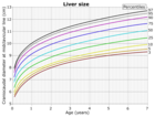
Onabdominal ultrasonography,the liver can be measured by themaximum dimensionon asagittal planeview through themidclavicular line,which is normally up to 18 cm in adults.[2]It is also possible to measure thecranio-caudaldimension,which is normally up to 15 cm in adults.[2]This can be measured together with theventro-dorsaldimension(ordepth), which is normally up to 13 cm.[2]Also, thecaudate lobeis enlarged in many diseases. In theaxial plane,the caudate lobe should normally have a cross-section of less than 0.55 of the rest of the liver.[2]
Otherultrasoundstudies have suggested hepat Omega ly as being defined as a longitudinal axis > 15.5 cm at the hepatic midline, or > 16.0 cm at themidclavicular line.[17][18]
Workup
[edit]Blood testsshould be done, especiallyliver-function tests,which give a good impression of the patient's broad metabolic picture.[medical citation needed]
A complete blood test can help distinguish intrinsic liver disease from extrahepaticbile-duct obstruction.[19]Anultrasoundof the liver can reliably detect a dilatedbiliary-ductsystem,[20] it can also detect the characteristics of acirrhotic liver.[21]
Computerized tomography(CT) can give accurateanatomicalinformation for a complete diagnosis.[22]
Treatment
[edit]
Treatment of hepat Omega ly varies with the cause, so accurate diagnosis is the first concern. In auto-immune liver disease,prednisoneandazathioprinemay be used for treatment.[3]
In lymphoma the treatment options include single-agent (or multi-agent)chemotherapyand regional radiotherapy, and surgery is an option in specific situations. Meningococcal group C conjugatevaccineis used in some cases.[23]
In primary biliary cirrhosis,ursodeoxycholic acidhelps the bloodstream remove bile, which may increase survival.[24]
See also
[edit]References
[edit]- ^abcdefghijklmnopqrs"Hepat Omega ly. Read about Hepat Omega ly (enlarged liver) | Patient".Patient.Archivedfrom the original on 2022-02-01.Retrieved2016-02-27.
- ^abcdefChristoph F. Dietrich, Carla Serra, Maciej Jedrzejczyk (2010-07-28)."Ultrasound of the liver - EFSUMB – European Course Book"(PDF).European federation of societies for ultrasound in medicine and biology (EFSUMB).Archived fromthe original(PDF)on 2017-08-12.Retrieved2017-12-21.
{{cite web}}:CS1 maint: multiple names: authors list (link) - ^ab"Cirrhosis: Practice Essentials, Overview, Etiology".Medscape.22 July 2021.Retrieved16 May2024.
- ^abcdefghi"Hepat Omega ly: MedlinePlus Medical Encyclopedia".nlm.nih.gov.Archivedfrom the original on 2016-07-05.Retrieved2016-02-27.
- ^Lang, Florian (2009-03-19).Encyclopedia of Molecular Mechanisms of Disease: With 213 Tables.Springer Science & Business Media. p. 824.ISBN9783540671367.Archivedfrom the original on 2023-01-12.Retrieved2016-03-11.
- ^Prevention, CDC - Centers for Disease Control and."CDC - Echinococcosis - Resources for Health Professionals".cdc.gov.Archivedfrom the original on 2016-03-11.Retrieved2016-03-11.
- ^"Leptospirosis (Weil's Disease) | Doctor | Patient".Patient.Archivedfrom the original on 2016-03-11.Retrieved2016-03-11.
- ^Banfalvi, Gaspar (2013-10-16).Homeostasis - Tumor - Metastasis.Springer Science & Business Media. p. 145.ISBN9789400773356.
- ^"Cholesteryl Ester Storage Disease - NORD (National Organization for Rare Disorders)".NORD (National Organization for Rare Disorders).Archivedfrom the original on 2016-03-12.Retrieved2016-03-11.
- ^Sherigar, JM; Castro, J; Yin, YM; Guss, D; Mohanty, SR (27 February 2018)."Glycogenic hepatopathy: A narrative review".World Journal of Hepatology.10(2): 172–185.doi:10.4254/wjh.v10.i2.172.PMC5838438.PMID29527255.
- ^"Hunter's Syndrome. MPS II information; symptoms | Patient".Patient.20 August 2014.Archivedfrom the original on 2016-03-12.Retrieved2016-03-11.
- ^"OMIM Entry - # 214100 - PEROXISOME BIOGENESIS DISORDER 1A (ZELLWEGER); PBD1A".omim.org.Archived fromthe originalon 2015-02-14.Retrieved2016-03-11.
- ^"CPT I deficiency".Genetics Home Reference.2016-03-07.Archivedfrom the original on 2020-09-19.Retrieved2016-03-11.
- ^"Sarcoidosis | Doctor | Patient".Patient.Archivedfrom the original on 2016-03-11.Retrieved2016-03-11.
- ^Dennis, Mark; Bowen, William Talbot; Cho, Lucy (2012-01-01).Mechanisms of Clinical Signs.Elsevier Australia. p. 469.ISBN9780729540759.Archivedfrom the original on 2023-01-12.Retrieved2020-10-25.
- ^Rocha, Silvia Maria Sucena da; Ferrer, Ana Paula Scoleze; Oliveira, Ilka Regina Souza de; Widman, Azzo; Chammas, Maria Cristina; Oliveira, Luiz Antonio Nunes de; Cerri, Giovanni Guido (2009)."Determinação do tamanho do fígado de crianças normais, entre 0 e 7 anos, por ultrassonografia".Radiologia Brasileira.42(1): 7–13.doi:10.1590/S0100-39842009000100004.ISSN0100-3984.
- ^Gosink, BB; Leymaster, CE (January 1981). "Ultrasonic determination of hepat Omega ly".Journal of Clinical Ultrasound.9(1): 37–44.doi:10.1002/jcu.1870090110.PMID6792230.S2CID22827636.
- ^Kratzer, W; Fritz, V; Mason, RA; Haenle, MM; Kaechele, V; Roemerstein Study, Group. (November 2003)."Factors affecting liver size: a sonographic survey of 2080 subjects".Journal of Ultrasound in Medicine.22(11): 1155–61.doi:10.7863/jum.2003.22.11.1155.PMID14620885.S2CID29904060.
- ^Goldman, Lee; Schafer, Andrew I. (2015-04-21).Goldman-Cecil Medicine.Elsevier Health Sciences. p. 991.ISBN9780323322850.
- ^Meacock, L M; Sellars, M E; Sidhu, P S (2010-07-01)."Evaluation of gallbladder and biliary duct disease using microbubble contrast-enhanced ultrasound".The British Journal of Radiology.83(991): 615–627.doi:10.1259/bjr/60619911.ISSN0007-1285.PMC3473688.PMID20603412.
- ^Murray, Karen F.; Horslen, Simon (2013-12-11).Diseases of the Liver in Children: Evaluation and Management.Springer Science & Business Media. p. 199.ISBN9781461490050.
- ^Mirvis, Stuart E.; Soto, Jorge A.; Shanmuganathan, Kathirkamanathan; Yu, Joseph; Kubal, Wayne S. (2014-08-19).Problem Solving in Emergency Radiology.Elsevier Health Sciences. p. 442.ISBN9781455758395.
- ^"Non-Hodgkin's Lymphoma | Doctor | Patient".Patient.Archivedfrom the original on 2018-02-08.Retrieved2016-03-11.
- ^"Primary biliary cirrhosis: MedlinePlus Medical Encyclopedia".nlm.nih.gov.Archivedfrom the original on 2016-07-05.Retrieved2016-03-12.
Further reading
[edit]- Hoffmann, Georg F.; Zschocke, Johannes; Nyhan, William L. (2009-11-21).Inherited Metabolic Diseases: A Clinical Approach.Springer Science & Business Media.ISBN9783540747239.
- Kim, Sun Bean; Kim, Do Kyung; Byun, Sun Jeong; Park, Ji Hye; Choi, Jin Young; Park, Young Nyun; Kim, Do Young (2015-12-01)."Peliosis hepatis presenting with massive hepat Omega ly in a patient with idiopathic thrombocytopenic purpura".Clinical and Molecular Hepatology.21(4): 387–392.doi:10.3350/cmh.2015.21.4.387.ISSN2287-2728.PMC4712167.PMID26770928.
