
Levator palpebrae superioris muscle
| Levator palpebrae superioris | |
|---|---|
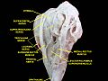
 Rectus muscles: 2 =superior,3 =inferior,4 =medial,5 =lateral Oblique muscles:6 =superior,8 =inferior Other muscle:9 =levator palpebrae superioris Other structures:1 =Annulus of Zinn,7 =Trochlea,10 =Superior tarsus,11 =Sclera,12 =Optic nerve | |
 The levator palebrae superioris can be seen here, travelling above the superior rectus muscle, and ending at the upper eyelid. | |
| Details | |
| Origin | Inferior surface of lesser wing ofsphenoid |
| Insertion | Superiortarsal plateand skin of uppereyelid |
| Artery | Muscular branches ofophthalmic arteryandsupraorbital artery |
| Nerve | Superior division ofoculomotor nerve |
| Actions | Elevation of upper eyelid |
| Antagonist | Palpebral part oforbicularis oculi muscle |
| Identifiers | |
| Latin | musculus levator palpebrae superioris |
| TA98 | A15.2.07.020 |
| TA2 | 2052 |
| FMA | 49041 |
| Anatomical terms of muscle | |
Thelevator palpebrae superioris(Latin:elevating muscle of upper eyelid) is themusclein theorbitthat elevates the uppereyelid.[1][2]
Structure[edit]
The levator palpebrae superioris originates from inferior surface of the lesser wing of thesphenoid bone,just above theoptic foramen.It broadens and decreases in thickness (becomes thinner) and becomes thelevator aponeurosis.This portion inserts on the skin of the upper eyelid, as well as the superiortarsal plate.It is askeletal muscle.Thesuperior tarsal muscle,a smooth muscle, is attached to the levator palpebrae superioris, and inserts on the superior tarsal plate as well.
Blood supply[edit]
The levator palebrae superioris receives its blood supply from branches of theophthalmic artery,specifically, muscular branches and thesupraorbital artery.Blood is drained into thesuperior ophthalmic vein.
Nerve supply[edit]
The levator palpebrae superioris receives motor innervation from the superior division of theoculomotor nerve.[1][2][3]The smooth muscle that originates from its undersurface, called thesuperior tarsal muscleis innervated by postganglionic sympathetic axons from thesuperior cervical ganglion.[2]
Function[edit]
The levator palpebrae superioris elevates the upper eyelid.[1][2]
Clinical significance[edit]
Damage to this muscle or its innervation can causeptosis,which is drooping of the eyelid.[4][5]Lesions in CN III can cause ptosis,[5]because without stimulation from the oculomotor nerve the levator palpebrae cannot oppose the force of gravity, and the eyelid droops.
Ptosis can also result from damage to the adjoiningsuperior tarsal muscleor its sympathetic innervation. Such damage to the sympathetic supply occurs inHorner's syndromeand presents as a partial ptosis. It is important to distinguish between these two very different causes of ptosis. This can usually be done clinically without issue, as each type of ptosis is accompanied by other distinct clinical findings.
The ptosis seen in paralysis of the levator palpebrae superioris is usually more pronounced than that seen due to paralysis of the superior tarsal muscle.
Additional images[edit]
-
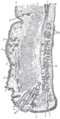 Sagittal section through the upper eyelid.
 -
 Levator palpebrae superioris muscle
 -
 Levator palpebrae superioris muscle
 -
 Extrinsic eye muscle. Nerves of orbita. Deep dissection.
 -
 Extrinsic eye muscle. Nerves of orbita. Deep dissection.


See also[edit]
References[edit]
- ^abcLiu, Grant T. (2003-01-01), Samuels, Martin A.; Feske, Steven K. (eds.),"Chapter 6 - Disorders of the Eyes and Eyelids",Office Practice of Neurology (Second Edition),Philadelphia: Churchill Livingstone, pp. 35–69,doi:10.1016/b0-44-306557-8/50008-3,ISBN978-0-443-06557-6,retrieved2020-11-11
- ^abcdStandring, Susan, ed. (2016). ""Extraocular muscles: levator palpebrae superioris"".Gray's anatomy: the anatomical basis of clinical practice(41st ed.). Philadelphia. p. 670.ISBN9780702052309.OCLC920806541.
{{cite book}}:CS1 maint: location missing publisher (link) - ^Jackson, Timothy L., ed. (2008-01-01),"Chapter 1 - OCULOPLASTICS",Moorfields Manual of Ophthalmology,Edinburgh: Mosby, pp. 1–54,doi:10.1016/b978-1-4160-2572-6.50006-x,ISBN978-1-4160-2572-6,S2CID241607885,retrieved2020-11-11
- ^Trobe, Jonathan D. (2008-01-01), Trobe, Jonathan D. (ed.), "Section 13 - Eyelid Disorders",Neuro-ophthalmology,Edinburgh: Mosby, pp. 229–239,doi:10.1016/b978-0-323-04456-1.50016-9,ISBN978-0-323-04456-1
- ^abHejtmancik, J. F.; Cabrera, P.; Chen, Y.; M’Hamdi, O.; Nickerson, J. M. (2017-01-01), Conn, P. Michael (ed.),"Chapter 19 - Vision",Conn's Translational Neuroscience,San Diego: Academic Press, pp. 399–438,doi:10.1016/b978-0-12-802381-5.00031-2,ISBN978-0-12-802381-5,retrieved2020-11-11
External links[edit]
- Anatomy figure: 29:01-01at Human Anatomy Online, SUNY Downstate Medical Center
- lesson3at The Anatomy Lesson by Wesley Norman (Georgetown University) (orbit2)