
Meninges
This articleneeds additional citations forverification.(June 2014) |
| Meninges | |
|---|---|
 Image depicting meninges around the brain | |
 The meninges: dura mater, arachnoid mater and pia mater | |
| Details | |
| Artery | Middle meningeal artery,meningeal branches of the ascending pharyngeal artery,accessory meningeal artery,branch ofanterior ethmoidal artery,meningeal branches of vertebral artery |
| Nerve | Middle meningeal nerve,nervus spinosus |
| Identifiers | |
| Latin | meninges |
| MeSH | D008578 |
| TA98 | A14.1.01.001 |
| TA2 | 5369 |
| FMA | 231572 |
| Anatomical terminology | |
Inanatomy,themeninges(/məˈnɪndʒiːz/,[1][2]sg.:meninx(/ˈmiːnɪŋks/or/ˈmɛnɪŋks/[3]), fromAncient Greekμῆνιγξ(mēninx)'membrane'[4]) are the threemembranesthat envelop thebrainandspinal cord.Inmammals,the meninges are thedura mater,thearachnoid mater,and thepia mater.Cerebrospinal fluidis located in the subarachnoid space between the arachnoid mater and the pia mater. The primary function of the meninges is to protect thecentral nervous system.[5]
Structure
[edit]Dura mater
[edit]The dura mater (Latin:tough mother),[6][a]is a thick, durable membrane, closest to theskulland vertebrae. The dura mater, the outermost part, is a loosely arranged, fibroelastic layer of cells, characterized by multiple interdigitating cell processes, no extracellular collagen,[citation needed]and significant extracellular spaces. The middle region is a mostly fibrous portion. It consists of two layers: theendosteallayer, which lies closest to the skull, and the inner meningeal layer, which lies closer to the brain.[7]It contains larger blood vessels that split into the capillaries in thepia mater.It is composed of dense fibrous tissue, and its inner surface is covered by flattened cells like those present on the surfaces of the pia mater and arachnoid mater. The dura mater is a sac that envelops the arachnoid mater and surrounds and supports the largedural sinusescarrying blood from the brain toward the heart.
The dura has four areas of infolding:
- Falx cerebri,the largest, sickle-shaped; separates thecerebral hemispheres.Starts from the frontal crest offrontal boneand thecrista gallirunning to theinternal occipital protuberance.
- Tentorium cerebelli,the second largest, crescent-shaped; separates theoccipital lobesfromcerebellum.The falx cerebri attaches to it giving a tentlike appearance.
- Falx cerebelli,vertical infolding; lies inferior to the tentorium cerebelli, separating thecerebellar hemispheres.
- Diaphragma sellae,smallest infolding; covers thepituitary glandandsella turcica.

Arachnoid mater
[edit]
The middle element of the meninges is thearachnoid mater,or arachnoid membrane, so named because of its resemblance to aspider web.It cushions thecentral nervous system.This thin, transparent membrane is composed of fibrous tissue and, like the pia mater, has an outer layer of tightly packed flat cells, forming thearachnoid barrier.[8]
The shape of the arachnoid does not follow the convolutions of the surface of the brain and so looks like a loosely fitting sac. In particular, in the region of the brain a large number of fine filaments called arachnoid trabeculae pass from the arachnoid through the subarachnoid space to blend with the tissue of the pia mater. The arachnoid barrier has no extracellular collagen and is considered to represent an effective morphological and physiological meningeal barrier between the cerebrospinal fluid in the subarachnoid space and the blood circulation in the dura.
The arachnoid barrier layer is characterized by a distinct continuous basal lamina on its inner surface toward the innermost collagenous portion of the arachnoid reticular layer.
Pia mater
[edit]The pia mater (Latin:tender mother)[9]is a very delicate membrane. It is the meningeal envelope that firmly adheres to the surfaces of the brain and spinal cord, following all of the brain's contours (gyriandsulci). It is a very thin membrane composed of fibrous tissue covered on its outer surface by a sheet of flat cells thought to be impermeable to fluid. The pia mater is pierced by blood vessels to the brain and spinal cord, and itscapillariesnourish the brain.
Subarachnoidal lymphatic-like membrane
[edit]Thesubarachnoid lymphatic-like membrane(SLYM) is a possible fourthmeningeallayer that was proposed in 2023 in thebrainofhumansandmice.[10]
The SLYM is located in thesubarachnoid space,the space between the middle reticular meninges and the innermost tender meninges that lie close to the brain.[10]It divides the subarachnoid space into an outer, superficial compartment and an inner, deeper area surrounding the brain.[10]Leptomeninges
[edit]Thearachnoidandpia materare sometimes together called theleptomeninges,[11]literally "thin meninges" (Greek:λεπτός"leptos" — "thin" ). Acutemeningococcal meningitiscan lead to anexudatewithin the leptomeninges along the surface of the brain.[12]Because the arachnoid is connected to the pia by cob-web like strands, it is structurally continuous with the pia, hence the name pia-arachnoid or leptomeninges. They are responsible for the production of beta-trace protein (prostaglandin D2 synthase), a major cerebrospinal fluid protein.[13]
Subarachnoid space
[edit]
Thesubarachnoid spaceis the space that normally exists between thearachnoidand thepia mater.It is filled withcerebrospinal fluidand continues down thespinal cord.Spaces are formed from openings at different points along the subarachnoid space; these are thesubarachnoid cisterns,which are filled with cerebrospinal fluid.
The dura mater is attached to theskull,whereas in the spinal cord, the dura mater is separated from thevertebraeby a space called theepidural space,which contains fat and blood vessels. The arachnoid is attached to the dura mater, while the pia mater is attached to the central nervous system tissue. When the dura mater and the arachnoid separate through injury or illness, the space between them is thesubdural space.There is asubpial spaceunderneath the pia mater that separates it from theglia limitans.
Clinical significance
[edit]Injuries involving the meninges, can result in ahemorrhageand two types ofhematoma.[14]
- Asubarachnoid hemorrhageis acute bleeding under the arachnoid; it may occur spontaneously or as a result of trauma.[15]
- Asubdural hematomais a hematoma (collection of blood) located in a separation of thearachnoidfrom the dura mater. Thebridging veinsthat connect the dura mater and thearachnoidare torn, usually during an accident, and blood leaks into this area.
- Anepidural hematoma,bleeding between the dura mater and theskull,may arise after an accident or spontaneously.
Other medical conditions that affect the meninges includemeningitis(usually from afungal,bacterial,orviralinfection) andmeningiomasthat arise from the meninges, or frommeningeal carcinomatoses(tumors) that form elsewhere in the body andmetastasizeto the meninges.
Other animals
[edit]Infish,there is a single membrane known as the primitive meninx.[16]Amphibiansandreptileshave two meninges, andbirdsand mammals have three.[16]In the early 1900s,Giuseppe Sterzi,an Italian anatomist, carried out comparative studies on the meninges from thelanceletto the human. Contrary to previous reports, the spinal meninges were seen to be very simple, both in the adult lower vertebrates and in the early developmental stages of the more advanced vertebrates. From themesenchymesurrounding theneural tube,only a single leaflet forms the primitive meninx. In the followingphylogeneticandontogeneticstages, the latter divides into an internal leaflet: the secondary meninx, and into an external one: the dura mater. Finally, in higher vertebrates, even the secondary meninx divides into the arachnoid and the pia. In the same animals, Sterzi demonstrated that, while in the spinal medulla the dura keeps its identity, in the skull it fuses with theperiosteum.He also demonstrated the continuity of all meninges with the envelopes of nerves and with thefilum terminale.
Mammals(as higher vertebrates) retain the dura mater, and the secondary meninx divides into thearachnoidandpia mater.[17]
Additional images
[edit]-
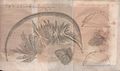 Illustration of the work by Antonio PacchioniDisquisitio anatomicae de durae meningis...published inActa Eruditorum,1703

See also
[edit]Notes
[edit]- ^Also rarely calledmeninx fibrosaorpachymeninx
References
[edit]- ^"meninges".Oxford Learner's Dictionaries.
- ^"Definition of meninges".Merriam-Webster Online Dictionary.Retrieved28 July2012.
- ^"Definition of meninx".Merriam-Webster Online Dictionary.Retrieved28 July2012.
- ^μῆνιγξ.Liddell, Henry George;Scott, Robert;A Greek–English Lexiconat thePerseus Project
- ^Castillero Mimenza, Oscar (January 2017)."Meninges: anatomía, partes y funciones en el cerebro".
- ^"Definition of dura mater".Merriam-WebsterOnline Dictionary.Retrieved22 June2022.
- ^"Scalp Anatomy: Structure, Nerve Supply, Arterial Supply".20 June 2017.
- ^Abbott, NJ; Patabendige, AA; Dolman, DE; Yusof, SR; Begley, DJ (January 2010). "Structure and function of the blood-brain barrier".Neurobiology of Disease.37(1): 13–25.doi:10.1016/j.nbd.2009.07.030.PMID19664713.S2CID14753395.
- ^"Definition of pia mater".Merriam-WebsterOnline Dictionary.Retrieved27 November2015.
- ^abcMøllgård, Kjeld; Beinlich, Felix R. M.; Kusk, Peter; et al. (2023)."A mesothelium divides the subarachnoid space into functional compartments".Science.379(6627): 84–88.Bibcode:2023Sci...379...84M.doi:10.1126/science.adc8810.PMID36603070.S2CID255440992.
- ^"leptomeninges".Oxford Dictionaries | English.Archived fromthe originalon November 4, 2016.
- ^Kumar, Vinay (2015).Robbins and Cotran Pathologic Mechanisms of Disease(9th ed.). Philadelphia: Elsevier Saunders. p. 1273.OCLC892583347.
In acute meningitis, an exudate is evident within the leptomeninges over the surface of the brain (Fig. 28-21).
- ^Yamashima, Tetsumori; Sakuda, Kazushige; Tohma, Yasuo; Yamashita, Junkoh; Oda, Hiroshi; Irikura, Daisuke; Eguchi, Naomi; Beuckmann, Carsten T.; Kanaoka, Yoshihide; Urade, Yoshihiro; Hayaishi, Osamu (1 April 1997)."Prostaglandin D Synthase (β-Trace) in Human Arachnoid and Meningioma Cells: Roles as a Cell Marker or in Cerebrospinal Fluid Absorption, Tumorigenesis, and Calcification Process".Journal of Neuroscience.17(7): 2376–2382.doi:10.1523/JNEUROSCI.17-07-02376.1997.PMC6573504.PMID9065498.S2CID15404074.
- ^"Overview of Adult Traumatic Brain Injuries"(PDF).Orlando Regional Healthcare, Education and Development. 2004. Archived fromthe original(PDF)on February 27, 2008.
- ^van Gijn J, Kerr RS, Rinkel GJ (2007). "Subarachnoid haemorrhage".Lancet.369(9558): 306–18.doi:10.1016/S0140-6736(07)60153-6.PMID17258671.S2CID29126514.
- ^abOstrander, Gary (12 September 2000).The Laboratory Fish.Elsevier.ISBN9780125296502.
- ^Kardong, Kenneth V. (1995).Vertebrates: Comparative Anatomy, Function, Evolution.Dubuque, Iowa: Wm. C. Brown Publishers. p. 539.ISBN0-697-21991-7.
External links
[edit] Media related toMeningesat Wikimedia Commons