
Pubis (bone)
| Pubis (bone) | |
|---|---|
 Pelvic girdle | |
 Male pelvis with pubis at bottom | |
| Details | |
| Part of | Pelvis |
| Identifiers | |
| Latin | os pubis |
| MeSH | D011630 |
| TA98 | A02.5.01.301 |
| TA2 | 1346 |
| FMA | 16595 |
| Anatomical terms of bone | |
Invertebrates,thepubisorpubic bone(Latin:os pubis) forms the lower and anterior part of each side of thehip bone.The pubis is the most forward-facing (ventralandanterior) of the three bones that make up the hip bone. The left and right pubic bones are each made up of three sections; a superior ramus, an inferior ramus, and a body.
Structure[edit]
The pubic bone is made up of abody,superior ramus,andinferior ramus(Latin:branch). The left and right coxal bones join at thepubic symphysis.It is covered by a layer offat– themons pubis.The pubis is the lower limit of thesuprapubic region.In the female, the pubis is anterior to theurethral sponge.
Body[edit]
The body of pubis has:
- a superior border or the pubic crest
- a pubic tubercle at the lateral end of the pubic crest
- three surfaces (anterior, posterior and medial).
The body forms the wide, strong, middle and flat part of the pubic bone. The bodies of the left and right pubic bones join at thepubic symphysis.[1]The rough upper edge is thepubic crest,[2]ending laterally in thepubic tubercle.Thistubercle,found roughly 3 cm from the pubic symphysis, is a distinctive feature on the lower part of theabdominal wall;important when localizing thesuperficial inguinal ringand thefemoral canalof theinguinal canal.[3]The inner surface of the body forms part of the wall of thelesser pelvisand joints to the origin of a part of theobturator internusmuscle.
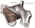
Superior pubic ramus[edit]
The superior pubic ramus is the upper of the two rami. It forms the upper edge of theobturator foramen.[4]It extends from the body to the median plane where it joins with the ramus of the opposite side. It consists of an inner flattened part and a narrow outer prismoid portion.
Medial surface[edit]
Surfaces[edit]
- The anterior surface is rough, directed downward and outward, and serves for the origin of various muscles. Theadductor longusarises from the upper and medial angle, immediately below the crest; lower down, theobturator externus,theadductor brevisand the upper part of thegracilistake origin.
- The posterior surface, convex from above downward, concave from side to side, is smooth, and forms part of the anterior wall of the pelvis. It gives origin to thelevator aniandobturator internus,and attachment to thepuboprostatic ligamentsand to a few muscular fibers prolonged from the bladder.
Borders[edit]
The upper border presents a prominent tubercle, thepubic tubercle(pubic spine), which projects forward; theinferior crusof thesubcutaneous inguinal ring(external abdominal ring), and theinguinal ligament(Poupart's ligament) are attached to it.
Passing upward and laterally from the pubic tubercle is a well-defined ridge, forming a part of thepectineal linewhich marks the brim of thelesser pelvis:to it are attached a portion of theinguinal falx(conjoined tendon ofobliquus internusandtransversus), thelacunar ligament(Gimbernat's ligament), and thereflected inguinal ligament(triangular fascia).
Medial to the pubic tubercle is the crest, which extends from this process to the medial end of the bone. It affords attachment to the inguinal falx, and to therectus abdominisandpyramidalis.
The point of junction of the crest with the medial border of the bone is called the angle; to it, as well as to thesymphysis,thesuperior crusof the subcutaneous inguinal ring is attached.
The medial border is articular; it is oval, and is marked by eight or nine transverse ridges, or a series of nipple-like processes arranged in rows, separated by grooves; they serve for the attachment of a thin layer of cartilage, which intervenes between it and the interpubic fibrocartilaginous lamina.
The lateral border presents a sharp margin, the obturator crest, which forms part of the circumference of theobturator foramenand affords attachment to theobturator membrane.
Lateral portion[edit]
Surfaces[edit]
- The superior surface presents a continuation of thepectineal line,already mentioned as commencing at thepubic tubercle.In front of this line, the surface of bone is triangular in form, wider laterally than medially, and is covered by thepectineus.The surface is bounded, laterally, by a rough eminence, theiliopectineal eminence,which serves to indicate the point of junction of theiliumand pubis, and below by a prominent ridge which extends from theacetabular notchto the pubic tubercle.
- The inferior surface forms the upper boundary of theobturator foramen,and presents, laterally, a broad and deep, oblique groove, for the passage of the obturator vessels and nerve; and medially, a sharp margin, theobturator crest,forming part of the circumference of the obturator foramen, and giving attachment to theobturator membrane.
- The posterior surface constitutes part of the anterior boundary of thelesser pelvis.It is smooth, convex from above downward, and affords origin to some fibers of theobturator internus.
Inferior pubic ramus[edit]
The inferior pubic ramus is a part of the pelvis and is thin and flat. It passes laterally and downward from the medial end of the superior ramus; it becomes narrower as it descends and joins with theinferior ramus of the ischiumbelow theobturator foramen.
Surfaces[edit]
- Itsanterior surfaceis rough, for the origin of muscles—thegracilisalong its medial border, a portion of theobturator externuswhere it enters into the formation of theobturator foramen,and between these two, theadductores brevisand magnus, the former being the more medial.
- Theposterior surfaceis smooth, and gives origin to theobturator internus,and, close to the medial margin, to theconstrictor urethrae.
Borders[edit]
- In the female pelvis, themedial borderis thick, rough, and everted, and presents two ridges, separated by an intervening space. The ridges extend downwards, and are continuous with similar ridges on theinferior ramus of the ischium;
- to the external ridge is attached thefascia of Colles
- to the internal ridge is attached the inferior fascia of theurogenital diaphragm
- Thelateral borderis thin and sharp, forms part of the circumference of theobturator foramen,and gives attachment to theobturator membrane.
Other animals[edit]
Mammals[edit]
Non-placentalmammals possess osteological projections of the pubis known asepipubicbones. These evolved first among derivedcynodonts,and evolved as a means of support for muscles fle xing the thigh, facilitating the development of an erect gait.[5]However, these prevent the expansion of the torso, preventing pregnancy and forcing the animal to give birth to larval young (the modernmarsupial"joeys" andmonotreme"puggles" ).[6]
Placentals are unique among all mammals, including othereutherians,in having lost epipubic bones and having been able to develop properpregnancy.In some groups, remnants of these pre-pubic bones can be found asos penises.[7]
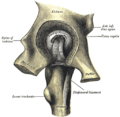
Dinosaurs[edit]
ThecladeDinosauriais divided into theSaurischiaandOrnithischiabased onhip structure,including importantly that of the pubis.[8]Anopisthopubicpelvis is a condition where the pubic bone extends back towards the tail of the animal, a trait that is also present in birds.[9]In apropubicpelvis, however, the pubic bone extends forward towards the head of the animal, as can be seen in the typical saurischian pelvic structure pictured below. Theacetabulum,which can be thought of as a "hip-socket", is an opening on each side of the pelvic girdle formed where theischium,ilium,and pubis all meet, and into which the head of the femur inserts. The orientation and position of the acetabulum is one of the main morphological traits that caused dinosaurs to walk in an upright posture with their legs directly underneath their bodies.[10]Theprepubic processis a bony extension of the pubis that extends forward from the hip socket and toward the front of the animal. This adaptation is thought to have played a role in supporting the abdominal muscles.
-
 Ornithischian pelvic structure (left side)
 -
 Saurischian pelvic structure (left side).


Additional images[edit]
-
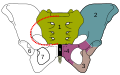 Thesacrumand pelvic bone, with parts labelled. The pubic bone consists of the body and superior pubic ramus (4), and the inferior pubic ramus (3), which join at thepubic symphysis.The gap between them is theobturator foramen.
 -
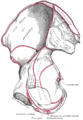
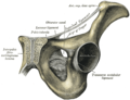
 Right hip bone. External surface.
 -
 Right hip bone. Internal surface.
 -
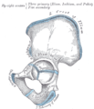 Plan of ossification of the hip bone.
 -
 Symphysis pubis exposed by a coronal section.
 -
 Left levator ani from within.
 -
 The obturator externus.
 -
 Left hip-joint, opened by removing the floor of the acetabulum from within the pelvis.
 -
 Pubis
Pubis


See also[edit]
References[edit]
![]() This article incorporates text in thepublic domainfrompage 236of the 20th edition ofGray's Anatomy(1918)
This article incorporates text in thepublic domainfrompage 236of the 20th edition ofGray's Anatomy(1918)
- ^"Definition: body of pubis from Online Medical Dictionary".Retrieved2008-10-18.
- ^"crista pubica"atDorland's Medical Dictionary
- ^Bojsen-Møller, Finn (2000).Rörelseapparatens anatomi(in Swedish). Liber. p. 239.ISBN91-47-04884-0.
- ^"Definition: superior pubic ramus from Online Medical Dictionary".Retrieved2008-10-18.
- ^White, T.D. (August 9, 1989). "An analysis of epipubic bone function in mammals using scaling theory". Journal of Theoretical Biology 139 (3): 343–57.doi:10.1016/S0022-5193(89)80213-9.PMID2615378.
- ^Michael L. Power, Jay Schulkin. The Evolution Of The Human Placenta. pp. 68–.
- ^Frederick S. Szalay (11 May 2006). Evolutionary History of the Marsupials and an Analysis of Osteological Characters. Cambridge University Press. pp. 293–.ISBN978-0-521-02592-8.
- ^Seeley, H.G. (1888). "On the classification of the fossil animals commonly named Dinosauria."Proceedings of the Royal Society of London,43:165-171.
- ^Barsbold, R., (1979) Opisthopubic pelvis in the carnivorous dinosaurs. Nature. 279, 792-793
- ^Martin, A.J. (2006). Introduction to the Study of Dinosaurs. Second Edition. Oxford, Blackwell Publishing. pg. 299-300.ISBN1-4051-3413-5.
External links[edit]
- Anatomy photo:44:st-0713at the SUNY Downstate Medical Center - "The Male Pelvis: Hip Bone"
| National | |
|---|---|
| Other | |