Rhinophore


Arhinophoreis one of a pair ofchemosensoryclub-shaped, rod-shaped or ear-like structures which are the most prominent part of the external head anatomy insea slugs,marinegastropodopisthobranchmolluskssuch as thenudibranchs,sea hares (Aplysiomorpha), and sap-sucking sea slugs (Sacoglossa).
Etymology
[edit]The name relates to the rhinophore's function as an organ of "smell".Rhino-meansnosefromAncient Greekῥίςrhisand from its genitive ῥινόςrhinos."Phore" means "to bear" fromNeo-Latin-phorusand from Greek -phoros (φορος) "bearing", a derivative ofphérein(φέρειν).
Function
[edit]Rhinophores arescentortastereceptors, also known aschemosensoryorgans situated on the dorsal surface of the head. They are primarily used for distancechemoreceptionandrheoreception(response to water current).[1]
The "scents" detected by rhinophores are chemicals dissolved in the sea water. The fine structure and hairs of the rhinophore provide a large surface area so that chemical detection is maximized.[2]This allows the nudibranchs to stay close to their food source (for example species ofsponges) and to find mates. In the sea hareAplysia californica,the rhinophores are able to detectpheromones.[1]
Protection
[edit]To protect the prominent rhinophores against nibbling by predators, including fish, most species of dorid nudibranchs are able to withdraw their rhinophores into a pocket beneath the skin.[2]
Structure
[edit]
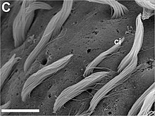
In reproductively matureAplysiaadults, the rhinophore is about 1 cm in length.[1]The neuroanatomical organization includes a rhinophore groove where most of the sensory cells appear to be concentrated. Its sensoryepitheliumcontainssensory neuronsthat projectaxonsback to rhinophoregangliaanddendritesthat end in either a surface-exposed cilium or a small protuberance.[1]
 Scale bar is 300 μm. rg - rhinophore groove tip - rhinophore tip. |
 Scale bar is 100 μm. f - folds |
 Scale bar is 10 μm. ci - numerous long cilia |
Comparison with oral tentacles
[edit]InA. californica,the oral tentacles, which are situated in a more ventral position, are possibly involved in contact chemoreception and mechanoreception.[1]
References
[edit]This article incorporatesCC-BY-2.0 text (but not underGFDL) from reference.[1]
- ^abcdefScott F Cummins, Dirk Erpenbeck, Zhihua Zou, Charles Claudianos, Leonid L Moroz, Gregg T Nagle & Bernard M Degnan. 2009.Candidate chemoreceptor subfamilies differentially expressed in the chemosensory organs of the mollusc Aplysia.BMC Biology2009, 7:28.doi:10.1186/1741-7007-7-28.
- ^abRhinophore in nudibranchsArchived2007-10-21 at theWayback Machine.Sea Slug Forum, accessed 8 July 2009.
Further reading
[edit]- Wertz A., Rössler W., Obermayer M. & Bickmeyer U. (6 April 2006) "Functional neuroanatomy of the rhinophore ofAplysia punctata".Frontiers in Zoology3:6.doi:10.1186/1742-9994-3-6
