
Rib cage
| Rib cage | |
|---|---|
 Human rib cage | |
 Animation of the rib cage | |
| Details | |
| Identifiers | |
| Latin | cavea thoracis |
| MeSH | D000070602 |
| TA98 | A02.3.04.001 |
| TA2 | 1096 |
| FMA | 7480 |
| Anatomical terminology | |
Therib cageorthoracic cageis anendoskeletalenclosure in thethoraxof mostvertebratesthat comprises theribs,vertebral columnandsternum,which protect thevital organsof thethoracic cavity,such as theheart,lungsandgreat vesselsand support theshoulder girdleto form thecorepart of theaxial skeleton.
A typicalhuman thoracic cageconsists of 12 pairs of ribs and the adjoiningcostal cartilages,the sternum (along with themanubriumandxiphoid process), and the 12thoracic vertebraearticulating with the ribs. The thoracic cage also provides attachments for extrinsicskeletal musclesof theneck,upper limbs,upper abdomenandback,and together with the overlyingskinand associatedfasciaandmuscles,makes up thethoracic wall.
Intetrapods,the rib cage intrinsically holds themuscles of respiration(diaphragm,intercostal muscles,etc.) that are crucial for activeinhalationand forcedexhalation,and therefore has a majorventilatoryfunction in therespiratory system.
Structure
[edit]There are thirty-three vertebrae in the human vertebral column. The rib cage is associated with TH1−TH12. Ribs are described based on their location and connection with the sternum. All ribs are attached posteriorly to thethoracic vertebraeand are numbered accordingly one to twelve. Ribs that articulate directly with the sternum are calledtrue ribs,whereas those that do not articulate directly are termedfalse ribs.Thefalse ribsinclude thefloating ribs(eleven and twelve) that are not attached to the sternum at all.
Attachment
[edit]The termstrue ribsandfalse ribsdescribe rib pairs that are directly or indirectly attached to thesternumrespectively. The first seven rib pairs known as thefixedorvertebrosternalribs are thetrue ribs(Latin:costae verae) as they connect directly to the sternum via their own individualcostal cartilages.The next five pairs (eighth to twelfth) are thefalse ribs(Latin:costae spuriae) orvertebrochondralribs, which do not connect directly to the sternum. The first three pairs of vertebrochondral ribs (eighth to tenth) connect indirectly to the sternum via the costal cartilages of the ribs above them,[1][2]and the overall elasticity of their articulations allows thebucket handle movementsof the rib cage essential for respiratory activity.
The phrasefloating rib(Latin:costae fluctuantes) orvertebralrib refers to the two lowermost (the eleventh and twelfth) rib pairs; so-called because they are attached only to thevertebraeand not to the sternum or any of the costal cartilages. These ribs are relatively small and delicate, and include a cartilaginous tip.[3]
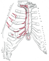
The spaces between the ribs are known asintercostal spaces;they contain the instrinsicintercostal musclesand theneurovascular bundlescontainingintercostal nerves,arteriesandveins.[4]The superficial surface of the rib cage is covered by thethoracolumbar fascia,which provides external attachments for theneck,back,pectoralandabdominal muscles.
-
 Human rib cage - CT scan (parallel projection (left) and perspective projection (right))
 -
 true / fixed ribsfalse ribsfalse and floating ribs


Parts of rib
[edit]
Each rib consists of a head, neck, and a shaft. All ribs are attached posteriorly to thethoracic vertebrae.They are numbered to match the vertebrae they attach to – one to twelve, from top (T1) to bottom. Theheadof the rib is the end part closest to thevertebrawith which itarticulates.It is marked by a kidney-shaped articular surface which is divided by a horizontal crest into two articulating regions. The upper region articulates with theinferior costal faceton the vertebra above, and the larger region articulates with thesuperior costal faceton the vertebra with the same number. Thetransverse processof a thoracic vertebra also articulates at thetransverse costal facetwith the tubercle of the rib of the same number. The crest gives attachment to theintra-articular ligament.[5]
Theneckof the rib is the flattened part that extends laterally from the head. The neck is about 3 cm long. Its anterior surface is flat and smooth, whilst its posterior is perforated by numerous foramina and its surface rough, to give attachment to the ligament of the neck. Its upper border presents a rough crest (crista colli costae) for the attachment of the anteriorcostotransverse ligament;its lower border is rounded.
On the posterior surface at the neck, is an eminence—thetuberclethat consists of an articular and a non-articular portion. The articular portion is the lower and more medial of the two and presents a small, oval surface for articulation with thetransverse costal faceton the end of the transverse process of the lower of the two vertebrae to which the head is connected. The non-articular portion is a rough elevation and affords attachment to the ligament of the tubercle. The tubercle is much more prominent in the upper ribs than in the lower ribs.
Theangleof arib(costal angle) may both refer to the bending part of it, and a prominent line in this area, a little in front of the tubercle. This line is directed downward and laterally; this gives attachment to a tendon of theiliocostalis muscle.At this point, the rib is bent in two directions, and at the same time twisted on its long axis.
The distance between the angle and the tubercle is progressively greater from the second to the tenth ribs. The area between the angle and the tubercle is rounded, rough, and irregular, and serves for the attachment of thelongissimus dorsi muscle.
Bones
[edit]Ribs and vertebrae
[edit]Thefirst rib(the topmost one) is the most curved and usually the shortest of all the ribs; it is broad and flat, its surfaces looking upward and downward, and its borders inward and outward.
-
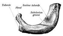 First rib seen from above
 -
 Costal groove position on a central rib


Theheadis small and rounded, and possesses only a single articular facet, for articulation with the body of the firstthoracic vertebra.Theneckis narrow and rounded. Thetubercle,thick and prominent, is placed on the outer border. It bears a small facet for articulation with thetransverse costal faceton the transverse process of T1. There is noangle,but at the tubercle, the rib is slightly bent, with the convexity upward, so that the head of the bone is directed downward. The upper surface of the body is marked by two shallow grooves, separated from each other by a slight ridge prolonged internally into a tubercle, thescalene tubercle,for the attachment of theanterior scalene;theanterior groovetransmits thesubclavian vein,theposteriorthesubclavian arteryand the lowest trunk of thebrachial plexus.Behind the posterior groove is a rough area for the attachment of themedial scalene.Theunder surfaceis smooth and without a costal groove. Theouter borderis convex, thick, and rounded, and at its posterior part gives attachment to the first digitation of theserratus anterior.Theinner borderis concave, thin, and sharp, and marked about its center by the scalene tubercle. Theanterior extremityis larger and thicker than that of any of the other ribs.
Thesecond ribis the second uppermost rib in humans or second most frontal in animals that walk on four limbs. In humans, the second rib is defined as a true rib since it connects with the sternum through the intervention of thecostal cartilageanteriorly (at the front). Posteriorly, the second rib is connected with thevertebral columnby thesecond thoracic vertebra.The second rib is much longer than thefirst rib,but has a very similar curvature. The non-articular portion of the tubercle is occasionally only feebly marked. The angle is slight and situated close to the tubercle. The body is not twisted so that both ends touch any plane surface upon which it may be laid; but there is a bend, with its convexity upward, similar to, though smaller than that found in the first rib. The body is not flattened horizontally like that of the first rib. Its external surface is convex, and looks upward and a little outward; near the middle of it is a rough eminence for the origin of the lower part of the first and the whole of the second digitation of the serratus anterior; behind and above this is attached theposterior scalene.The internal surface, smooth, and concave, is directed downward and a little inward: on its posterior part there is a short costal groove between the ridge of the internal surface of the rib and the inferior border. It protects the intercostal space containing theintercostal veins,intercostal arteries,andintercostal nerves.[6][4]
Theninth ribhas a frontal part at the same level as thefirst lumbar vertebra.This level is called thetranspyloric plane,since thepylorusis also at this level.[7]
Thetenth ribattaches directly to the body of vertebra T10 instead of between vertebrae like the second through ninth ribs. Due to this direct attachment, vertebra T10 has a complete costal facet on its body.[3]

Theeleventh and twelfth ribs,thefloating ribs,have a singlearticular faceton the head, which is of rather large size. They have no necks or tubercles, and are pointed at their anterior ends. The eleventh has a slight angle and a shallow costal groove, whereas the twelfth does not. The twelfth rib is much shorter than the eleventh rib, and only has a one articular facet.[8]
Sternum
[edit]The sternum is a long,flat bonethat forms the front of the rib cage. The cartilages of the top seven ribs (thetrue ribs) join with the sternum at the sternocostal joints. The costal cartilage of the second rib articulates with the sternum at the sternal angle making it easy to locate.[9]
The manubrium is the wider, superior portion of the sternum. The top of the manubrium has a shallow, U-shaped border called the jugular (suprasternal) notch. The clavicular notch is the shallow depression located on either side at the superior-lateral margins of the manubrium. This is the site of thesternoclavicular joint,between the sternum and clavicle. The first ribs also attach to the manubrium.[10]
Thetransversus thoracismuscle is innervated by one of theintercostal nervesand superiorly attaches at the posterior surface of the lower sternum. Its inferior attachment is the internal surface of costal cartilages two through six and works to depress the ribs.[11]
Development
[edit]Expansion of the rib cage in males is caused by the effects oftestosteroneduring puberty.[12]Thus, males generally have broad shoulders and expanded chests, allowing them to inhale more air to supply their muscles with oxygen.

Variation
[edit]Variations in the number of ribs occur. About 1 in 200–500 people have an additionalcervical rib,and there is a female predominance.[13]Intrathoracic supernumerary ribs are extremely rare.[14]The rib remnant of the7th cervical vertebraon one or both sides is occasionally replaced by a free extra rib called acervical rib,which can mechanically interfere with the nerves (brachial plexus) going to the arm.
In several ethnic groups, most significantly the Japanese, the tenth rib is sometimes afloating rib,as it lacks a cartilaginous connection to the seventh rib.[3]
Function
[edit]
The human rib cage is a component of the humanrespiratory system.It encloses the thoracic cavity, which contains the lungs. An inhalation is accomplished when the musculardiaphragm,at the floor of the thoracic cavity, contracts and flattens, while the contraction ofintercostal muscleslift the rib cage up and out.
Expansion of the thoracic cavity is driven in three planes; the vertical, the anteroposterior and the transverse. The vertical plane is extended by the help of the diaphragm contracting and the abdominal muscles rela xing to accommodate the downward pressure that is supplied to the abdominal viscera by the diaphragm contracting. A greater extension can be achieved by the diaphragm itself moving down, rather than simply the domes flattening. The second plane is the anteroposterior and this is expanded by a movement known as the 'pump handle'. The downward sloping nature of the upper ribs are as such because they enable this to occur. When the external intercostal muscles contract and lift the ribs, the upper ribs are able also to push the sternum up and out. This movement increases the anteroposterior diameter of the thoracic cavity, and hence aids breathing further. The third, transverse, plane is primarily expanded by the lower ribs (some say it is the 7th to 10th ribs in particular), with the diaphragm's central tendon acting as a fixed point. When the diaphragm contracts, the ribs are able to evert (meaning turn outwards or inside out) and produce what is known as thebucket handle movement,facilitated by gliding at thecostovertebral joints.In this way, the transverse diameter is expanded and the lungs can fill.
The circumference of the normal adult human rib cage expands by 3 to 5 cm during inhalation.[15]
Clinical significance
[edit]Rib fracturesare the most common injury to the rib cage. These most frequently affect the middle ribs. When several adjacent ribs incur two or more fractures each, this can result in aflail chestwhich is a life-threatening condition.
A dislocated rib can be painful and can be caused simply by coughing, or for example by trauma or lifting heavy weights.[16]
One or more costal cartilages can become inflamed – a condition known ascostochondritis;the resulting pain is similar to that of a heart attack.
Abnormalities of the rib cage includepectus excavatum( "sunken chest" ) andpectus carinatum( "pigeon chest" ). Abifid ribis a bifurcated rib, split towards the sternal end, and usually just affecting one of the ribs of a pair. It is acongenital defectaffecting about 1.2% of the population. It is often without symptoms though respiratory difficulties and other problems can arise.
Rib removalis the surgical removal of one or more ribs for therapeutic or cosmetic reasons.
Rib resection is the removal of part of a rib.
Regeneration
[edit]The ability of the human rib to regenerate itself has been appreciated for some time.[2][5]However, the repair has only been described in a few case reports. The phenomenon has been appreciated particularly by craniofacial surgeons, who use both cartilage and bone material from the rib for ear, jaw, face, and skull reconstruction.[6][8]
The perichondrium and periosteum are fibrous sheaths of vascular connective tissue surrounding the rib cartilage and bone respectively. These tissues containing a source of progenitor stem cells that drive regeneration.[1][17][18]
Society and culture
[edit]The position of ribs can be permanently altered by a form ofbody modificationcalledtightlacing,which uses acorsetto compress and move the ribs.
The ribs, particularly their sternal ends, are used as a way of estimating age inforensic pathologydue to their progressive ossification.[19]
Biblical Story
[edit]The number of ribs as 24 (12 pairs) was noted by theFlemishanatomistVesaliusin his key work of anatomyDe humani corporis fabricain 1543, setting off a wave of controversy, as it was traditionally assumed from the Biblical story ofAdam and Evethat men's ribs would number one fewer than women's.[20][21]However, thirteenth or “cervical rib” occurs in 1% of humans[12]and this is more common in females than in males.[13]
Other animals
[edit]
Inherpetology,costal grooves refer to lateral indents along the integument ofsalamanders.The grooves run between theaxillato thegroin.Each groove overlies the myotomal septa to mark the position of the internal rib.[22][23]
Birdsandreptileshave bonyuncinate processeson their ribs that project caudally from the vertical section of each rib.[24]These serve to attachsacralmuscles and also aid in allowing greater inspiration.Crocodileshave cartilaginous uncinate processes.
Additional images
[edit]-
 Anterior surface of sternum and costal cartilages
 -
 X-ray image of a human chest, with ribs labelled
 -
 3D model of rib cage
 -
 Surface projections of the trunk, including each rib, and the costal margin


See also
[edit]Notes
[edit]![]() This article incorporates text in thepublic domainfrom the 20th edition ofGray's Anatomy(1918)
This article incorporates text in thepublic domainfrom the 20th edition ofGray's Anatomy(1918)
- ^ab"The Thoracic Cage · Anatomy and Physiology".Retrieved10 March2018.
- ^abHyman, Libbie Henrietta (1992).Hyman's Comparative Vertebrate Anatomy.University of Chicago Press. p. 230.ISBN9780226870137.Retrieved10 March2018.
- ^abcSaladin, Kenneth (2010).Anatomy and Physiology: The Unity of Form and Function.USA:The McGraw-Hill Companies, Inc.p. 485.ISBN978-0-07-352569-3.
- ^abSmith, Sarah."Intercostal spaces | Radiology Reference Article | Radiopaedia.org".radiopaedia.org.
- ^ab"Osteology of the Thorax".TeachMeAnatomy.2013-05-02. Archived fromthe originalon 2013-05-02.Retrieved2024-05-28.
- ^abMoore, Dalley & Agur. 2009.Clinically Oriented Anatomy,6th Edition. 90 Pp. Lippincott, Williams & Wilkins,ISBN0-7817-7525-6,ISBN978-0-7817-7525-0
- ^Bålens ytanatomi (surface anatomy). Godfried Roomans, Mats Hjortberg and Anca Dragomir. Institution for Anatomy, Uppsala. 2008.
- ^abJung, Jaewoong; Lee, Misoon; Choi, Dasom (2020-09-04)."Twelfth rib syndrome: a case report".The Journal of International Medical Research.48(9): 0300060520952651.doi:10.1177/0300060520952651.ISSN0300-0605.PMC7479855.PMID32883133.
- ^Agur, Anne M.R.; Dalley, Arthur F. II (2009).Grant's Atlas of Anatomy, Twelfth Edition.Philadelphia, PA: Lippincott Williams and Wilkins. p.10.ISBN978-0-7817-7055-2.
- ^
 This article incorporatestextavailable under theCC BY 4.0license.Betts, J Gordon; Desaix, Peter; Johnson, Eddie; Johnson, Jody E; Korol, Oksana; Kruse, Dean; Poe, Brandon; Wise, James; Womble, Mark D; Young, Kelly A (May 14, 2023).Anatomy & Physiology.Houston: OpenStax CNX. 7.4 The Thoracic Cage.ISBN978-1-947172-04-3.
- ^Agur, Anne M.R.; Dalley, Arthur F. II (2009).Grant's Atlas of Anatomy, Twelfth Edition.Philadelphia, PA: Lippincott Williams and Wilkins. p.21.ISBN978-0-7817-7055-2.
- ^abTestosterone causes expansion of rib cage during puberty as one of secondary sex characteristics."Archived copy".Archived fromthe originalon 2015-09-11.Retrieved2013-12-31.
{{cite web}}:CS1 maint: archived copy as title (link) - ^abKurihara Y; Yakushiji YK; Matsumoto J; Ishikawa T; Hirata K (Jan–Feb 1999)."The Ribs: Anatomic and Radiologic Considerations"(PDF).RadioGraphics.19(1). Radiological Society of North America: 105–119.doi:10.1148/radiographics.19.1.g99ja02105.ISSN1527-1323.PMID9925395.RetrievedAugust 13,2009.
- ^Kamano H; Ishihama T; Ishihama H; Kubota Y; Tanaka T; Satoh K (June 1, 2006)."Bifid intrathoracic rib: a case report and classification of intrathoracic ribs".Internal Medicine.45(9). The Japanese Society of Internal Medicine: 627–630.doi:10.2169/internalmedicine.45.1502.PMID16755094.
- ^Respiratory system examinationArchived2012-03-23 at theWayback Machineciting:Health & Physical Assessment,Mosby-Year Book, inc. School of Nursing, Peking University, 2003
- ^"Anatomy of the Human ribs - Dislocated Rib".Dislocated Rib.2 February 2016. Archived fromthe originalon 12 August 2016.
- ^Srour, MK; Fogel, Jl; Yamaguchi, KT; Montgomery, AP; Izuhara, AK; Misakian, AL; Lam, S; Lakeland, DL; Urata, MM; Lee, JS; Mariani, FV (2015)."Natural large-scale regeneration of rib cartilage in a mouse model".JBMR.30(2): 297–308.doi:10.1002/jbmr.2326.PMC8253918.PMID25142306.
- ^Kuwahara, ST; Serowoky, MA; Vakhshori, V; Tripuraneni, N; Hegde, NV; Lieberman, JR; Crump, JG; Mariani, FV (2019)."Sox9+ messenger cells orchestrate large-scale skeletal regeneration in the mammalian rib".eLife.8.doi:10.7554/eLife.40715.PMC6464605.PMID30983567.
- ^Franklin, D (Jan 2010). "Forensic age estimation in human skeletal remains: current concepts and future directions".Legal Medicine (Tokyo, Japan).12(1): 1–7.doi:10.1016/j.legalmed.2009.09.001.PMID19853490.
- ^"Chapter 19 On the Bones of the Thorax".Archived fromthe originalon 2007-07-06.Retrieved2007-08-23.
- ^Dresden, Danielle (2020-03-12)."How many ribs do humans have? Men, women, and anatomy".Medical News Today.Retrieved2022-06-05.
Although many people might think that males have fewer ribs than females — most likely sparked by the biblical story of Adam and Eve — there is no factual evidence.
- ^Duellman, W.E., Trueb, L. (1986). Biology of Amphibians. 670 Pp. McGraw - Hill Book Company, New York, New York,ISBN0-8018-4780-X,9780801847806
- ^J. W. Petranka. 1998. Salamanders of the United States and Canada. 587 Pp. Smithsonian Institution Press,ISBN1-56098-828-2,ISBN978-1-56098-828-1
- ^Kardong, Kenneth V. (1995).Vertebrates: comparative anatomy, function, evolution.McGraw-Hill. pp. 55, 57.ISBN0-697-21991-7.
References
[edit]- Orientation of the intercostal muscle fibers in the human rib cage,Subit D., Glacet A., Hamzah M., Crandall J., Computer Methods in Biomechanics and Biomedical Engineering, 2015, 18, pp. 2064–2065
- Clinically Oriented Anatomy,4th ed. Keith L. Moore and Robert F. Dalley. pp. 62–64
- Principles of Anatomy Physiology,Tortora GJ and Derrickson B. 11th ED. John Wiley and Sons, 2006.ISBN0-471-68934-3
- De Humani Corporis Fabrica:online English translation of Vesalius' books on human anatomy.