
Small intestine
| Small intestine | |
|---|---|
 Small intestine and surrounding structures | |
| Details | |
| Part of | Gastrointestinal tract |
| System | Digestive system |
| Artery | Superior mesenteric artery,jejunal arteries,ileal arteries |
| Vein | Hepatic portal vein,superior mesenteric vein |
| Nerve | Celiac ganglia,vagus[1] |
| Lymph | Intestinal lymph trunk |
| Identifiers | |
| Latin | intestinum tenue |
| MeSH | D007421 |
| TA98 | A05.6.01.001 |
| TA2 | 2933 |
| FMA | 7200 |
| Anatomical terminology | |
 |
| Major parts of the |
| Gastrointestinal tract |
|---|
Thesmall intestineorsmall bowelis anorganin thegastrointestinal tractwhere most of theabsorptionofnutrientsfrom food takes place. It lies between thestomachandlarge intestine,and receivesbileandpancreatic juicethrough thepancreatic ductto aid indigestion.The small intestine is about 5.5 metres (18 feet) long and folds many times to fit in the abdomen. Although it is longer than the large intestine, it is called the small intestine because it is narrower in diameter.
The small intestine has three distinct regions – theduodenum,jejunum,andileum.The duodenum, the shortest, is where preparation for absorption through small finger-like protrusions calledvillibegins.[2]The jejunum is specialized for the absorption through its lining byenterocytes:small nutrient particles which have been previously digested byenzymesin the duodenum. The main function of the ileum is to absorbvitamin B12,bile salts,and whatever products of digestion that were not absorbed by the jejunum.
Structure
[edit]Size
[edit]The length of the small intestine can vary greatly, from as short as 3 metres (10 feet) to as long as10.5 m (34+1⁄2ft), also depending on the measuring technique used.[3]The typical length in a living person is3–5 m (10–16+1⁄2ft).[4][5]The length depends both on how tall the person is and how the length is measured.[3]Taller people generally have a longer small intestine and measurements are generally longer after death and when the bowel is empty.[3]
| <2.5 cm | Non-dilated |
| 2.5-2.9 cm | Mildly dilated |
| 3–4 cm | Moderately dilated |
| >4 cm | Severely dilated |
It is approximately1.5 centimetres (5⁄8inch) in diameter innewbornsafter 35 weeks ofgestational age,[7]and2.5–3 cm (1–1+1⁄8in) in diameter in adults. Onabdominal X-rays,the small intestine is considered to be abnormally dilated when the diameter exceeds 3 cm.[8][9]OnCT scans,a diameter of over 2.5 cm is considered abnormally dilated.[8][10]The surface area of the humansmall intestinal mucosa,due to enlargement caused by folds, villi and microvilli, averages 30 square metres (320 sq ft).[11]
Parts
[edit]
The small intestine is divided into three structural parts.
- Theduodenumis a short structure ranging from 20–25 cm (8–10 in) in length, and shaped like a "C".[12]It surrounds the head of the pancreas. It receives gastricchymefrom the stomach, together with digestive juices from thepancreas(digestive enzymes) and theliver(bile). The digestive enzymes break down proteins and bileemulsifiesfats intomicelles.TheduodenumcontainsBrunner's glands,which produce a mucus-rich alkaline secretion containingbicarbonate.These secretions, in combination with bicarbonate from the pancreas, neutralize the stomach acids contained in gastric chyme.
- Thejejunumis the midsection of the small intestine, connecting the duodenum to the ileum. It is about 2.5 m (8 ft) long, and contains thecircular folds,andintestinal villithat increase its surface area. Products of digestion (sugars, amino acids, and fatty acids) are absorbed into the bloodstream here. Thesuspensory muscle of duodenummarks the division between the duodenum and the jejunum.
- Theileum:The final section of the small intestine. It is about 3 m long, and contains villi similar to the jejunum. It absorbs mainlyvitamin B12andbile acids,as well as any other remaining nutrients. The ileum joins to thececumof thelarge intestineat theileocecal junction.[citation needed]
The jejunum and ileum are suspended in theabdominal cavitybymesentery.The mesentery is part of theperitoneum.Arteries, veins, lymph vessels and nerves travel within the mesentery.[13]
Blood supply
[edit]The small intestine receives a blood supply from theceliac trunkand thesuperior mesenteric artery.These are both branches of theaorta.The duodenum receives blood from thecoeliac trunkvia thesuperior pancreaticoduodenal arteryand from the superior mesenteric artery via theinferior pancreaticoduodenal artery.These two arteries both haveanterior and posteriorbranches that meet in the midline andanastomose.The jejunum and ileum receive blood from the superior mesenteric artery.[14]Branches of the superior mesenteric artery form a series of arches within the mesentery known asarterial arcades,which may be several layers deep. Straight blood vessels known asvasa rectatravel from the arcades closest to the ileum and jejunum to the organs themselves.[14]
Microanatomy
[edit]
The three sections of the small intestine look similar to each other at a microscopic level, but there are some important differences. The parts of the intestine are as follows:

| Layer | Duodenum | Jejunum | Ileum |
|---|---|---|---|
| Serosa | 1st part serosa, 2nd–4th adventitia | Normal | Normal |
| Muscularis externa | Longitudinal and circular layers, withAuerbach's (myenteric) plexusin between | Same as duodenum | Same as duodenum |
| Submucosa | Brunner's glandsandMeissner's (submucosal) plexus | No BG | No BG |
| Mucosa:muscularis mucosae | Normal | Normal | Normal |
| Mucosa:lamina propria | No PP | No PP | Peyer's patches |
| Mucosa:intestinal epithelium | Simple columnar.Containsgoblet cells,Paneth cells | Similar toduodenum,but theintestinalvillus is long | Similar toduodenum,but theintestinal villusis short |
Gene and protein expression
[edit]About 20,000 protein coding genes areexpressedin human cells and 70% of these genes are expressed in the normal duodenum.[15][16]Some 300 of these genes are more specifically expressed in the duodenum with very few genes expressed only in the small intestine. The corresponding specific proteins are expressed in glandular cells of the mucosa, such asfatty acid binding proteinFABP6.Most of the more specifically expressed genes in the small intestine are also expressed in the duodenum, for exampleFABP2and theDEFA6protein expressed in secretory granules ofPaneth cells.[17]
Development
[edit]The small intestine develops from themidgutof theprimitive gut tube.[18]By the fifth week ofembryologicallife, theileumbegins to grow longer at a very fast rate, forming a U-shaped fold called theprimary intestinal loop.The loop grows so fast in length that it outgrows the abdomen and protrudes through theumbilicus.By week 10, the loop retracts back into the abdomen. Between weeks six and ten the small intestine rotates anticlockwise, as viewed from the front of the embryo. It rotates a further 180 degrees after it has moved back into the abdomen. This process creates the twisted shape of thelarge intestine.[18]
-
 First stage of the development of the intestinal canal and the peritoneum, seen from the side (diagrammatic). From colon 1 the ascending and transverse colon will be formed and from colon 2 the descending and sigmoid colons and the rectum.
 -
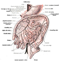
 Second stage of development of the intestinal canal and peritoneum, seen from in front (diagrammatic). The liver has been removed and the two layers of the ventral mesogastrium (lesser omentum) have been cut. The vessels are represented in black and the peritoneum in the reddish tint.
 -
 Third state of the development of the intestinal canal and peritoneum, seen from in front (diagrammatic). The mode of preparation is the same as inFig 400


Function
[edit]Food from the stomach is allowed into the duodenum through thepylorusby a muscle called the pyloric sphincter.
Digestion
[edit]The small intestine is where most chemical digestion takes place. Many of thedigestive enzymesthat act in the small intestine are secreted by thepancreasandliverand enter the small intestine via thepancreatic duct.Pancreatic enzymes andbilefrom the gallbladder enter the small intestine in response to the hormonecholecystokinin,which is produced in the response to the presence of nutrients.Secretin,another hormone produced in the small intestine, causes additional effects on the pancreas, where it promotes the release ofbicarbonateinto the duodenum in order to neutralize the potentially harmful acid coming from the stomach.
The three major classes of nutrients that undergo digestion areproteins,lipids(fats) andcarbohydrates:
- Proteins are degraded into smallpeptidesandamino acidsbefore absorption.[19]Chemical breakdown begins in the stomach and continues in the small intestine. Proteolytic enzymes, includingtrypsinandchymotrypsin,are secreted by thepancreasand cleave proteins into smaller peptides. Carboxypeptidase, which is a pancreaticbrush borderenzyme, splits one amino acid at a time.Aminopeptidaseanddipeptidasefree the end amino acid products.
- Lipids (fats) are degraded intofatty acidsandglycerol.Pancreatic lipase breaks downtriglyceridesinto free fatty acids andmonoglycerides.Pancreatic lipase works with the help of the salts from thebilesecreted by theliverand stored in thegall bladder.Bile salts attach to triglycerides to helpemulsifythem, which aids access by pancreatic lipase. This occurs because the lipase is water-soluble but the fatty triglycerides are hydrophobic and tend to orient towards each other and away from the watery intestinal surroundings. The bile salts emulsify the triglycerides in the watery surroundings until the lipase can break them into the smaller components that are able to enter the villi for absorption.
- Some carbohydrates are degraded into simple sugars, ormonosaccharides(e.g.,glucose). Pancreatic amylase breaks down some carbohydrates (notablystarch) into oligosaccharides. Other carbohydrates pass undigested into the large intestine for further handling byintestinal bacteria.Brush border enzymes take over from there. The most important brush border enzymes are dextrinase and glucoamylase, which further break down oligosaccharides. Other brush border enzymes are maltase, sucrase and lactase. Lactase is absent in some adult humans and, for them, lactose (a disaccharide), as well as most polysaccharides, is not digested in the small intestine. Some carbohydrates, such ascellulose,are not digested at all, despite being made of multipleglucoseunits. This is because the cellulose is made out of beta-glucose, making the inter-monosaccharidal bindings different from the ones present in starch, which consists of Alpha -glucose. Humans lack the enzyme for splitting the beta-glucose-bonds, something reserved for herbivores and bacteria from the large intestine.
Absorption
[edit]Digested food is now able to pass into the blood vessels in the wall of the intestine through eitherdiffusionor active transport. The small intestine is the site where most of the nutrients from ingested food are absorbed. The inner wall, or mucosa, of the small intestine, is lined withintestinal epithelium,asimple columnar epithelium.Structurally, the mucosa is covered in wrinkles or flaps calledcircular folds,which are considered permanent features in the mucosa. They are distinct fromrugaewhich are considered non-permanent or temporary allowing for distention and contraction. From the circular folds project microscopic finger-like pieces of tissue calledvilli(Latinfor "shaggy hair" ). The individual epithelial cells also have finger-like projections known asmicrovilli.The functions of the circular folds, the villi, and the microvilli are to increase the amount of surface area available for the absorption ofnutrients,and to limit the loss of said nutrients to intestinal fauna.
Each villus has a network ofcapillariesand fine lymphatic vessels calledlactealsclose to its surface. The epithelial cells of the villi transport nutrients from the lumen of the intestine into these capillaries (amino acids and carbohydrates) and lacteals (lipids). The absorbed substances are transported via the blood vessels to different organs of the body where they are used to build complex substances such as the proteins required by our body. The material that remains undigested and unabsorbed passes into the large intestine.

Absorption of the majority of nutrients takes place in thejejunum,with the following notable exceptions:
- Ironis absorbed in the duodenum.
- Folate(Vitamin B9) is absorbed in the duodenum and jejunum.
- Vitamin B12andbile saltsare absorbed in theterminal ileum.Vitamin B12 will only be absorbed by the ileum after binding to a protein known asintrinsic factor.
- Water is absorbed byosmosisandlipidsby passive diffusion throughout the small intestine.
- Sodium bicarbonateis absorbed by active transport andglucoseandamino acidco-transport
- Fructoseis absorbed byfacilitated diffusion.
Immunological
[edit]The small intestine supports the body'simmune system.[20]The presence ofgut floraappears to contribute positively to the host's immune system. Peyer's patches,located within the ileum of the small intestine, are an important part of the digestive tract's local immune system. They are part of the lymphatic system, and provide a site for antigens from potentially harmful bacteria or other microorganisms in the digestive tract to be sampled, and subsequently presented to the immune system.[21]
Clinical significance
[edit]The small intestine is a complex organ, and as such, there are a very large number of possible conditions that may affect the function of the small bowel. A few of them are listed below, some of which are common, with up to 10% of people being affected at some time in their lives, while others are vanishingly rare.
- Small intestine obstruction or obstructive disorders
- Meconium ileus
- Paralyticileus
- Volvulus
- Hernia
- Intussusception
- Adhesions
- Obstruction from external pressure
- Obstruction by masses in the lumen (foreign bodies,bezoar,gallstones)
- Infectious diseases
- Giardiasis
- Ascariasis
- Tropical sprue
- Tape worm (Diphyllobothrium latum,Taenia solium,Hymenolepis nana)
- Hookworm (e.g.Necator americanus,Ancylostoma duodenale)
- Nematodes (e.g.Ascaris lumbricoides)
- Other Protozoa (e.g.Cryptosporidium parvum,Cyclospora,Microsporidia,Entamoeba histolytica)
- Bacterial infections
- EnterotoxigenicEscherichia coli
- Salmonella enterica
- Campylobacter
- Shigella
- Yersinia
- Clostridium difficile(antibiotic-associated colitis,Pseudomembranous colitis)
- Mycobacterium(Mycobacterium avium paratuberculosis,disseminatedMycobacterium tuberculosis)
- Whipple's disease
- Vibrio(cholera)
- Enteric (typhoid) fever(Salmonella entericavar. typhii) andparatyphoid fever
- Bacillus cereus
- Clostridium perfringens(gas gangrene)
- Viral infections
- Neoplasms(cancers)
- Adenocarcinoma
- Carcinoid
- Gastrointestinal stromal tumor(GIST)
- Lymphoma
- Sarcoma
- Leiomyoma
- Metastatic tumors, especiallySCLCormelanoma
- Small intestine cancer
- Developmental, congenital or genetic conditions
- Duodenal (intestinal) atresia
- Hirschsprung's disease
- Meckel's diverticulum
- Pyloric stenosis
- Pancreas divisum
- Ectopic pancreas
- Enteric duplication cyst
- Situs inversus
- Cystic fibrosis
- Malrotation
- Persistenturachus
- Omphalocele
- Gastroschisis
- Disaccharidase(lactase) deficiencies
- Primarybile acid malabsorption
- Gardner syndrome
- Familial adenomatous polyposissyndrome (FAP)
- Other conditions
- Crohn's disease,and the more generalinflammatory bowel disease
- Typhlitis(neutropeniccolitisin theimmunosuppressed
- Coeliac disease(sprue or non-tropical sprue)
- Mesenteric ischemia
- Embolusorthrombusof thesuperior mesenteric arteryor thesuperior mesenteric vein
- Arteriovenous malformation
- Gastric dumping syndrome
- Irritable bowel syndrome
- Duodenal (peptic) ulcers
- Gastrointestinal perforation
- Hyperthyroidism
- Diverticulitis
- Radiation enterocolitis
- Mesenteric cysts
- PeritonealInfection
- Sclerosingretroperitonitis
- Small intestinal bacterial overgrowth
- Endometriosis
Other animals
[edit]The small intestine is found in alltetrapodsand also inteleosts,although its form and length vary enormously between species. In teleosts, it is relatively short, typically around one and a half times the length of the fish's body. It commonly has a number ofpyloric caeca,small pouch-like structures along its length that help to increase the overall surface area of the organ for digesting food. There is no ileocaecal valve in teleosts, with the boundary between the small intestine and therectumbeing marked only by the end of the digestive epithelium.[22]
In tetrapods, theileocaecal valveis always present, opening into the colon. The length of the small intestine is typically longer in tetrapods than in teleosts, but is especially so inherbivores,as well as in mammals andbirds,which have a highermetabolic ratethanamphibiansorreptiles.The lining of the small intestine includes microscopic folds to increase its surface area in all vertebrates, but only in mammals do these develop into true villi.[22]
The boundaries between the duodenum, jejunum, and ileum are somewhat vague even in humans, and such distinctions are either ignored when discussing the anatomy of other animals, or are essentially arbitrary.[22]
There is no small intestine as such in non-teleost fish, such assharks,sturgeons,andlungfish.Instead, the digestive part of the gut forms aspiral intestine,connecting the stomach to the rectum. In this type of gut, the intestine itself is relatively straight but has a long fold running along the inner surface in a spiral fashion, sometimes for dozens of turns. This valve greatly increases both the surface area and the effective length of the intestine. The lining of the spiral intestine is similar to that of the small intestine in teleosts and non-mammalian tetrapods.[22]
Inlampreys,the spiral valve is extremely small, possibly because their diet requires little digestion.Hagfishhave no spiral valve at all, with digestion occurring for almost the entire length of the intestine, which is not subdivided into different regions.[22]
Society and culture
[edit]Intraditional Chinese medicine,thesmall intestineis ayangorgan.[23]
Additional images
[edit]-
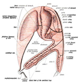
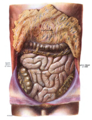 Small intestinein situ,greater omentum folded upwards.
 -
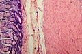 Tissue layers (mucosa, submucosa & muscularis)


References
[edit]- ^Nosek, Thomas M."Section 6/6ch2/s6ch2_30".Essentials of Human Physiology.Archived fromthe originalon 2016-03-24.
- ^human body | Britannica
- ^abcDiBaise, John K.; Parrish, Carol Rees; Thompson, Jon S. (2016).Short Bowel Syndrome: Practical Approach to Management.CRC Press. p. 31.ISBN9781498720809.
- ^Tortora, Gerard (2014).Principles of Anatomy & Physiology.USA: Wiley. pp.913.ISBN978-1-118-34500-9.
..its length is about 3m in a living person and about 6.5m in a cadaver due to loss of smooth muscle tone after death.
- ^Standring, Susan (2016).Gray's Anatomy.UK: Elsevier. p. 1124.ISBN978-0-7020-5230-9.
..and has a mean length of 5 metres (3 - 8.5 metres) when measured intraoperatively in the living adult (Tietelbaum et al 2013).
- ^Jacobs, S.L.; Rozenblit, A.; Ricci, Z.; Roberts, J.; Milikow, D.; Chernyak, V.; Wolf, E. (2007). "Small bowel faeces sign in patients without small bowel obstruction".Clinical Radiology.62(4): 353–357.doi:10.1016/j.crad.2006.11.007.ISSN0009-9260.PMID17331829.
- ^Debora Duro, Daniel Kamin (2007). "Overview of short bowel syndrome and intestinal transplantation".Colombia Médica.38(1).
- ^abAli Nawaz Khan (2016-09-22)."Small-Bowel Obstruction Imaging".Medscape.Retrieved2017-02-07.
- ^"Abdominal X-ray - Abnormal bowel gas pattern".radiologymasterclass.co.uk.Retrieved2017-02-07.
- ^Gazelle, G S; Goldberg, M A; Wittenberg, J; Halpern, E F; Pinkney, L; Mueller, P R (1994)."Efficacy of CT in distinguishing small-bowel obstruction from other causes of small-bowel dilatation".American Journal of Roentgenology.162(1): 43–47.doi:10.2214/ajr.162.1.8273687.ISSN0361-803X.PMID8273687.
- ^Helander, Herbert F; Fändriks, Lars (2015). "Surface area of the digestive tract – revisited".Scandinavian Journal of Gastroenterology.49(6): 681–689.doi:10.3109/00365521.2014.898326.ISSN0036-5521.PMID24694282.S2CID11094705.
- ^Drake, Richard L.; Vogl, Wayne; Tibbitts, Adam W.M. Mitchell; illustrations by Richard; Richardson, Paul (2005).Gray's anatomy for students.Philadelphia: Elsevier/Churchill Livingstone. p. 273.ISBN978-0-8089-2306-0.
- ^Drake, Richard L.; Vogl, Wayne; Tibbitts, Adam W.M. Mitchell; illustrations by Richard; Richardson, Paul (2005).Gray's anatomy for students.Philadelphia: Elsevier/Churchill Livingstone. p. 271.ISBN978-0-8089-2306-0.
- ^abDrake, Richard L.; Vogl, Wayne; Tibbitts, Adam W.M. Mitchell; illustrations by Richard; Richardson, Paul (2005).Gray's anatomy for students.Philadelphia: Elsevier/Churchill Livingstone. pp. 295–299.ISBN978-0-8089-2306-0.
- ^"The human proteome in small intestine - The Human Protein Atlas".proteinatlas.org.Retrieved2017-09-26.
- ^Uhlén, Mathias; Fagerberg, Linn; Hallström, Björn M.; Lindskog, Cecilia; Oksvold, Per; Mardinoglu, Adil; Sivertsson, Åsa; Kampf, Caroline; Sjöstedt, Evelina (2015-01-23). "Tissue-based map of the human proteome".Science.347(6220): 1260419.doi:10.1126/science.1260419.ISSN0036-8075.PMID25613900.S2CID802377.
- ^Gremel, Gabriela; Wanders, Alkwin; Cedernaes, Jonathan; Fagerberg, Linn; Hallström, Björn; Edlund, Karolina; Sjöstedt, Evelina; Uhlén, Mathias; Pontén, Fredrik (2015-01-01). "The human gastrointestinal tract-specific transcriptome and proteome as defined by RNA sequencing and antibody-based profiling".Journal of Gastroenterology.50(1): 46–57.doi:10.1007/s00535-014-0958-7.ISSN0944-1174.PMID24789573.S2CID21302849.
- ^abSchoenwolf, Gary C.; Bleyl, Steven B.; Brauer, Philip R.; Francis-West, Philippa H. (2009). "Development of the Urogenital system".Larsen's human embryology(4th ed.). Philadelphia: Churchill Livingstone/Elsevier. p. 237.ISBN9780443068119.
- ^Silk DB (1974)."Progress report. Peptide absorption in man".Gut.15(6): 494–501.doi:10.1136/gut.15.6.494.PMC1413009.PMID4604970.
- ^"Intestinal immune cells play an unexpected role in immune surveillance of the bloodstream".Massachusetts General Hospital. 13 December 2012. Archived fromthe originalon 16 October 2018.Retrieved28 August2013.
- ^Canny, G. O.; McCormick, B. A. (2008)."Bacteria in the Intestine, Helpful Residents or Enemies from Within?".Infection and Immunity.76(8): 3360–3373.CiteSeerX10.1.1.596.7265.doi:10.1128/IAI.00187-08.ISSN0019-9567.PMC2493210.PMID18474643.
- ^abcdeRomer, Alfred Sherwood; Parsons, Thomas S. (1977).The Vertebrate Body.Philadelphia, PA: Holt-Saunders International. pp. 349–353.ISBN978-0-03-910284-5.
- ^Porter [ed.], Roy (1997).Medicine: a history of healing.[S.l.]: Diane Pub Co. p. 104.ISBN9780756751432.
{{cite book}}:|last1=has generic name (help)
Bibliography
[edit]- Sherwood, Lauralee (2006).Fundamentals of physiology: a human perspective(Third ed.). Florence, KY: Cengage Learning. p. 768.ISBN978-0-534-46697-8.
- Solomon et al. (2002) Biology Sixth Edition, Brooks-Cole/Thomson LearningISBN0-03-033503-5
- Townsend et al. (2004) Sabiston Textbook of Surgery, ElsevierISBN0-7216-0409-9
- Thomson A, Drozdowski L, Iordache C, Thomson B, Vermeire S, Clandinin M, Wild G (2003). "Small bowel review: Normal physiology, part 1".Dig Dis Sci.48(8): 1546–64.doi:10.1023/A:1024719925058.PMID12924651.S2CID37494914.
- Thomson A, Drozdowski L, Iordache C, Thomson B, Vermeire S, Clandinin M, Wild G (2003). "Small bowel review: Normal physiology, part 2".Dig Dis Sci.48(8): 1565–81.doi:10.1023/A:1024724109128.PMID12924652.S2CID42442830.