
Testicle
| Testicle | |
|---|---|
 Diagram of inner structures of a human testicle (the labelling "seminal vesicle lobules" is incorrect and should be "testicular lobules" instead) | |
 Diagram of the external features and surrounding structures of the testicles of an adult male | |
| Details | |
| Artery | Testicular artery |
| Vein | Testicular vein,pampiniform plexus |
| Nerve | Spermatic plexus |
| Lymph | Lumbar lymph nodes |
| Identifiers | |
| Latin | testis |
| MeSH | D013737 |
| TA98 | A09.3.01.001 |
| TA2 | 3576 |
| FMA | 7210 |
| Anatomical terminology | |

Atesticleortestis(pl.:testes) is the malegonadin allbilaterians,including humans. It ishomologousto the femaleovary.The functions of the testicles are to produce bothspermandandrogens,primarilytestosterone.Testosterone release is controlled by the anterior pituitaryluteinizing hormone,whereas sperm production is controlled both by theanterior pituitaryfollicle-stimulating hormoneand gonadal testosterone.
Structure
Appearance

Males have two testicles of similar size contained within thescrotum,which is an extension of theabdominal wall.[1]Scrotal asymmetry, in which one testicle extends farther down into the scrotum than the other, is common. This is because of the differences in the vasculature's anatomy.[1]For 85% of men, the right testis hangs lower than the left one.[1]
Measurement and volume
The volume of the testicle can be estimated by palpating it and comparing it toellipsoids(anorchidometer) of known sizes. Another method is to use calipers, a ruler, or anultrasoundimage to obtain the three measurements of the x, y, and z axes (length, depth and width). These measurements can then be used to calculate the volume, using the formula for the volume of an ellipsoid:


However, the most accurate calculation of actual testicular volume is gained from the formula:[2]

An average adult testicle measures up to5 cm × 2 cm × 3 cm (2 in ×3⁄4in ×1+1⁄4in). TheTanner scale,which is used to assess the maturity of the male genitalia, assigns a maturity stage to the calculated volume ranging from stage I, a volume of less than 1.5 cm3;to stage V, a volume greater than 20 cm3.Normal volume is 15 to 25 cm3;the average is 18 cm3per testis (range 12–30 cm3).[1]
The number of spermatozoa an adult human male produces is directly proportional to testicular volume, as larger testicles contain more seminiferous tubules andSertoli cellsas a result.[3]As such, men with larger testicles produce on average more sperm cells in eachejaculate,as testicular volume is positively correlated with semen profiles.[4]
Internal structure

Duct system
The testes are covered by a tough fibrous shell called thetunica albuginea.[5]Under the tunica albuginea, the testes contain very fine-coiled tubes calledseminiferous tubules.[5]The tubules are lined with a layer of cells (germ cells) that develop frompubertythrough old age intospermcells(also known asspermatozoaor malegametes).[5]The developing sperm travel through the seminiferous tubules to therete testislocated in themediastinum testis,to theefferent ducts,and then to theepididymiswhere newly created sperm cells mature (spermatogenesis).[6]The sperm move into thevas deferens,and are eventually expelled through theurethraand out of theurethral orificethrough muscular contractions.[6]
Primary cell types
Within the seminiferous tubules, the germ cells develop intospermatogonia,spermatocytes,spermatidsand spermatozoa through the process of spermatogenesis. The gametes contain DNA for fertilization of anovum.[7]Sertoli cells – the true epithelium of the seminiferous epithelium, critical for the support of germ cell development into spermatozoa. Sertoli cells secreteinhibin.[8]Peritubular myoid cellssurround the seminiferous tubules.[9]
Between tubules (interstitial cells) existLeydig cells[10]– cells localized between seminiferous tubules that produce and secretetestosteroneand otherandrogensimportant forpuberty(includingsecondary sexual characteristicslike facial hair),sexual behavior,andlibido.Sertoli cells support spermatogenesis.[11]Testosterone controls testicular volume.
Immature Leydig cells and interstitialmacrophagesandepithelial cellsare also present.
Blood supply and lymphatic drainage
The testis has three sources of arterial blood supply: thetesticular artery,thecremasteric artery,and theartery to the ductus deferens.[12]Blood supply andlymphatic drainageof the testes and scrotum are distinct:
- The paired testicular arteries arise directly from theabdominal aortaand descend through theinguinal canal,while the scrotum and the rest of the external genitalia is supplied by theinternal pudendal artery(a branch of theinternal iliac artery).[13][14]
- The testis has collateral blood supply from the cremasteric artery (a branch of theinferior epigastric artery,which is a branch of theexternal iliac artery), and the artery to the ductus deferens (a branch of theinferior vesical artery,which is a branch of the internal iliac artery).[15][16]Therefore, if the testicular artery is ligated, e.g., during a Fowler-Stevensorchiopexyfor a high undescended testis, the testis will usually survive on these other blood supplies.[17]
- Lymphatic drainage of the testes follows the testicular arteries back to theparaaortic lymph nodes,while lymph from the scrotum drains to theinguinal lymph nodes.[13][16]
Layers
Many anatomical features of the adult testis reflect its developmental origin in theabdomen.The layers of tissue enclosing each testicle are derived from the layers of the anteriorabdominal wall.[1]Thecremasteric musclearises from theinternal oblique muscle.[1][18]
The blood–testis barrier
Large molecules cannot pass from the blood into the lumen of a seminiferous tubule due to the presence oftight junctionsbetween adjacent Sertoli cells.[13]The spermatogonia occupy the basal compartment (deep to the level of the tight junctions) and the more mature forms, such as primary and secondary spermatocytes and spermatids, occupy the adluminal compartment.[13]
The function of the blood–testis barrier may be to prevent anauto-immunereaction.[13]Mature sperm (and theirantigens) emerge significantly after immune tolerance is set in infancy.[13]Since sperm are antigenically different from self-tissue, a male animal can react immunologically to his own sperm. The male can make antibodies against them.[13]
Injection of sperm antigens causes inflammation of the testis (auto-immune orchitis) and reduced fertility.[13]The blood–testis barrier may reduce the likelihood that sperm proteins will induce an immune response.[19]
Temperature regulation and responses
Carl Richard Moorein 1926[20]proposed that testicles were external due tospermatogenesisbeing enhanced at temperatures slightly less than core body temperature outside the body. The spermatogenesis is less efficient at lower and higher temperatures than 33 °C. Because the testes are located outside the body, the smooth tissue of the scrotum can move them closer or further away from the body.[5]The temperature of the testes is maintained at 34.4 °C, a little below body temperature, as temperatures above 36.7 °C impede spermatogenesis.[1][5]There are a number of mechanisms to maintain the testes at the optimum temperature.[21]
The cremasteric muscle covers the testicles and thespermatic cord.[22]When this muscle contracts, the cord shortens and the testicles move closer up toward the body, which provides slightly more warmth to maintain optimal testicular temperature.[22]When cooling is required, the cremasteric muscle relaxes and the testicles lower away from the warm body and are able to cool.[22]Contraction also occurs in response tophysical stress,such as blunt trauma; the testicles withdraw and the scrotum shrinks very close to the body in an effort to protect them.[23]
Thecremasteric reflexwill reflexively raise the testicles. The testicles can also be lifted voluntarily using thepubococcygeusmuscle, which partially activates related muscles.
Gene and protein expression
Thehuman genomeincludes approximately 20,000 protein coding genes: 80% of thesegenes are expressedin adult testes.[24]The testes have the highest fraction of tissue type-specific genes compared to other organs and tissues.[25]About 1000 of them are highly specific for the testes,[24]and about 2,200 show an elevated pattern of expression. A majority of these genes encode for proteins that are expressed in the seminiferous tubules and have functions related to spermatogenesis.[25]Sperm cells express proteins that result in the development offlagella;these same proteins are expressed in the female in cells lining thefallopian tubeand cause the development ofcilia.Sperm cell flagella and fallopian tube cilia arehomologousstructures. The testis-specific proteins that show the highest level of expression areprotamines.[26]
Development
There are two phases in which the testes grow substantially. These are the embryonic and pubertal phases. During mammalian development, the gonads are at first capable of becoming eitherovariesor testes.[27]In humans, starting at about week 4, the gonadal rudiments are present within theintermediate mesodermadjacent to the developing kidneys. At about week 6,sex cordsdevelop within the forming testes.[1][28]These are made up of early Sertoli cells that surround and nurture the germ cells that migrate into the gonads shortly before sex determination begins.[1]In males, the sex-specific geneSRYthat is found on theY chromosomeinitiates sex determination by downstream regulation of sex-determining factors (such asGATA4,SOX9andAMH), which lead to development of the male phenotype, including directing development of the early bipotential gonad toward the male path of development.[1]
Testes follow the path of descent, from high in the posterior fetal abdomen to the inguinal ring and beyond to theinguinal canaland into thescrotum.[29]In most cases (97% full-term, 70%preterm), both testes have descended by birth.[29][30]In most other cases, only one testis fails to descend. This is calledcryptorchidism.In most cases of cryptorchidism, the issue will mostly resolve itself within the first half year of life. However, if the testes do not descend far enough into the scrotum, surgical anchoring in the scrotum is required due to risks of infertility andtesticular cancer.[30]
The testes grow in response to the start of spermatogenesis. Size depends on lytic function, sperm production (amount of spermatogenesis present in testis),interstitial fluid,and Sertoli cell fluid production. The testicles are fully descended before the male reaches puberty.
Clinical significance
Protection and injury
- The testicles are very sensitive to impact and injury. The pain involved travels up from each testicle into the abdominal cavity, via thespermatic plexus,which is the primary nerve of each testicle.[31]This will cause pain in the hip and the back. The pain usually fades within a few minutes.
- Testicular torsionis a medical emergency. This is because the longer it takes to access medical intervention with respect to extending ischemia, the higher the chance that the testicle will be lost. There is a 90% chance to save the testicle if de-torsion surgery is performed within six hours of testicular torsion onset.[32]
- Testicular ruptureis severe trauma affecting the tunica albuginea.[33]
- Penetrating injuries to the scrotum may causecastration,or physical separation or destruction of the testes, possibly along with part or all of the penis, which results in total sterility if the testicles are not reattached quickly. In an effort to avoid severe infection, ample application ofsalineandbacitracinhelp remove debris and foreign objects from the wound.[34]
- Jockstrapssupport and protect the testicles.
Diseases and conditions
| Testicular disease | |
|---|---|
| Specialty | Urology,Reproductive medicine |
- To improve the chances of catching cases oftesticular cancer,otherneoplasms,or other health issues early, regulartesticular self-examinationis recommended.
- Varicocele,swollen vein(s) from the testes, usually affecting the left side, the testis usually being normal.[35][36]
- Hydrocele testisis swelling around testes caused by accumulation of clear liquid within a membranous sac, the testis usually being normal. It is the most common cause of scrotal swelling.[37]
- Spermatoceleis a retention cyst of a tubule of therete testisor the head of theepididymisdistended with barely watery fluid that contains spermatozoa.[36]
- Endocrine disorderscan also affect the size and function of the testis.
- Certain inherited conditions involving mutations in key developmental genes also impair testicular descent, resulting in abdominal or inguinal testes, which remain nonfunctional and may become cancerous.[38]Other genetic conditions can result in the loss of theWolffian ductsand allow for the persistence ofMüllerian ducts.Both excess and deficient levels ofestrogenscan disruptspermatogenesisand causeinfertility.[39]
- Bell-clapper deformityis a deformity in which the testicle is not attached to the scrotal walls, and can rotate freely on the spermatic cord within the tunica vaginalis. Those with Bell-clapper are at a higher risk of testicular torsion.[40][41]
- Orchitisis inflammation of the testicles
- Epididymitisis a painful inflammation of the epididymis or epididymides, frequently caused by bacterial infection but sometimes of unknown origin.
- Anorchiais the absence of one or both testicles.
- Cryptorchidism,or "undescended testicles", is when the testicle does not descend into the scrotum of an infant boy.[30]
- Testicular enlargement is anunspecificsign of various testicular diseases, and can be defined as a testicular size of more than 5 cm (long axis) × 3 cm (short axis).[42]
- Blue ballsis a condition concerning temporary fluid congestion in the testicles andprostateregion, caused by prolongedsexual arousal.
Testicular prosthesesare available to mimic the appearance and feel of one or both testicles, when absent as from injury or as treatment in association togender dysphoria.There have also been some instances of their implantation in dogs.
Scientists are working on developing lab-grown testicles that might help infertile men in the future.[43]
Effects of exogenous hormones
To some extent, it is possible to change testicular size. Short of direct injury or subjecting them to adverse conditions, e.g., higher temperature than they are normally accustomed to, they can be shrunk by competing against their intrinsic hormonal function through the use of externally administeredsteroidalhormones. Steroids taken for muscle enhancement (especiallyanabolic steroids) often have the undesired side effect of testicular shrinkage.
Stimulation of testicular functions viagonadotropic-like hormonesmay enlarge their size. Testes may shrink or atrophy duringhormone replacement therapyor throughchemical castration.
In all cases, the loss in testes volume corresponds with a loss of spermatogenesis.
Society and culture

The testicles ofcalves,lambs,roosters,turkeys,and other animals are eaten in many parts of the world, often undereuphemisticculinary names.Testicles are a by-product of the castration of young animals raised for meat, so they might have been a late-spring seasonal specialty.[44]In modern times, they are generally frozen and available year-round.
In theMiddle Ages,men who wanted a boy sometimes had their left testicle removed. This was because people believed that the right testicle made "boy" sperm and the left made "girl" sperm.[45]As early as 330 BC,Aristotleprescribed the ligation (tying off) of the left testicle in men wishing to have boys.[46]
Etymology and slang
One theory about the etymology of the wordtestisis based onRoman law.The originalLatinwordtestis,"witness", was used in the firmly established legal principle "Testis unus, testis nullus"(one witness [equals] no witness), meaning that testimony by any one person in court was to be disregarded unless corroborated by the testimony of at least another. This led to the common practice of producing two witnesses, bribed to testify the same way in cases of lawsuits with ulterior motives. Since such witnesses always came in pairs, the meaning was accordingly extended, often in the diminutive (testiculus, testiculi).[citation needed]
Another theory says thattestisis influenced by a loan translation, from Greekparastatēs"defender (in law), supporter" that is "two glands side by side".[47]
There are multiple slang terms for the testes. They may be referred to as "balls". Frequently, "nuts"(sometimes intentionally misspelled as" nutz ") are also a slang term for the testes due to the geometric resemblance. One variant of the term includes" Deez Nuts ", which was used for asatirical political candidate in 2016.
InSpanish,the termhuevosis used, which is Spanish foreggs.
Other animals

External appearance
Inseasonal breeders,the weight of the testes often increases during the breeding season.[48]Thetesticles of a dromedary camelare 7–10 cm (2.8–3.9 in) long, 4.5 cm (1.8 in) deep and 5 cm (2.0 in) in width. The right testicle is often smaller than the left.[49]
Insharks,the testicle on the right side is usually larger. In many bird and mammal species, the left may be larger.Fish usually have two testesof a similar size. The primitivejawless fishhave only a single testis, located in the midline of the body, although this forms from the fusion of paired structures in the embryo.[50]
Location
Internal
The basal condition for mammals is to have internal testes.[51]The testes ofmonotremes,[52][53]xenarthrans,[53]andafrotherians[54]remain within the abdomen (testicondy). There are also somemarsupialswith external testes[55][56][57]andboreoeutherianmammals with internal testes, such as the rhinoceros.[58]Cetaceanssuch as whales and dolphins also have internal testes.[59][60]As external testes would increase drag in the water, they have internal testes, which are kept cool by special circulatory systems that cool the arterial blood going to the testes by placing the arteries near veins bringing cooled venous blood from the skin.[61][62]Inodobenidsandphocids,the location of the testes is para-abdominal, thoughotariidshave scrotal testes.[63]
External
Boreoeutherianland mammals, the large group of mammals that includes humans, have externalized testes.[64]Their testes function best at temperatures lower than their core body temperature. Their testes are located outside of the body and are suspended by the spermatic cord within the scrotum.
There are several hypotheses as to why most boreotherian mammals have external testes that operate best at a temperature that is slightly less than the core body temperature. One view is that it is stuck with enzymes evolved in a colder temperature due to external testes evolving for different reasons. Another view is that the lower temperature of the testes simply is more efficient for sperm production.
The classic hypothesis is that cooler temperature of the testes allows for more efficient fertile spermatogenesis. There are no possible enzymes operating at normal core body temperature that are as efficient as the ones evolved.
Early mammals had lower body temperatures and thus their testes worked efficiently within their body. However, boreotherian mammals may have higher body temperatures than the other mammals and had to develop external testes to keep them cool. One argument is that mammals with internal testes, such as the monotremes, armadillos, sloths, elephants, and rhinoceroses, have a lower core body temperatures than those mammals with external testes.[citation needed]
Researchers have wondered why birds, despite having very high core body temperatures, have internal testes and did not evolve external testes.[65]It was once theorized that birds used theirair sacsto cool the testes internally, but later studies revealed that birds' testes are able to function at core body temperature.[65]
Some mammals with seasonal breeding cycles keep their testes internal until the breeding season. After that, their testes descend and increase in size and become external.[66]
The ancestor of the boreoeutherian mammals may have been a small mammal that required very large testes forsperm competitionand thus had to place its testes outside the body.[67]This might have led to enzymes involved in spermatogenesis, spermatogenicDNA polymerase betaandrecombinaseactivities evolving a unique temperature optimum that is slightly less than core body temperature. When the boreoeutherian mammals diversified into forms that were larger or did not require intense sperm competition, they still produced enzymes that operated best at cooler temperatures and had to keep their testes outside the body. This position is made less parsimonious because thekangaroo,a non-boreoeutherian mammal, has external testicles. Separately from boreotherian mammals, the ancestors of kangaroos might have also been subject to heavy sperm competition and thus developed external testes; however, kangaroo external testes are suggestive of a possible adaptive function for external testes in large animals.
One argument for theevolution of external testesis that it protects the testes from abdominal cavity pressure changes caused by jumping and galloping.[68]
Mild, transient scrotal heat stress causes DNA damage,reduced fertilityand abnormal embryonic development in mice.[69]DNA strand breaks were found in spermatocytes recovered from testicles subjected to 40 °C or 42 °C for 30 minutes.[69]These findings suggest that the external location of the testicles provides the adaptive benefit of protecting spermatogenic cells from heat-induced DNA damage that could otherwise lead to infertility andgermline mutation.
Size

Therelative size of the testesis often influenced bymating systems.[70]Testicular size as a proportion of body weight varies widely. In the mammalian kingdom, there is a tendency for testicular size to correspond with multiple mates (e.g., harems,polygamy). Production of testicular output sperm and spermatic fluid is also larger in polygamous animals, possibly aspermatogenic competitionfor survival. The testes of theright whaleare likely to be the largest of any animal, each weighing around 500 kg (1,100 lb).[71]
Among theHominidae,gorillashave littlefemale promiscuityand sperm competition and the testes are small compared to body weight (0.03%).Chimpanzeeshave high promiscuity and large testes compared to body weight (0.3%). Human testicular size falls between these extremes (0.08%).[72]
Testis weight also varies in seasonal breeders likered foxes,[73]golden jackals,[74]andcoyotes.[48]
Internal structure
Amphibiansand most fish do not possess seminiferous tubules. Instead, the sperm are produced in spherical structures calledsperm ampullae.These are seasonal structures, releasing their contents during the breeding season, and then being reabsorbed by the body. Before the next breeding season, new sperm ampullae begin to form and ripen. The ampullae are otherwise essentially identical to the seminiferous tubules inhigher vertebrates,including the same range of cell types.[50]
Gallery
-
 Testicle
 -
 Testicle
 -
 Testicle hanging on cremaster muscle. These are two healthy testicles. Heat causes them to descend, allowing cooling.
 -
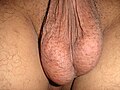
 A healthyscrotumcontaining normal size testes. The scrotum is in tight condition. The image also shows the texture.
 -
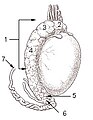
 Testicle of a cat: 1: Extremitas capitata, 2: Extremitas caudata, 3: Margo epididymalis, 4: Margo liber, 5: Mesorchium, 6: Epididymis, 7: testicular artery and vene, 8: Ductus deferens
 -
 Testis surface
 -
 Testis cross section
 -
 The right testis, exposed by laying open the tunica vaginalis.
 -
 Microscopic view of rabbit testis 100×
 -
 Testicle


See also
General and cited references
- Heptner, V. G.; Naumov, N. P. (1998).Mammals of the Soviet Union Vol. II Part 1a, SIRENIA AND CARNIVORA (Sea cows; Wolves and Bears).Enfield, NH: Science Publishers.ISBN978-1-886106-81-9.OCLC490089621.Retrieved9 November2013.
Citations
- ^abcdefghijSteger, Klaus; Weidner, Wolfgang (2011)."Anatomy of the Male Reproductive System".Practical Urology: Essential Principles and Practice.Springer Science & Business Media. pp. 57–59.ISBN978-1-84-882034-0.Archivedfrom the original on 2023-06-29.Retrieved2022-06-01.
- ^Lao, Michael; Smith, Shannon; Gilbert, Bruce R. (2020)."Male Reproductive Ultrasound".Practical Urological Ultrasound.Springer Nature. p. 298.ISBN978-3-03-052309-1.Archivedfrom the original on 2023-06-29.Retrieved2022-07-05.
- ^Rhoades, Rodney A.; Bell, David R. (2012).Medical Physiology: Principles for Clinical Medicine.Lippincott Williams & Wilkins. p. 681.ISBN978-1-60-913427-3.Archivedfrom the original on 2023-06-29.Retrieved2022-07-05.
- ^Condorelli, Rosita; Calogero, Aldo E.; La Vignera, Sandro (2013)."Relationship between Testicular Volume and Conventional or Nonconventional Sperm Parameters".International Journal of Endocrinology.2013:1–6.doi:10.1155/2013/145792.PMC3780703.PMID24089610.
- ^abcdeCho, S; Bae, J.H. (2017)."Penis and Testis".Clinical Regenerative Medicine in Urology.Springer. p. 281.ISBN978-9-81-102723-9.Archivedfrom the original on 2023-06-29.Retrieved2022-06-01.
- ^abPocock, Gillian; Richards, Christopher D.; Richards, David A. (2018).Human Physiology.Oxford University Press. p. 766.ISBN978-0-19-873722-3.Archivedfrom the original on 2023-06-29.Retrieved2022-06-02.
- ^Histology, A Text and AtlasArchived2023-06-29 at theWayback Machineby Michael H. Ross and Wojciech Pawlina, Lippincott Williams & Wilkins, 5th ed, 2006[page needed]
- ^Huhtaniemi, Ilpo (2018).Encyclopedia of Endocrine Diseases.Academic Press. p. 667.ISBN978-0-12-812200-6.Archivedfrom the original on 2023-06-29.Retrieved2022-06-02.
- ^Schlegel, P.N.; Katzovitz, M.A. (2020)."Male Reproductive Physiology".Urologic Principles and Practice.Springer Nature. p. 50.ISBN978-3-03-028599-9.Archivedfrom the original on 2023-06-29.Retrieved2022-06-02.
- ^Bitzer, Johannes; Mahmood, Tahir A. (2022).Handbook of Contraception and Sexual Reproductive Healthcare.Cambridge University Press. p. 16.ISBN978-1-10-895863-9.Archivedfrom the original on 2023-06-29.Retrieved2022-07-05.
- ^Miell, John; Davies, Zoe (2014)."Reproductive function in the male".Clinical Biochemistry: Metabolic and Clinical Aspects.Elsevier Health Sciences. p. 451.ISBN978-0-70-205478-5.Archivedfrom the original on 2023-06-29.Retrieved2022-07-05.
- ^Goldenberg, Etai; Benjamin, Tavya G.R; Gilbert, Bruce R. (2020)."Scrotal Ultrasound".Practical Urological Ultrasound.Springer Nature. p. 80.ISBN978-3-03-052309-1.Archivedfrom the original on 2023-06-29.Retrieved2022-07-06.
- ^abcdefghSteger, Klaus; Weidner, Wolfgang (2011)."Anatomy of the Male Reproductive System".Practical Urology: Essential Principles and Practice.Springer Science & Business Media. p. 63.ISBN978-1-84-882034-0.Archivedfrom the original on 2023-06-29.Retrieved2022-06-05.
- ^Tortora, Gerard J.; Nielsen, Mark (2017).Principles of Human Anatomy.John Wiley & Sons. p. 486.ISBN978-1-11-944446-6.Archivedfrom the original on 2023-06-29.Retrieved2022-07-06.
- ^Pua, Bradley B.; Covey, Anne M.; Madoff, David C. (2018).Interventional Radiology: Fundamentals of Clinical Practice.Oxford University Press. p. 533.ISBN978-0-19-027625-6.Archivedfrom the original on 2023-06-29.Retrieved2022-07-06.
- ^abBerney, Daniel M; Ulbright, Thomas M. (2015)."Anatomy of the Testis and Staging of its Cancers: Implications for Diagnosis".Genitourinary Pathology: Practical Advances.Springer. p. 436.ISBN978-1-49-392044-0.Archivedfrom the original on 2023-06-29.Retrieved2022-07-06.
- ^Aboumarzouk, Omar M. (2019).Blandy's Urology.John Wiley & Sons. p. 747.ISBN978-1-11-886336-7.Archivedfrom the original on 2023-06-29.Retrieved2022-07-06.
- ^Tubbs, R. Shane; Shoja, Mohammadali M.; Loukas, Marios (2016).Bergman's Comprehensive Encyclopedia of Human Anatomic Variation.John Wiley & Sons. p. 1393.ISBN978-1-11-843068-2.Archivedfrom the original on 2023-06-29.Retrieved2022-06-03.
- ^Wiser, Herbert J.; Sandlow, Jay; Kohler, Tobias S. (2012)."Causes of Male Infertility".Male Infertility: Contemporary Clinical Approaches, Andrology, ART & Antioxidants.Springer Science & Business Media. p. 8.ISBN978-1-46-143335-4.Archivedfrom the original on 2023-06-29.Retrieved2022-07-10.
- ^Moore, Carl R. (1926). "The Biology of the Mammalian Testis and Scrotum".The Quarterly Review of Biology.1(1): 4–50.doi:10.1086/394235.ISSN0033-5770.
- ^Coad, Jane; Pedley, Kevin; Dunstall, Melvyn (2019).Anatomy and Physiology for Midwives E-Book.Elsevier Health Sciences. pp. 53–54.ISBN978-0-70-206665-8.Archivedfrom the original on 2023-06-29.Retrieved2022-06-17.
- ^abcde Jong, M. Robert (2020).Sonography Scanning E-Book: Principles and Protocols.Elsevier Health Sciences. p. 343.ISBN978-0-32-376425-4.Archivedfrom the original on 2023-06-29.Retrieved2022-06-05.
- ^Song, David H; Neligan, Peter C (2017).Plastic Surgery E-Book: Volume 4: Trunk and Lower Extremity.Elsevier Health Sciences. p. 293.ISBN978-0-32-335707-4.Archivedfrom the original on 2023-06-29.Retrieved2022-06-10.
- ^abUhlén, Mathias; Fagerberg, Linn; Hallström, Björn M.; Lindskog, Cecilia; Oksvold, Per; Mardinoglu, Adil; Sivertsson, Åsa; Kampf, Caroline; Sjöstedt, Evelina (2015-01-23). "Tissue-based map of the human proteome".Science.347(6220): 1260419.doi:10.1126/science.1260419.ISSN0036-8075.PMID25613900.S2CID802377.
- ^abDjureinovic, D.; Fagerberg, L.; Hallström, B.; Danielsson, A.; Lindskog, C.; Uhlén, M.; Pontén, F. (2014-06-01)."The human testis-specific proteome defined by transcriptomics and antibody-based profiling".MHR: Basic Science of Reproductive Medicine.20(6): 476–488.doi:10.1093/molehr/gau018.ISSN1360-9947.PMID24598113.
- ^Hammoud, S; Carrell, D.T. (2011)."The Emerging Role of the Sperm Epigenome and its Potential Role in Development".Biennial Review of Infertility: Volume 2, 2011, Volume 2;Volume 2011.Springer Science & Business Media. p. 184.ISBN978-1-44-198456-2.Archivedfrom the original on 2023-06-29.Retrieved2022-06-16.
- ^Online textbook: "Developmental BiologyArchived2018-04-05 at theWayback Machine"6th ed. By Scott F. Gilbert (2000) published by Sinauer Associates, Inc. of Sunderland (MA).
- ^Khurana, Indu; Khurana, Arushi (2015).Textbook of Medical Physiology - E-book.Elsevier Health Sciences. p. 807.ISBN978-8-13-124254-4.Archivedfrom the original on 2023-06-29.Retrieved2022-06-14.
- ^abMoore, Keith L.; Persaud, T. V. N.; Torchia, Mark G. (2018).The Developing Human - E-Book: Clinically Oriented Embryology.Elsevier Health Sciences. p. 259.ISBN978-0-32-361156-5.Archivedfrom the original on 2023-06-29.Retrieved2022-06-11.
- ^abcWinkler, Stephan; Dalkowski, Katja; Mair, Jörg; Klebe, Sonja (2018).Sobotta Anatomy Textbook: English Edition with Latin Nomenclature.Elsevier Health Sciences. p. 374.ISBN978-0-72-067617-4.Archivedfrom the original on 2023-06-29.Retrieved2022-06-13.
- ^Kulkarni, Neeta V (2015).Clinical Anatomy: A Problem Solving Approach Author.JP Medical Ltd. p. 621.ISBN978-9-35-152966-8.Archivedfrom the original on 2023-06-29.Retrieved2022-07-07.
- ^Ovadia, Aaron E; Yang, Hailiu; Neiderberger, Craig S.; Ho, Christina; Sabia, Michael; Seftel, Allen D. (2017)."Scrotal Pain".Urogenital Pain: A Clinicians Guide to Diagnosis and Interventional Treatments.Springer. p. 108.ISBN978-3-3-1945794-9.Archivedfrom the original on 2023-06-29.Retrieved2022-07-12.
- ^Bucci, Stefano; Rizzo, Michele; Liguori, Giovanni; Umari, Paolo; Chiriaco, Giovanni; Bertolotto, Michele (2017)."The Testicles: Trauma, Inflammation and Testicular Torsion".Atlas of Ultrasonography in Urology, Andrology, and Nephrology.Springer. p. 500.ISBN978-3-31-940782-1.Archivedfrom the original on 2023-06-29.Retrieved2022-07-07.
- ^Wessells, Hunter (2013).Urological Emergencies: A Practical Approach.Springer Science & Business Media. p. 96.ISBN978-1-62-703423-4.Archivedfrom the original on 2023-06-29.Retrieved2022-07-12.
- ^Medical Symptoms.Penguin. 2022. p. 211.ISBN978-0-74-406302-8.Archivedfrom the original on 2023-06-29.Retrieved2022-07-13.
- ^abKamaya, Aya; Wong-You-Cheong, Jade (2021).Diagnostic Ultrasound: Abdomen and Pelvis E-Book Diagnostic Ultrasound.Elsevier Health Sciences. p. 938.ISBN978-0-32-379403-9.Archivedfrom the original on 2023-06-29.Retrieved2022-07-13.
- ^Kumar, Vinay; Abbas, Abul K.; Aster, Jon C. (2017).Robbins Basic Pathology E-Book Robbins Pathology.Elsevier Health Sciences. p. 692.ISBN978-0-32-339413-0.Archivedfrom the original on 2023-06-29.Retrieved2022-07-13.
- ^Jameson, J. Larry; De Groot, Leslie J. (2015).Endocrinology: Adult and Pediatric.Elsevier Health Sciences. p. 2363.ISBN978-0-32-332195-2.Archivedfrom the original on 2023-06-29.Retrieved2022-07-14.
- ^Sierens, J. E.; Sneddon, S. F.; Collins, F.; Millar, M. R.; Saunders, P. T. (2005). "Estrogens in Testis Biology".Annals of the New York Academy of Sciences.1061(1): 65–76.Bibcode:2005NYASA1061...65S.doi:10.1196/annals.1336.008.PMID16467258.S2CID24905596.
- ^Soto, Jorge A; Lucey, Brian (2016).Emergency Radiology: The Requisites E-Book.Elsevier Health Sciences. p. 202.ISBN978-0-32-339008-8.Archivedfrom the original on 2023-06-29.Retrieved2022-07-14.
- ^Melmed, Shlomo; Koenig, Ronald; Rosen, Clifford; Auchus, Richard; Goldfine, Allison (2019).Williams Textbook of Endocrinology E-Book.Elsevier Health Sciences. p. 669.ISBN978-0-32-371154-8.Archivedfrom the original on 2023-06-29.Retrieved2022-07-14.
- ^Page 559in:John Pellerito, Joseph F Polak (2012).Introduction to Vascular Ultrasonography(6 ed.). Elsevier Health Sciences.ISBN9781455737666.
- ^Dewan, Pandora (2024-02-19)."Scientists create lab-grown testicles".Newsweek.Retrieved2024-02-20.
- ^Mason, Laura (2014). Davidson, Alan (ed.).The Oxford Companion to Food.Oxford University Press. p. 816.ISBN9780199677337.Archivedfrom the original on 2023-06-29.Retrieved2021-12-05.
- ^"Understanding Genetics".Genetics.TheTech.org.Archivedfrom the original on 9 November 2013.Retrieved25 January2018.
- ^Hoag, Hannah.I'll take a girl, please...Cherry-picking from the dish of life.Drexel University Publication.ArchivedAugust 31, 2011, at theWayback Machine
- ^The American Heritage Dictionary of the English Language,Fourth Edition
- ^abA.D. Johnson (2 December 2012).Development, Anatomy, and Physiology.Elsevier.ISBN978-0-323-14323-3.Archivedfrom the original on 29 June 2023.Retrieved19 October2020.
- ^Mukasa-Mugerwa, E.The Camel (Camelus dromedarius): A Bibliographical Review.pp. 11–3.
- ^abRomer, Alfred Sherwood; Parsons, Thomas S. (1977).The Vertebrate Body.Philadelphia: Holt-Saunders International. pp. 385–386.ISBN978-0-03-910284-5.
- ^Kleisner, Karel; Ivell, Richard; Flegr, Jaroslav (March 2010)."The evolutionary history of testicular externalization and the origin of the scrotum".Journal of Biosciences.35(1). Indian Academy of Sciences: 27–37.doi:10.1007/s12038-010-0005-7.PMID20413907.S2CID11962872.Archived fromthe originalon 25 February 2018.Retrieved8 December2018.
- ^Mervyn Griffiths (2 December 2012).The Biology of the Monotremes.Elsevier Science.ISBN978-0-323-15331-7.Archivedfrom the original on 29 June 2023.Retrieved19 October2020.
- ^abRonald M. Nowak; Ernest Pillsbury Walker (29 July 1999).Walker's Mammals of the World.JHU Press.ISBN978-0-8018-5789-8.
testes.
- ^Murray Fowler; Susan K. Mikota (9 January 2008).Biology, Medicine, and Surgery of Elephants.John Wiley & Sons.ISBN978-0-470-34411-8.Archivedfrom the original on 29 June 2023.Retrieved19 October2020.
- ^Don II Hunsaker (2 December 2012).The Biology of Marsupials.Elsevier Science.ISBN978-0-323-14620-3.Archivedfrom the original on 29 June 2023.Retrieved19 October2020.
- ^C. Hugh Tyndale-Biscoe (2005).Life of Marsupials.Csiro Publishing.ISBN978-0-643-06257-3.Archivedfrom the original on 2023-06-29.Retrieved2020-10-19.
- ^Hugh Tyndale-Biscoe; Marilyn Renfree (30 January 1987).Reproductive Physiology of Marsupials.Cambridge University Press.ISBN978-0-521-33792-2.Archivedfrom the original on 29 June 2023.Retrieved19 October2020.
- ^Schaffer, N. E., et al. "Ultrasonography of the reproductive anatomy in the Sumatran rhinoceros (Dicerorhinus sumatrensis)Archived2017-09-23 at theWayback Machine."Journal of Zoo and Wildlife Medicine (1994): 337-348.
- ^Bernd Würsig; J.G.M. Thewissen; Kit M. Kovacs (27 November 2017).Encyclopedia of Marine Mammals.Elsevier Science.ISBN978-0-12-804381-3.Archivedfrom the original on 29 June 2023.Retrieved19 October2020.
- ^Rommel, S.A.; Pabst, D.A.; McLellan, W.A. (2007)."Functional anatomy of the cetacean reproductive system, with comparisons to the domestic dog".In Miller, D.L. (ed.).Reproductive Biology and Phylogeny of Cetacea: Whales, Porpoises and Dolphins.pp. 127–145.doi:10.1201/b11001.ISBN9780429063626.
- ^Rommel, S.A.; Pabst, D.A.; McLellan, W.A. (1998)."Reproductive Thermoregulation in Marine Mammals"(PDF).American Scientist.Vol. 86, no. 5. pp. 440–448.JSTOR27857097.Archived(PDF)from the original on 22 November 2021.
- ^Pabst, D.A.; Sentiel, A.R; McLellan, W.A. (1998). "Evolution of thermoregulatory function in cetacean reproductive systems". In Thewissen, J.G.M. (ed.).The Emergence of Whales.Advances in Vertebrate Paleobiology. Springer US. pp. 379–397.doi:10.1007/978-1-4899-0159-0_13.ISBN978-1-4899-0161-3.
- ^Frances M.D. Gulland; Leslie A. Dierauf; Karyl L. Whitman (20 March 2018).CRC Handbook of Marine Mammal Medicine.CRC Press.ISBN978-1-351-38416-2.Archivedfrom the original on 29 June 2023.Retrieved23 January2020.
- ^D. S. Mills; Jeremy N. Marchant-Forde (2010).The Encyclopedia of Applied Animal Behaviour and Welfare.CABI. pp. 293–.ISBN978-0-85199-724-7.Archivedfrom the original on 2023-06-29.Retrieved2020-10-19.
- ^abBIOLOGY OF REPRODUCTION 56, 1570–1575 (1997)- Determination of Testis Temperature Rhythms and Effects of Constant Light on Testicular Function in the Domestic Fowl (Gallus domesticus)Archived2015-09-23 at theWayback Machine
- ^"Ask a Biologist Q&A / Human sexual physiology – good design?".Askabiologist.org.uk. 4 September 2007.Archivedfrom the original on 27 October 2010.Retrieved25 October2010.
- ^"'The Human Body as an Evolutionary Patchwork' by Alan Walker, Princeton.edu ".RichardDawkins.net. 2007-04-10. Archived fromthe originalon 9 November 2013.Retrieved25 October2010.
- ^"Science: Bumpy lifestyle led to external testes".Archivedfrom the original on 2015-02-22.Retrieved2017-09-15.
- ^abPaul C, Murray AA, Spears N, Saunders PT (2008)."A single, mild, transient scrotal heat stress causes DNA damage, subfertility and impairs formation of blastocysts in mice".Reproduction.136(1): 73–84.doi:10.1530/REP-08-0036.PMID18390691.
- ^Pitcher, T.E.; Dunn, P.O.; Whittingham, L.A. (2005)."Sperm competition and the evolution of testes size in birds".Journal of Evolutionary Biology.18(3): 557–567.doi:10.1111/j.1420-9101.2004.00874.x.PMID15842485.S2CID18331398.
- ^Crane, J.; Scott, R. (2002)."Eubalaena glacialis".Animal Diversity Web.Archivedfrom the original on 28 March 2022.Retrieved1 May2009.
- ^Shackelford, T. K.; Goetz, A. T. (2007). "Adaptation to Sperm Competition in Humans".Current Directions in Psychological Science.16:47–50.doi:10.1111/j.1467-8721.2007.00473.x.S2CID6179167.
- ^Heptner & Naumov 1998,p. 537
- ^Heptner & Naumov 1998,pp. 154–155