
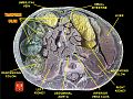
Transverse colon
| Transverse colon | |
|---|---|
 Drawing ofcolonseen from front (transverse colon coloured blue) | |
 | |
| Details | |
| Precursor | Midgut(first 2/3)Hindgut(last 1/3) |
| Artery | Middle colic artery |
| Vein | Middle colic vein |
| Identifiers | |
| Latin | colon transversum |
| MeSH | D044684 |
| TA98 | A05.7.03.004 |
| TA2 | 2984 |
| FMA | 14546 |
| Anatomical terminology | |
Inhuman anatomy,thetransverse colonis the longest and most movable part of thecolon.
Anatomical position
[edit]It crosses theabdomenfrom theascending colonat theright colic flexure(hepatic flexure) with a downward convexity to thedescending colonwhere it curves sharply on itself beneath the lower end of thespleenforming theleft colic flexure(splenic flexure). In its course, it describes an arch, the concavity of which is directed backward and a little upward. Toward its splenic end there is often an abrupt U-shaped curve which may descend lower than the main curve.
It is almost completely invested by theperitoneum,and is connected to the inferior border of thepancreasby a large and wide duplicature of that membrane, thetransverse mesocolon.
It is in relation, by its upper surface, with theliverandgall-bladder,thegreater curvature of the stomach,and the lower end of thespleen;by its under surface, with thesmall intestine;by its anterior surface, with the posterior layer of thegreater omentumand theabdominal wall;its posterior surface is in relation from right to left with the descending portion of theduodenum,thehead of the pancreas,and some of the convolutions of thejejunumandileum.
Function
[edit]The transverse colon absorbs water and salts.
Additional images
[edit]-

-
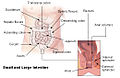
![Inner diameters of different sections of the large intestine, with transverse colon (at top) measuring on average 5.8 cm (range 5.0-6.5 cm).[1]](https://upload.wikimedia.org/wikipedia/commons/thumb/2/22/Diameters_of_the_large_intestine.svg/120px-Diameters_of_the_large_intestine.svg.png) Inner diameters of different sections of the large intestine, with transverse colon (at top) measuring on average 5.8 cm (range 5.0-6.5 cm).[1]
Inner diameters of different sections of the large intestine, with transverse colon (at top) measuring on average 5.8 cm (range 5.0-6.5 cm).[1] -
 Intestines
 -
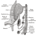 Schematic figure of the bursa omentalis, etc. The human embryo of eight weeks
 -
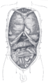 Front view of the thoracic and abdominal viscera
 -
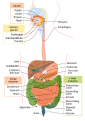 Digestive system
 -
 Transverse colon

![Inner diameters of different sections of the large intestine, with transverse colon (at top) measuring on average 5.8 cm (range 5.0-6.5 cm).[1]](/translate/en.wikipedia.org?u=https%3A%2F%2Fen.wikipedia.org%2Fwiki%2FFile%3ADiameters_of_the_large_intestine.svg&t=vi)


See also
[edit]References
[edit]- ^Nguyen H, Loustaunau C, Facista A, Ramsey L, Hassounah N, Taylor H, Krouse R, Payne CM, Tsikitis VL, Goldschmid S, Banerjee B, Perini RF, Bernstein C (2010)."Deficient Pms2, ERCC1, Ku86, CcOI in field defects during progression to colon cancer".J Vis Exp(41).doi:10.3791/1931.PMC3149991.PMID20689513.
![]() This article incorporates text in thepublic domainfrompage 1180of the 20th edition ofGray's Anatomy(1918)
This article incorporates text in thepublic domainfrompage 1180of the 20th edition ofGray's Anatomy(1918)
External links
[edit]- Anatomy figure: 37:06-03at Human Anatomy Online, SUNY Downstate Medical Center - "The large intestine."