Bak
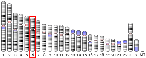
Bak(Bcl-2 homologous antagonist/killer)またはBAK1(BCL2 antagonist/killer 1)は、ヒトでは6番染色体のBAK1遺伝子にコードされるタンパク質である[4][5]。この遺伝子にコードされるタンパク質は、Bcl-2タンパク質ファミリーに属する。Bcl-2ファミリーのメンバーはオリゴマーまたはヘテロ二量体を形成し、さまざまな細胞活動に関与して抗アポトーシスまたはアポトーシス促進調節因子として作用する。このタンパク質はミトコンドリアに局在し、アポトーシスを誘導する機能を果たす。ミトコンドリアの電位依存性アニオンチャネル(VDAC)と相互作用して開口を促進し、膜電位の喪失とシトクロムcの放出を引き起こす。また、このタンパク質は細胞ストレスへの曝露後にがん抑制因子であるp53と相互作用する[6]。
構造
[編集]BAK1はアポトーシス促進性のBcl-2ファミリータンパク質であり、BH1、BH2、BH3、BH4の4つのBcl-2相同ドメイン(BHドメイン)を持つ。これらのドメインは9本のαヘリックスからなり、疎水的なαヘリックスのコアが両親媒性ヘリックスとC末端の膜貫通ヘリックスに囲まれた形状をしている。膜貫通ヘリックスはミトコンドリア外膜(MOM)に固定されている。α2ヘリックスのC末端からα5ヘリックスのN末端にかけての領域とα8ヘリックスの一部の残基によって形成される疎水的な溝は、他の活性型Bcl-2ファミリータンパク質のBH3ドメインを結合する[7]。
機能
[編集]BAK1はBcl-2タンパク質ファミリーのメンバーとして、さまざまな細胞活動に関与するアポトーシス促進性調節因子として機能する[6]。健康な哺乳類細胞では、BAK1は主にMOMに局在しているが、アポトーシスシグナルによって刺激されるまでは不活性型に維持される。BAK1は、VDAC2、Mtx2や他の抗アポトーシス性Bcl-2ファミリータンパク質との相互作用によって不活性型に維持される。一方で、VDAC2は新たに合成されたBAK1をミトコンドリアへリクルートし、アポトーシスを実行する機能も持つ[8]。さらに、BAK1はミトコンドリアのVDACの開口を誘導し、ミトコンドリアからのシトクロムcの放出を引き起こすと考えられている[6]。BAK1自身もMOMでMACと呼ばれるオリゴマーのポアを形成し、ミトコンドリア外膜透過化と呼ばれる過程でアポトーシス促進性因子を漏出させる[9][10][11]。
臨床的意義
[編集]一般的に、BAK1のアポトーシス促進機能は過剰発現した場合には神経変性疾患や自己免疫疾患に、阻害された場合にはがんに寄与する[8]。例えば、BAK1遺伝子の調節異常はヒトの消化器がんへの関与が示唆されており、一部のがんの発症に関与していることが示唆されている[12][13]。BAK1はHIVの複製経路にも関与しており、ウイルスはT細胞でCasp8p41を介してアポトーシスを誘導する。Casp8p41はBAKを活性化して膜を透過化し、細胞死を引き起こす[14]。したがって、BAK1の活性を調節する薬剤はこれらの疾患の治療法として有望である[7]。
腹部大動脈瘤(AAA)における遺伝的役割に関する研究では、AAAの疾患組織と非疾患組織の双方において、血液試料中とは異なるBAK1バリアントが存在することが示されている[15][16]。すべての細胞が同じゲノムDNAを持っているという現在のパラダイムに基づけば、このさまざまな組織でのBAK1遺伝子の多様性は、6番染色体上のBAK1遺伝子と20番染色体上に存在するプロセシングを受けたBAK1遺伝子のコピーからの発現によって説明が可能であるかもしれない[17]。
相互作用
[編集]BAK1は次に挙げる因子と相互作用することが示されている。
- BCL2[18][19]
- BCL2L1[20][21][22][23][24]
- MCL1[8][23][25][26][27]
- P53[25]
- Casp8p41[14]
- VDAC2[8]
- MTX2[8]
- BID[10]
- BIM[10]
- PUMA[10]
出典
[編集]- ^ a b c GRCh38: Ensembl release 89: ENSG00000030110 - Ensembl, May 2017
- ^ Human PubMed Reference:
- ^ Mouse PubMed Reference:
- ^ “Induction of apoptosis by the Bcl-2 homologue Bak”. Nature 374 (6524): 733–6. (April 1995). Bibcode: 1995Natur.374..733C. doi:10.1038/374733a0. PMID 7715730.
- ^ “Modulation of apoptosis by the widely distributed Bcl-2 homologue Bak”. Nature 374 (6524): 736–9. (April 1995). Bibcode: 1995Natur.374..736K. doi:10.1038/374736a0. PMID 7715731.
- ^ a b c “Entrez Gene: BAK1 BCL2-antagonist/killer 1”. 2021年9月27日閲覧。
- ^ a b “Building blocks of the apoptotic pore: how Bax and Bak are activated and oligomerize during apoptosis”. Cell Death and Differentiation 21 (2): 196–205. (February 2014). doi:10.1038/cdd.2013.139. PMC 3890949. PMID 24162660.
- ^ a b c d e “Metaxins 1 and 2, two proteins of the mitochondrial protein sorting and assembly machinery, are essential for Bak activation during TNF alpha triggered apoptosis”. Cellular Signalling 26 (9): 1928–34. (September 2014). doi:10.1016/j.cellsig.2014.04.021. PMID 24794530.
- ^ “Deficiency in apoptotic effectors Bax and Bak reveals an autophagic cell death pathway initiated by photodamage to the endoplasmic reticulum”. Autophagy 2 (3): 238–40. (2006). doi:10.4161/auto.2730. PMID 16874066.
- ^ a b c d “Bioactive lipids and the control of Bax pro-apoptotic activity”. Cell Death & Disease 5 (5): e1266. (May 2014). doi:10.1038/cddis.2014.226. PMC 4047880. PMID 24874738.
- ^ “BAK/BAX macropores facilitate mitochondrial herniation and mtDNA efflux during apoptosis”. Science 359 (6378): eaao6047. (February 2018). doi:10.1126/science.aao6047. PMID 29472455.
- ^ “BAK overexpression mediates p53-independent apoptosis inducing effects on human gastric cancer cells”. BMC Cancer 4: 33. (July 2004). doi:10.1186/1471-2407-4-33. PMC 481072. PMID 15248898.
- ^ “Suppression of apoptosis, crypt hyperplasia, and altered differentiation in the colonic epithelia of bak-null mice”. Gastroenterology 136 (3): 943–52. (March 2009). doi:10.1053/j.gastro.2008.11.036. PMID 19185578.
- ^ a b “Casp8p41 generated by HIV protease kills CD4 T cells through direct Bak activation”. The Journal of Cell Biology 206 (7): 867–76. (September 2014). doi:10.1083/jcb.201405051. PMC 4178959. PMID 25246614.
- ^ Michel Eduardo Beleza Yamagishi (2009). "A simpler explanation to BAK1 gene variation in Aortic and Blood tissues". arXiv:0909.2321 [q-bio.GN]。
- ^ “BAK1 gene variation and abdominal aortic aneurysms”. Human Mutation 30 (7): 1043–7. (July 2009). doi:10.1002/humu.21046. PMID 19514060.
- ^ “BAK1 gene variation and abdominal aortic aneurysms-variants are likely due to sequencing of a processed gene on chromosome 20”. Human Mutation 31 (1): 108–9; author reply 110–1. (January 2010). doi:10.1002/humu.21147. PMID 19847788.
- ^ “Conversion of Bcl-2 from protector to killer by interaction with nuclear orphan receptor Nur77/TR3”. Cell 116 (4): 527–40. (February 2004). doi:10.1016/s0092-8674(04)00162-x. PMID 14980220.
- ^ “Discovery of small-molecule inhibitors of Bcl-2 through structure-based computer screening”. Journal of Medicinal Chemistry 44 (25): 4313–24. (December 2001). doi:10.1021/jm010016f. PMID 11728179.
- ^ “Towards a proteome-scale map of the human protein-protein interaction network”. Nature 437 (7062): 1173–8. (October 2005). Bibcode: 2005Natur.437.1173R. doi:10.1038/nature04209. PMID 16189514.
- ^ “Development of a high-throughput fluorescence polarization assay for Bcl-x(L)”. Analytical Biochemistry 307 (1): 70–5. (August 2002). doi:10.1016/s0003-2697(02)00028-3. PMID 12137781.
- ^ “High-throughput methods to detect dimerization of Bcl-2 family proteins”. Analytical Biochemistry 322 (2): 170–8. (November 2003). doi:10.1016/j.ab.2003.07.014. PMID 14596824.
- ^ a b “Proapoptotic Bak is sequestered by Mcl-1 and Bcl-xL, but not Bcl-2, until displaced by BH3-only proteins”. Genes & Development 19 (11): 1294–305. (June 2005). doi:10.1101/gad.1304105. PMC 1142553. PMID 15901672.
- ^ “Death by design: the big debut of small molecules”. Nature Cell Biology 3 (2): E43–6. (February 2001). doi:10.1038/35055145. PMID 11175758.
- ^ a b “Fatal liaisons of p53 with Bax and Bak”. Nature Cell Biology 6 (5): 386–8. (May 2004). doi:10.1038/ncb0504-386. PMID 15122264.
- ^ “Specific cleavage of Mcl-1 by caspase-3 in tumor necrosis factor-related apoptosis-inducing ligand (TRAIL)-induced apoptosis in Jurkat leukemia T cells”. The Journal of Biological Chemistry 280 (11): 10491–500. (March 2005). doi:10.1074/jbc.M412819200. PMID 15637055.
- ^ “MCL-1S, a splicing variant of the antiapoptotic BCL-2 family member MCL-1, encodes a proapoptotic protein possessing only the BH3 domain”. The Journal of Biological Chemistry 275 (33): 25255–61. (August 2000). doi:10.1074/jbc.M909826199. PMID 10837489.
関連文献
[編集]- “Deficiency in apoptotic effectors Bax and Bak reveals an autophagic cell death pathway initiated by photodamage to the endoplasmic reticulum”. Autophagy 2 (3): 238–40. (2007). doi:10.4161/auto.2730. PMID 16874066.
- “Cloning of a bcl-2 homologue by interaction with adenovirus E1B 19K”. Nature 374 (6524): 731–3. (April 1995). Bibcode: 1995Natur.374..731F. doi:10.1038/374731a0. PMID 7715729.
- “A conserved domain in Bak, distinct from BH1 and BH2, mediates cell death and protein binding functions”. The EMBO Journal 14 (22): 5589–96. (November 1995). doi:10.1002/j.1460-2075.1995.tb00246.x. PMC 394673. PMID 8521816.
- “Structure of Bcl-xL-Bak peptide complex: recognition between regulators of apoptosis”. Science 275 (5302): 983–6. (February 1997). doi:10.1126/science.275.5302.983. PMID 9020082.
- “A common binding site mediates heterodimerization and homodimerization of Bcl-2 family members”. The Journal of Biological Chemistry 272 (17): 11350–5. (April 1997). doi:10.1074/jbc.272.17.11350. PMID 9111042.
- “The conserved N-terminal BH4 domain of Bcl-2 homologues is essential for inhibition of apoptosis and interaction with CED-4”. The EMBO Journal 17 (4): 1029–39. (February 1998). doi:10.1093/emboj/17.4.1029. PMC 1170452. PMID 9463381.
- “Genomic structure and domain organisation of the human Bak gene”. Gene 211 (1): 87–94. (April 1998). doi:10.1016/S0378-1119(98)00101-2. PMID 9573342.
- “Bax interacts with the permeability transition pore to induce permeability transition and cytochrome c release in isolated mitochondria”. Proceedings of the National Academy of Sciences of the United States of America 95 (25): 14681–6. (December 1998). Bibcode: 1998PNAS...9514681N. doi:10.1073/pnas.95.25.14681. PMC 24509. PMID 9843949.
- “Boo, a novel negative regulator of cell death, interacts with Apaf-1”. The EMBO Journal 18 (1): 167–78. (January 1999). doi:10.1093/emboj/18.1.167. PMC 1171112. PMID 9878060.
- “Cell damage-induced conformational changes of the pro-apoptotic protein Bak in vivo precede the onset of apoptosis”. The Journal of Cell Biology 144 (5): 903–14. (March 1999). doi:10.1083/jcb.144.5.903. PMC 2148192. PMID 10085290.
- “Bcl-2 family proteins regulate the release of apoptogenic cytochrome c by the mitochondrial channel VDAC”. Nature 399 (6735): 483–7. (June 1999). Bibcode: 1999Natur.399..483S. doi:10.1038/20959. PMID 10365962.
- “A novel adenovirus E1B19K-binding protein B5 inhibits apoptosis induced by Nip3 by forming a heterodimer through the C-terminal hydrophobic region”. Cell Death and Differentiation 6 (4): 314–25. (April 1999). doi:10.1038/sj.cdd.4400493. PMID 10381623.
- “Survival activity of Bcl-2 homologs Bcl-w and A1 only partially correlates with their ability to bind pro-apoptotic family members”. Cell Death and Differentiation 6 (6): 525–32. (June 1999). doi:10.1038/sj.cdd.4400519. PMID 10381646.
- “Characterization of the antiapoptotic Bcl-2 family member myeloid cell leukemia-1 (Mcl-1) and the stimulation of its message by gonadotropins in the rat ovary”. Endocrinology 140 (12): 5469–77. (December 1999). doi:10.1210/en.140.12.5469. PMID 10579309.
- “Proapoptotic BH3-only Bcl-2 family members induce cytochrome c release, but not mitochondrial membrane potential loss, and do not directly modulate voltage-dependent anion channel activity”. Proceedings of the National Academy of Sciences of the United States of America 97 (2): 577–82. (January 2000). Bibcode: 2000PNAS...97..577S. doi:10.1073/pnas.97.2.577. PMC 15372. PMID 10639121.
- “MCL-1S, a splicing variant of the antiapoptotic BCL-2 family member MCL-1, encodes a proapoptotic protein possessing only the BH3 domain”. The Journal of Biological Chemistry 275 (33): 25255–61. (August 2000). doi:10.1074/jbc.M909826199. PMID 10837489.
- “tBID, a membrane-targeted death ligand, oligomerizes BAK to release cytochrome c”. Genes & Development 14 (16): 2060–71. (August 2000). PMC 316859. PMID 10950869.
- “Identification of small-molecule inhibitors of interaction between the BH3 domain and Bcl-xL”. Nature Cell Biology 3 (2): 173–82. (February 2001). doi:10.1038/35055085. PMID 11175750.
- “Suppression of apoptosis, crypt hyperplasia, and altered differentiation in the colonic epithelia of bak-null mice”. Gastroenterology 136 (3): 943–52. (March 2009). doi:10.1053/j.gastro.2008.11.036. PMID 19185578.
外部リンク
[編集]- Human BAK1 genome location and BAK1 gene details page in the UCSC Genome Browser.


